seminar 2- gluteal region, posterior thigh & leg
 Call Kai
 Learn
 Practice Test
 Spaced Repetition
 Match
 Flashcards
 Knowt Play
1/66

There's no tags or description
Looks like no tags are added yet.
Name | Mastery | Learn | Test | Matching | Spaced | Call with Kai | Chat |
|---|
No analytics yet
Send a link to your students to track their progress
67 Terms

label anterior femur
femoral head
femoral neck
lesser trochanter
medial epicondyle
medial condyle
lateral condyle
lateral epicondyle
intertrochanteric line
greater trochanter
what condyles are in the joint capsule
epis aren’t. normal ones are

posterior femur labelling
intertrochanteric crest
linea aspera
lateral supracondylar ridge
medial supracondylar ridge
what movements does the knee do
flexion & extension, and a little rotation

muscles of anterior thigh
vastus medialis
vastus lateralis
vastus intermedialis
rectus femoris
sagitorus
illius
psoas
what doe the vastus muscles form
1 tendon that the patella sits inside
which condyle’s bigger & why
lateral’s larger than medial to prevent the patella from drifting to prevent the patella from drifting to the lateral side
what gender’s in more danger of a shifty patella and why
women. larger pelvis, femur’s at a bigger angle. patella’s more likely to drift to the lateral condyle.
how is the knee stabilised
by collateral ligaments

which collateral ligament’s thicker & why
medial, as you’re likely to suffer injury from the lateral side, pushing the knee towards the medial ligament and making it stretch.
what movement do collateral ligaments prevent
adduction & abduction
problem w bones in knee joint
tibia’s flat (tibial plateau). condyles of femur are round. not matching
what’s there to better the joint
menisci, lying around the edge of the tibia to add more shape to the tibia. C-shaped.

Label tibia (above) & femur (below)
tibia tuberosity
Lateral meniscus
articular surface of lateral condyle
meniscofemoral ligament
articular surface of medial condyle
medial meniscus
how’s rotation of the knee allowed
the medial’s attached to the medial ligament, so it moves less. lateral’s, like, not. so it can move a little

LABEL
medial condyle
lateral condyle
intercondylar notch
anterior cruciate ligament (ACL)
Posterior cruciate ligament (PCL)
intercondylar ridge
How’s ACL positioned
anterior on the tibia, anterior on femur
What’s the ACL prevent
tibia from moving anterior & femur posteriorly
Where’s the PCL positioned
posterior on the tibia and femur
the pcl prevent
femur form moving posteriorly and femur anteriorly
How does the knee ‘lock’
as the medial condyle’s longer, there’s more room for the tibia to move when the lateral stops, allowing internal rotation.
The ACL’s pulled up and tightened, pulling on the lateral condyle to help it move more

LABEL!
meniscofemoral ligament
lateral meniscus
popliteus
How’s the knee unlock
popliteal pulls on lateral meniscus so it unlocks
What’s a bursa
a structure allowing smooth movement of a tendon over a bone
what’s the bursa of the patella
suprapatellar bursa


LABEL
Joint capsule
infrapatellar fat pad
what’s vasculitis
inflammation of the tissue
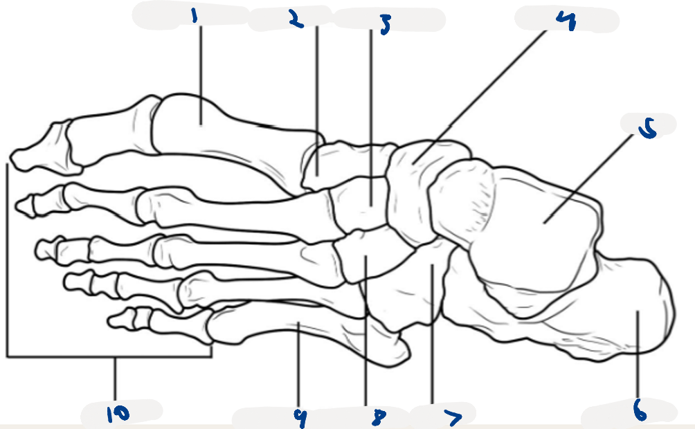
LABEL the foot
1st metatarsal
medial cuneiform
middle cuneiform
navicular
talus
calcaneous
cuboid…
lateral cuneiform
5th metatarsal
phalanges
What movements do ankle joints allow
plantarflexion
dorsiflexion
inversion
eversion
the 2 joints of the ankle
talocrural joint
subtalor joint
what does the talocural joint allow
flexion & extensions
what bones are in the talocural joint
talus, tibia & fibula
what does the subtalor joint allow
inversion and eversion
what bones are the subtalor joint
calcaneous, cuboid, navicular

Label anterior leg muscles
extensor hallucis longus
extensor digitorum longus
tibialis anterior
what’s the tibialis anterior control
dorsiflexion & inversion w a friend :3
what’s the extensor digitorum longus control
toe extension
what’s the extension hallucis longus control
big toe extension
what innervates the anterior leg muscles
deep peroneal nerve
what blood vessels run through anterior leg muscles
tibial artery
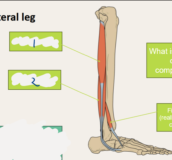
LABEL lateral leg
Fibularis longus
fibularis brevis
what actions do the lateral leg muscles control
eversion of the foot
what innervates the lateral leg
superficial fibular nerve
what blood vessels supply the lateral leg
anterior tibial and fibular artery
what are the 4 facial compartments of the leg
anterior, lateral, superficial posterior and deep posterior
what can the leg compartments suffer from
compartment syndrome

Label superficial posterior leg muscles
gastrocnemius
soleus
plantaris
where does the gastrocnemius proximally attach to
the posterior bit of femoral condyle
what does the gastrocnemius allow
knee flexion
what’s the solus attach to and where is it relative to the gastrocnemius
only tibia
it’s deep. (under it)
what do the gastrocnemius and soleus form distally? what’s attach to
achillies tendon, attaching to the calcaneus
what’s the achilles tendon control
plantarflexion
what can’t you do if the achillies is ruptured
loss of plantarflexion ofc
can’t stand on tiptoes
when’s achillies often tear
weekend athletes
what’s fibre type
how quickly the muscles contract & generate energy
gastrocnemius vs soleus on fibre type
fast twitch, slow-twitch
function of gastrocnemius vs soleus
power (running, jumping)
posture- standing
Plantaris is vestigial. what’s that mean
lost its functionality over evolution. so some have it and others don’t
what can plantaris be used for
grafts
what’s grafting
when a tissue’s taken out and moved to another part of the body to heal, grow or repair something
often used in achillies, and other tendons

LABEL deep posterior leg muscles
Flexor hallucis longus
flexor digitorum longus
tibialis posterior
what action does tibialis posterior allow
inversion
what actions do the flexor digitorum longus do
flexion of the toes
what action does the flexus hallucis longus do
flexion of the big toe
What’s the popliteal fossa
a space behind the knee joint containing popliteal vessels and sciatic nerve as it divides into fibular/peroneal and tibial branches
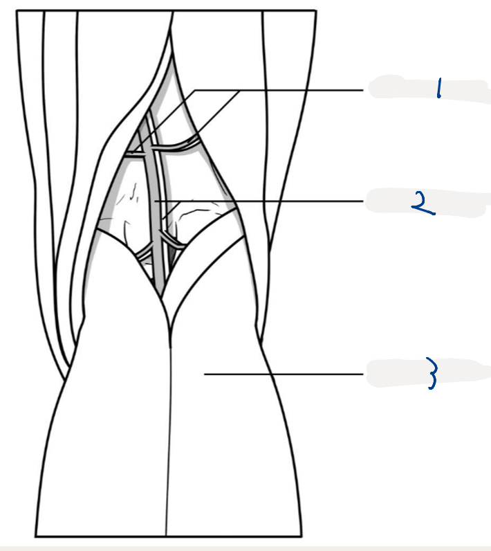
LABEL. What superiorly borders the fossa
genicular vessels
popliteal vessels
gastrocnemius
hamstring muscles
how the popliteal fossa can be used
you can find pulse on the popliteal artery