Exam 3 Study Guide for Nervous Tissue & Brain Nerves
 Call Kai
 Learn
Learn Practice Test
 Spaced Repetition
 Match
Match Flashcards
 Knowt Play
1/158

There's no tags or description
Looks like no tags are added yet.
Name | Mastery | Learn | Test | Matching | Spaced | Call with Kai | Chat |
|---|
No analytics yet
Send a link to your students to track their progress
159 Terms
NERVOUS TISSUE
What is the NERVOUS SYSTEM?
The body’s communication and control system

What are the main functions of the Nervous System?
Directs responses to stimuli and coordinates tissues and organs so they work together, including:
🏃 Movement
⚖ Balance
🤹 Coordination
💡 Learning
💭 Memory
❤ Feelings
😊😢 Emotions
👁👂👃👅✋ Senses
🌙💤 Sleep

What organs belong to the Nervous System?
Brain
Spinal Cord
Nerves

What is your Central Nervous System?
Brain
Spinal cord
Nucleus – a cluster of cell bodies
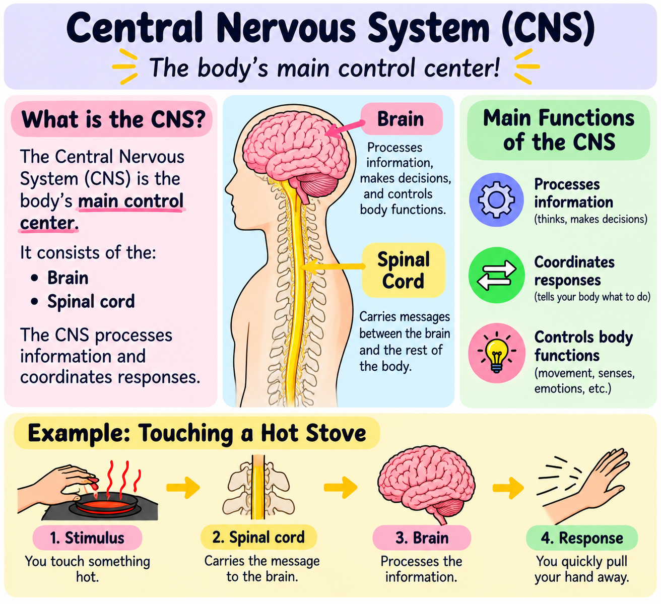
🔑Key idea: CNS = Brain + Spinal Cord = Processes information & coordinates responses.
CNS = CONTROL CENTER
C = Central
N = Nervous
S = System
Think: 🧠 BRAIN + 🦴 SPINAL CORD = CNS
What is your Peripheral Nervous System?
Nerves
Ganglion – a cluster of cell bodies

🔑Key idea:
📥 PNS (Nerves/Ganglion) carries sensory information TO the CNS
📤 PNS (Nerves/Ganglion)carries motor commands FROM the CNS
⭐ Easy memory:
PNS = OUTSIDE the brain & spinal cord
Think:
🌳PNS = Nerves + Ganglia branching OUTSIDE the CNS

Neuron

The CELL
🔑Key idea: The whole nerve cell = NEURON.
⭐ Easy memory:
NEURON = the whole nerve cell that receives and sends messages.
Think: 📥 Receive → ⚡ Respond → 📤 Send

Neurotransmitter

The CHEMICAL
🔑Key idea: Neurotransmitters are the CHEMICALS that carry the message.
⭐ Easy memory:
Neurotransmitters = chemical messengers 📩
Think: Neuron → releases CHEMICALS → chemicals carry the MESSAGE → next cell

Synapse

The Junction
🔑Key Idea: Synape is the small space where a neuron transmits a signal to another cell.
⭐ Easy memory:
SYNAPSE = SPACE between cells where the signal passes.
Think: Neuron → Synapse → Next Cell

Presynaptic Cell

🔑Key Idea: The presynaptic cell is the sending cell that releases neurotransmitters to communicate with another cell.
⭐ Easy memory:
PRE = BEFORE + SENDS 📤
Think: Presynaptic cell → releases neurotransmitters → synapse → next cell

Postsynaptic Cell

🔑Key Idea: The postsynaptic cell is the receiving cell that receives the signal from the presynaptic cell through neurotransmitters, received from the Dentrites.
⭐ Easy memory:
POST = AFTER + RECEIVES 📥
Think: Presynaptic = SENDS 📤 → Synapse → Postsynaptic = RECEIVES 📥
What makes Neurons different from other cells?
High Rate of Metabolism 20-30%
Excitable Cells that send and receive electrical signals
Long-Lasting Cells that do not divide
Use Neurotransmitters to communicate with other cells


Be able to Identify/Locate all of structures on a Neuron, and know what they do!
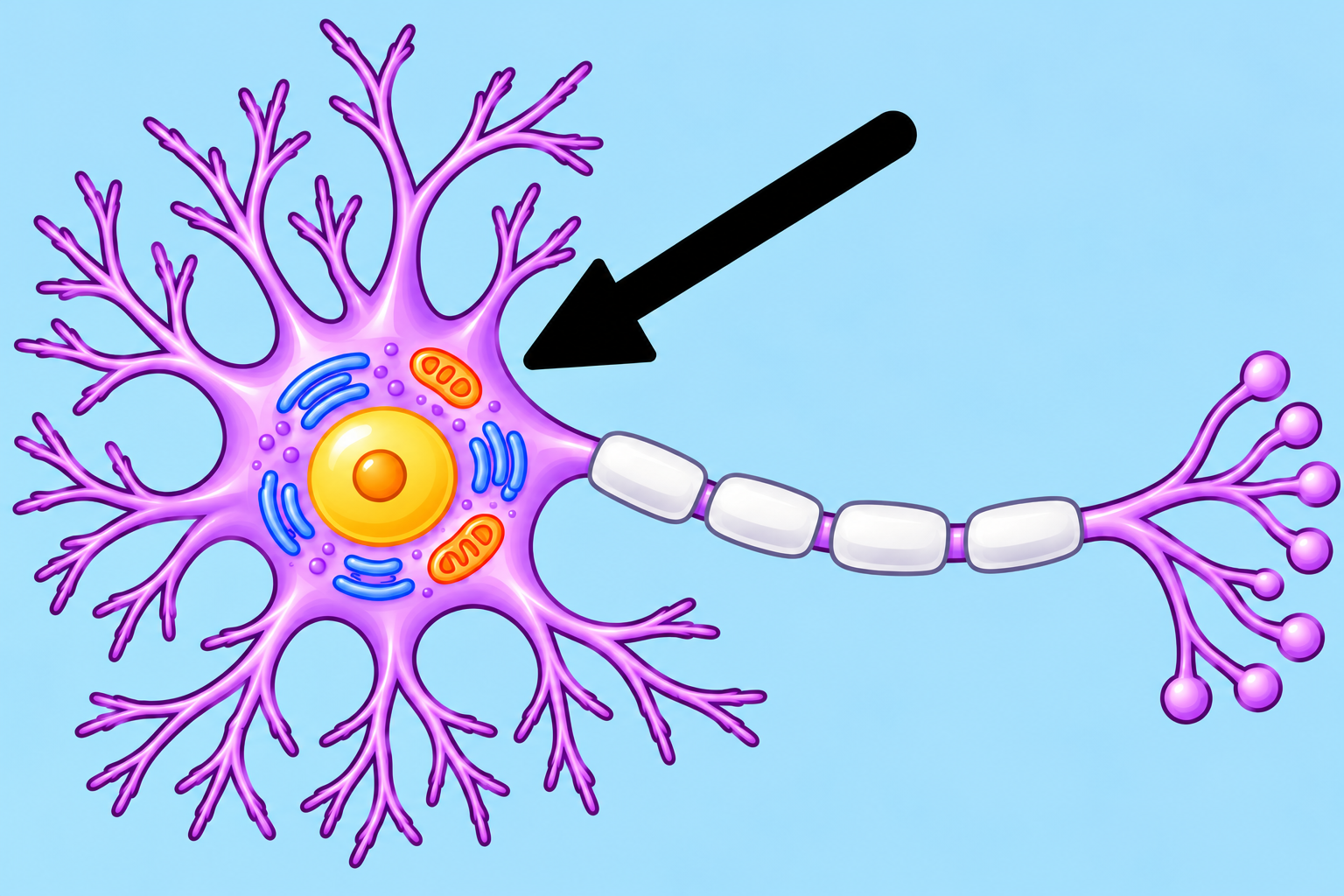
Cell Body
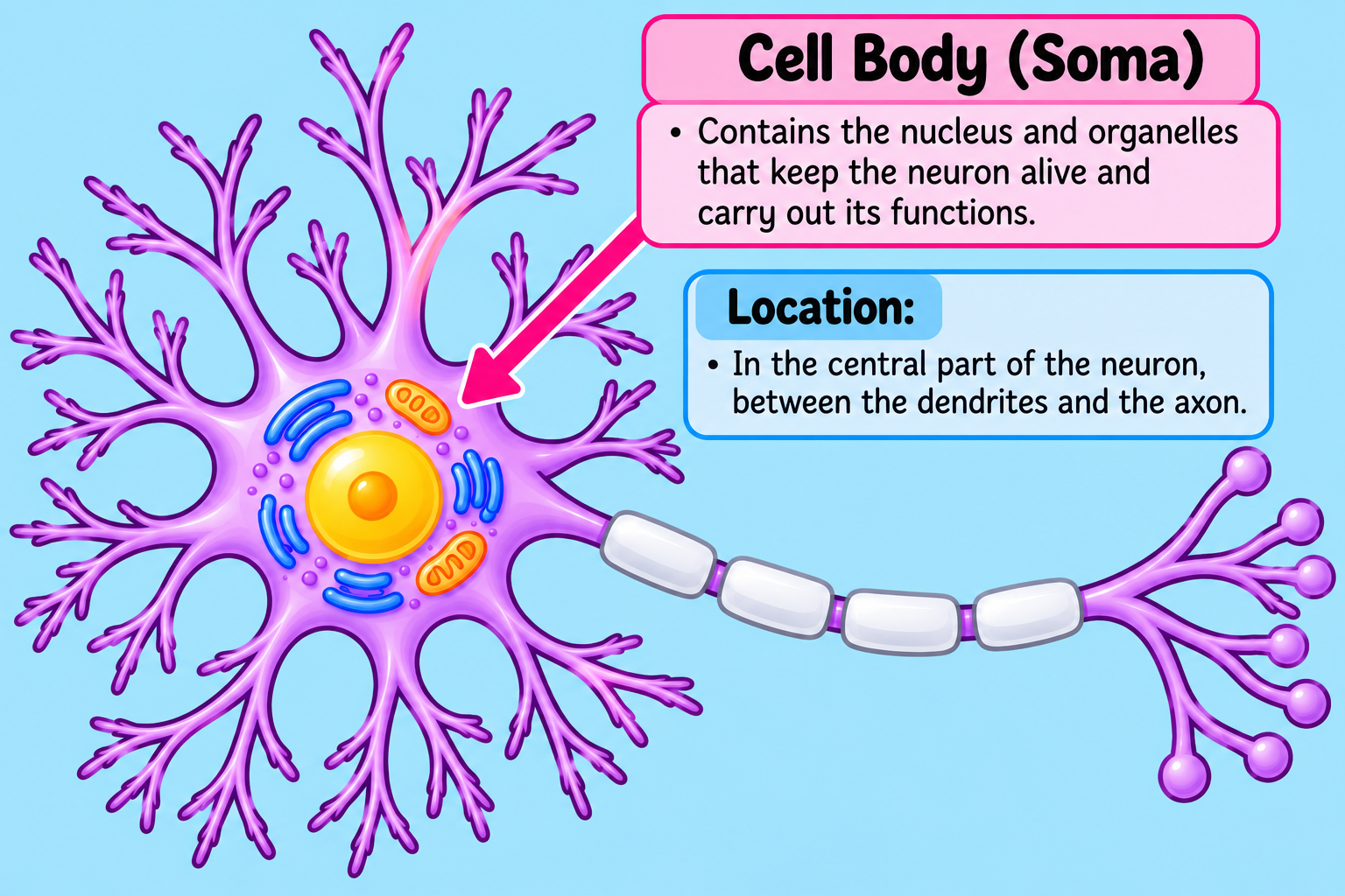
⭐ Memory trick:
Cell BODY = the main BODY of the neuron.
Think: Dendrites 🌳 → CELL BODY 🟣 → Axon ➡

Dendrites

⭐ Easy memory trick:
Dendrites = Detect & Receive 📥
Think: Tree branches 🌳 that catch incoming messages.

Axon

🔑 Key Idea: AXON = Carries electrical signals AWAY from the cell body.

Axon Terminal

🔑 Key Idea: Axon terminals are at the end🛑 of the axon and RELEASE neurotransmitters. 📤
⭐ Easy memory:
TERMINAL = END 🛑
Axon Terminal = END of the axon.

Myelin Sheath
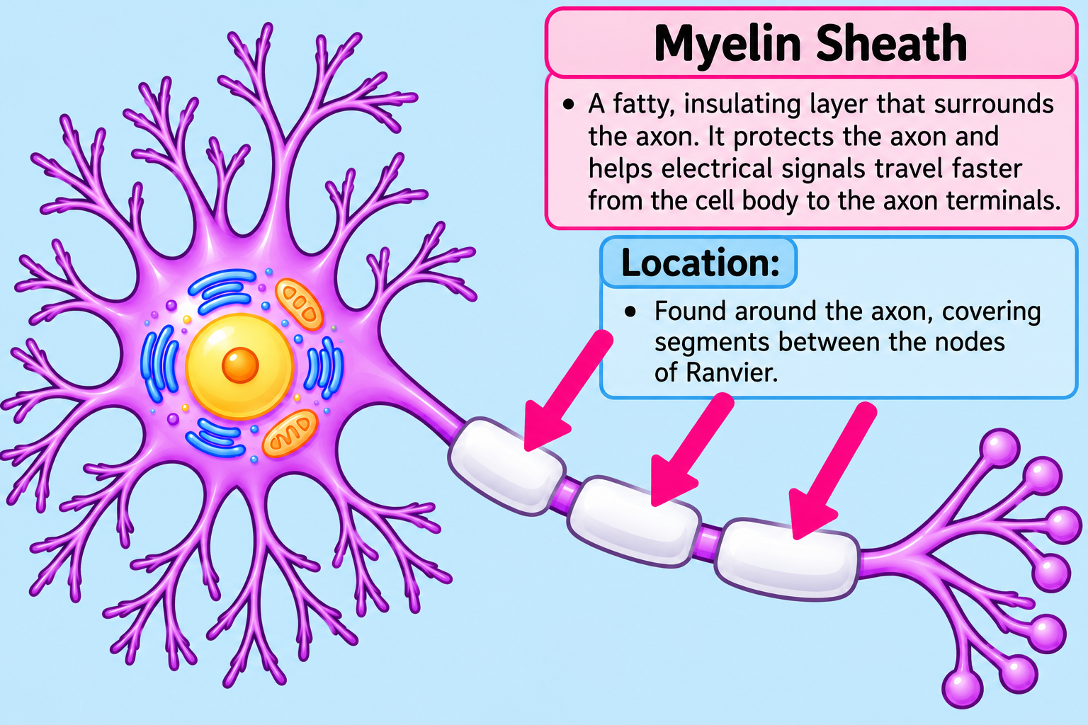
🔑 Key Idea: Myelin Sheath = wraps around the AXON → helps signals travel FASTER. ⚡➡
⭐ Easy memory:
MYELIN = insulation around a wire 🛡⚡
Think: Just like insulation helps protect a wire, myelin wraps around the axon and helps the signal travel faster.

Node of Ranvier
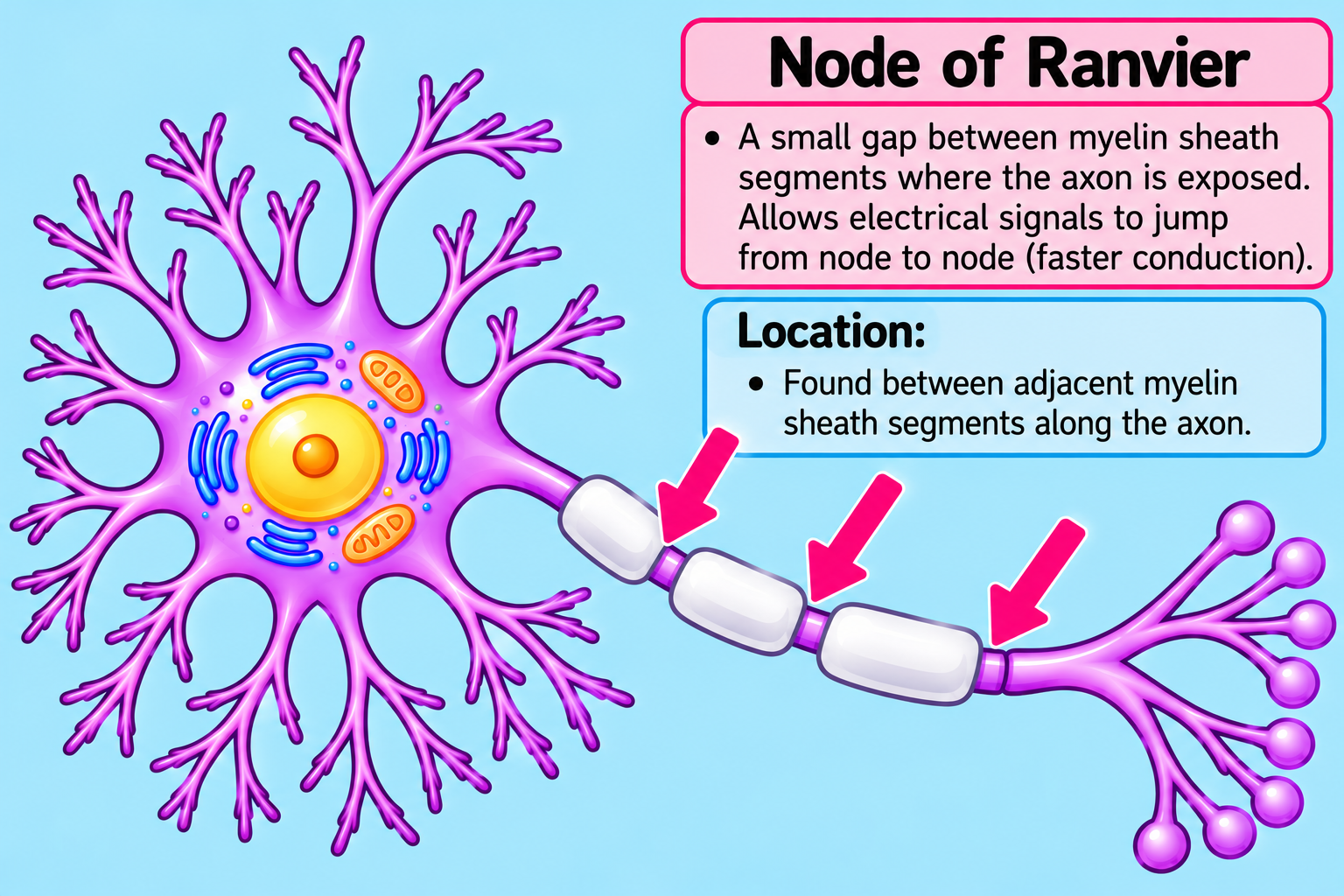
🔑 Key Idea: Node of Ranvier = GAP between myelin sheath pieces.
⭐ Easy memory:
NODE = GAP ↔
Think: Look for the tiny spaces between the yellow myelin pieces.
What parts of the Neuron are in the Grey Matter?
Glial Cells
Neuron Cell Bodies
Dendrites

🔑 Key Idea: Grey Matter = Glial Cells + Cell Bodies + Dendrites.
🧩 G = Glial cells
🟣 B = Cell Bodies
🌳 D = Dendrites Easy Memory:
Grey Matter = G + B + D
What parts of the Neuron are in the White Matter?
Axons
Myelin Sheath

White Matter Contains:
Axons and Myelin Sheath
Why is the White Matter colored White?
Myelin is lipid-Rich
Lipid is Fat
Fat is White colored


Know those Neurons and their Functions!
Sensory Neuron
Converts a stimulus (touch, pain, temperature, or pressure) into an electrical signal

Key Idea: Sensory Neuron = Stimulus → Electrical Signal → PNS TO CNS 📥🧠
Interneuron
Processes, stores, and retrieves information
Stays completely within the central nervous system
Responsible for quick reflexes

🔑 Key Idea: Interneuron = processes information + quick reflexes + stays completely in the CNS.
Think: INTER = INSIDE 🧠, Interneurons stay INSIDE the CNS.
And for the function:
🧠 Process • Store • Retrieve
⚡ Quick Reflexes
🏠 Stay in CNS
Motor Neuron
Carries messages from the CNS to muscles and glands, telling your muscles to move

Key Idea: Motor Neuron = FROM CNS→muscles & glands→tells muscles to move
🧠 CNS → ⚡ Motor Neuron → 💪 Muscles / Glands
Think: MOTOR = MOVEMENT 🚗💨, a motor makes something MOVE, a motor neuron tells your muscles to MOVE.
Motor = messages move OUT of the CNS 📤
🧠 CNS → 📤 OUT → 💪 Muscle moves
Glial Cells
Cells that support, protect, nourish, and maintain neurons in the nervous system

🔑 Key Idea: Glial Cells = HELP neurons→Provide+Support+Protect+Housekeep
🛠 Function: Glial Cells = Provide, Support, Protect, and Housekeeper of neurons.
📍 Location:
Glial cells are found in both:
🧠 CNS — brain & spinal cord
🌐 PNS — nerves outside the brain & spinal cord
Think of glial cells as the “HELPERS” of neurons. 🧹🛡

Oligodendrocytes
Ependymal Cells
Microglia
Astrocytes
Oligodendrocytes
Covers axons with myelin in the CNS

🔑 Key Idea: Oligodendrocytes = CNS → cover axons with myelin.
📍 Location: CNS only
⚙ Function: Covers axons with myelin.
Think: OLIGO = CNS myelin wrapper 🧠🧣
🧠 CNS → Oligodendrocyte → 🟡 Myelin around axons
Ependymal Cells
Produces cerebrospinal fluid in the CNS

🔑 Key Idea: Ependymal Cells = CNS → produce cerebrospinal fluid (CSF).
📍 Location: CNS — brain and spinal cord
⚙ Function: Produces cerebrospinal fluid (CSF).
Think: EPENDYMAL=makes the brain’s FLUID💧🧠; 💜 Ependymal Cells→💧 CSF
For your test, connect these two facts:
CNS 🧠 + CSF 💧 = Ependymal Cells
Microglia
Phagocytizes (Eats & Cleans) dead tissue and microorganisms in the CNS

🔑 Key Idea: Microglia are the CNS cleanup crew—they “eat” dead tissue and microorganisms.
📍 Location: CNS — brain and spinal cord
⚙ Function: Phagocytizes dead tissue and microorganisms.
Think: MICROglia = MICRObe cleaners 🦠🧹
Microglia are like the cleanup crew of the CNS. They eat/engulf
Astrocytes
Maintains the blood-brain barrier and chemical environment in the CNS

⭐ Protect → blood-brain barrier
⭐ Balance → chemical environment
⭐ Live in → CNS
🔑 Key Idea: Astrocytes = CNS → maintain the blood-brain barrier + chemical environment.
For your test, memorize this exact connection:
ASTROCYTES → CNS 🧠 → PROTECT 🛡 + BALANCE ⚖

Know the Glial Cells of the Peripheral Nervous System
Schwann cells
Covers the axons with myelin in the PNS

Think: SCHWANN = WRAP 🧻
Imagine a Schwann cell wrapping a blanket around an axon:
🟡 Axon → 🧻 Schwann cell WRAPS it → 🛡 Myelin covering
🔑 Key Idea: Schwann Cells = PNS + Myelin
Satellite cells
Supports and maintains the chemical environment in the PNS

🔑 Key Idea: Satellite Cells=PNS→SUPPORT+maintain the chemical environment
Satellite cells SURROUND & SUPPORT neurons. 🤝
They help keep the chemical environment stable ⚖ so neurons can function properly.
For your test, memorize:
🛰 SATELLITE = SURROUND & SUPPORT

Know the functions of these players in the CNS Circulation and what fluid flows through it, if applicable.
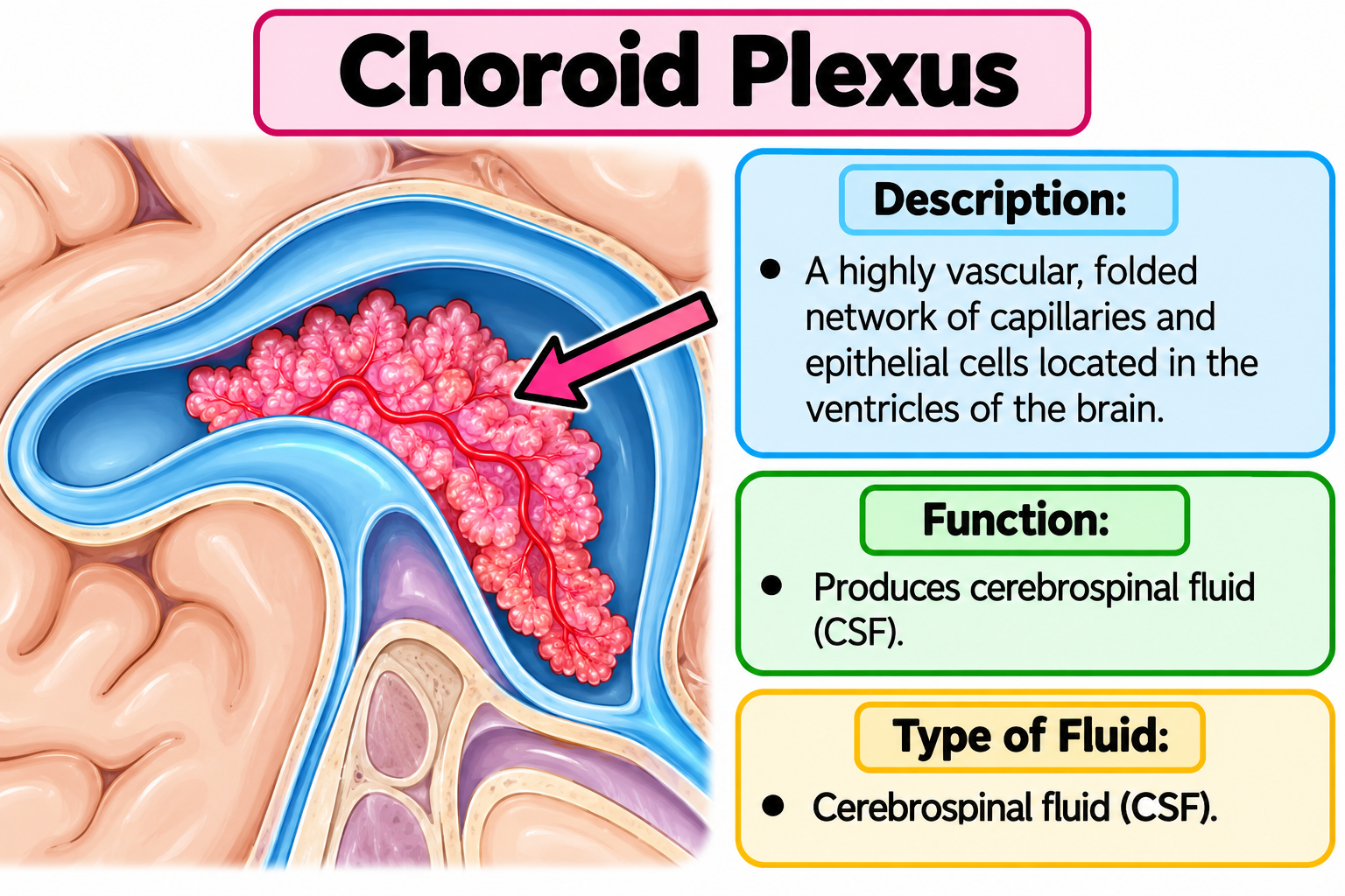
Choroid plexus
A network of blood vessels and specialized cells inside the brain's ventricles that produces cerebrospinal fluid (CSF).
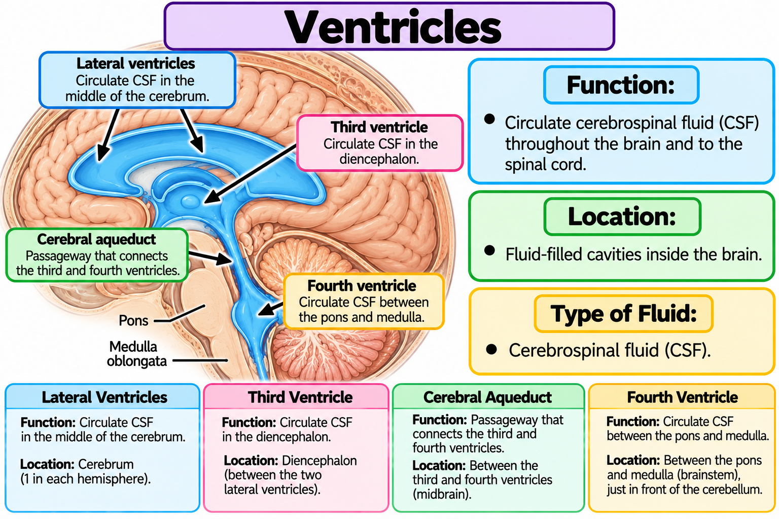
Ventricles
Fluid-filled cavities inside the brain that contain and circulate cerebrospinal fluid (CSF).
Cerebrospinal Fluid
Clear, colorless liquid, that cushions and removes metabolic waste from within the cranial cavity

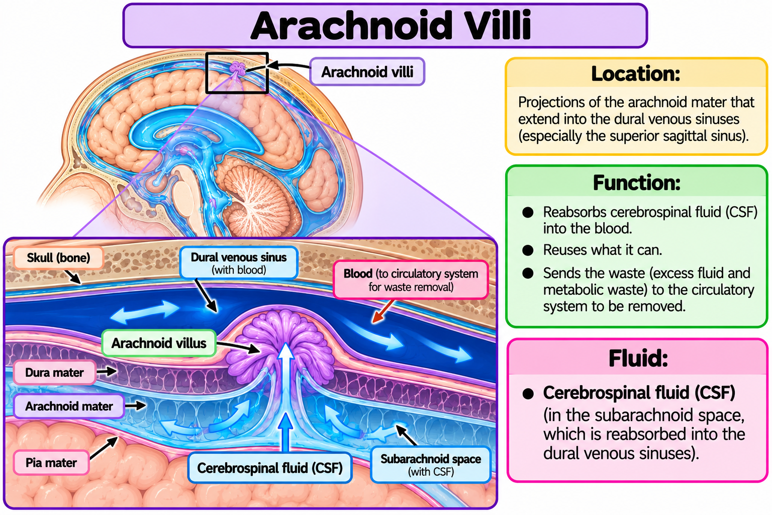
Arachnoid Villi
Reabsorbs cerebrospinal fluid, reuses what it can, sends the waste to the circulatory system to be removed
Dural Venous Sinuses
Spaces between the two dura mater layers, that drains blood from the brain and back to the circulatory system

Brain and Nerves

The 4 Main Regions of the Brain
Cerebrum: Controls thinking, memory, emotions, senses, and voluntary movements.
Diencephalon: Helps regulate body temperature, hunger, thirst, sleep, and sensory information. Includes the thalamus and hypothalamus.
Brainstem: Controls vital automatic functions like breathing, heart rate, and blood pressure. Includes the midbrain, pons, and medulla oblongata.
Cerebellum: Controls balance, posture, coordination, and smooth muscle movements.
Medulla Oblongata
Controls automatic, life-sustaining functions that you don't have to think about:
Breathing
Heart rate
Blood pressure
Swallowing
Reflexes
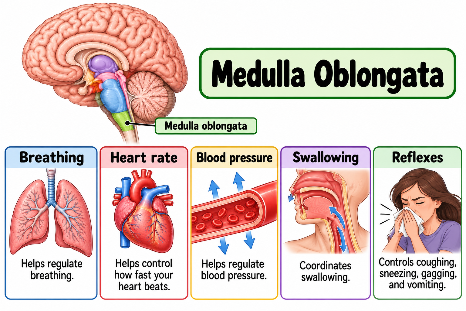
Breathing: regulates breathing
Heart rate: Controls how fast your heart beats
Blood pressure: Regulates blood pressure
Swallowing: Coordinates swallowing
Reflexes: Controls coughing, sneezing, gagging, and vomiting
Medulla Oblongata
Pons
Control automatic body functions:
Involuntary Actions
Respiration
Bladder Control

Involuntary Actions: Helps control actions your body does automatically, without thinking.
Respiration: Helps regulate your breathing rhythm.
Bladder Control: Helps coordinate urination (peeing).
Pons
Superior Colliculi
Help control eye movements and visual attention
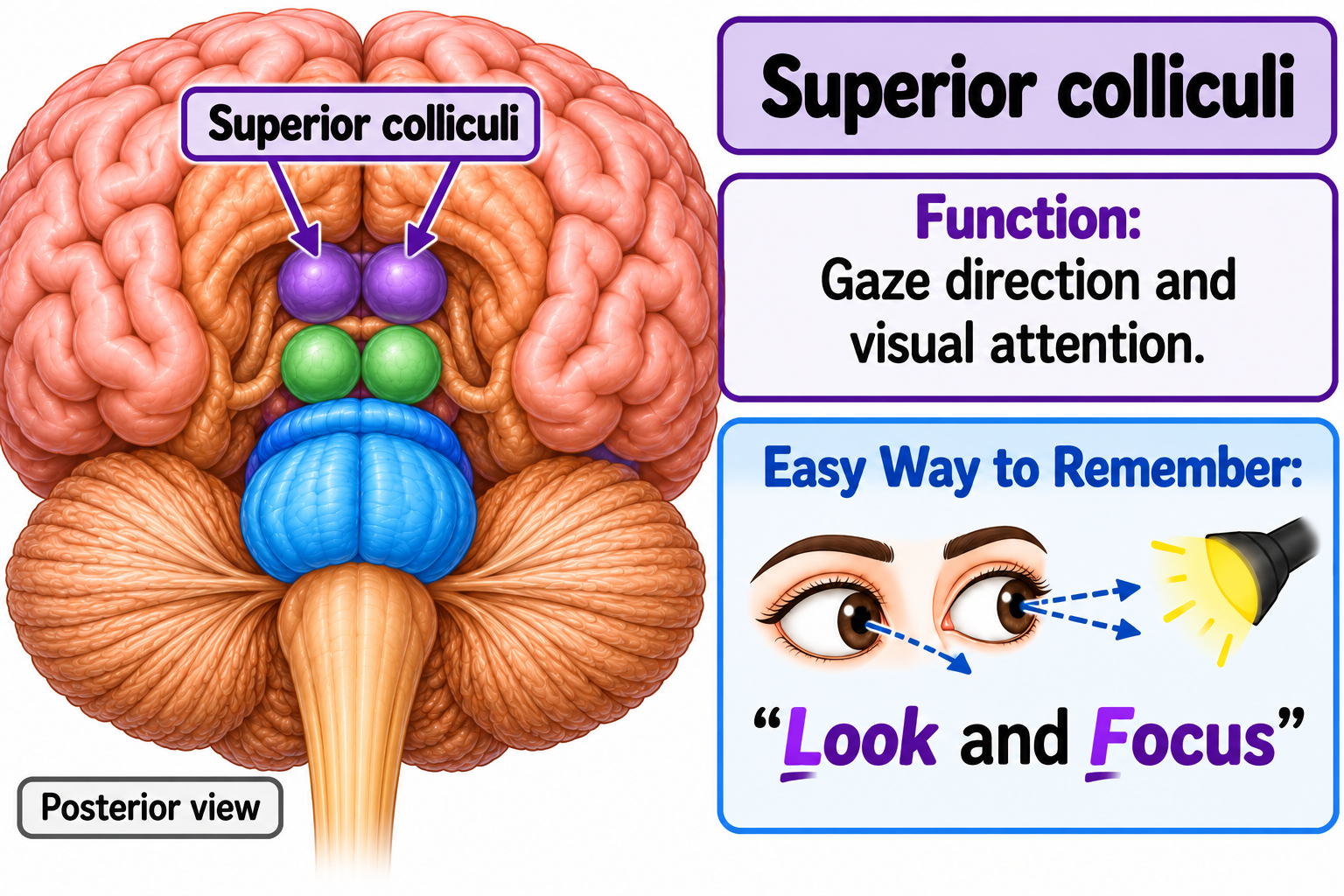
2 Main Functions to Memorize
Gaze Direction – Helps move your eyes toward something you want to look at.
Visual Attention – Helps direct your attention toward something you see.
Superior Colliculi
Inferior Colliculi
Determines the location of a sound and startle responses

Inferior Colliculi
Cerebellum
Inferior olive: compares information from the cerebrum with feedback from the spinal cord
Balance, coordination, smooth movements
Also responsible for motion sickness

Cerebellum
Thalamus
Filters all the sensory signals from the spinal cord - the important ones get passed up to the cerebrum, the unimportant ones do not

Thalamus
Hypothalamus
Maintains homeostasis
Regulates the anterior pituitary (to produce hormones)
Regulates the autonomic nervous system
Food and water intake, thermoregulation, sleep and circadian rhythms

Hypothalamus
Corpus Callosum
White matter, connects left and right hemispheres

Corpus Callosum
Frontal Lobe
Responsible for:
Thinking
Moving
Tasting

Frontal Lobe
Primary Motor Cortex
Initiates all voluntary body movements

Primary Motor Cortex
Parietal lobe
Controls General Senses
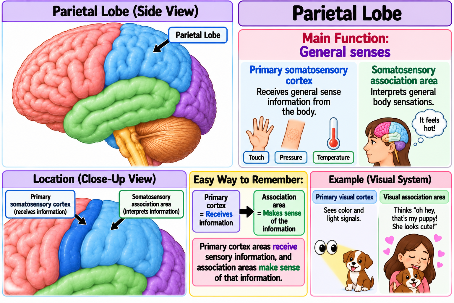
Parietal lobe
Occipital Lobe
Controls Vision