Anatomy I: Skull Development
 Call Kai
 Learn
 Practice Test
 Spaced Repetition
 Match
 Flashcards
 Knowt Play
1/92

There's no tags or description
Looks like no tags are added yet.
93 Terms
Development
What is a physiological phenomenon and qualitative in nature?
Growth
What is an anatomic phenomenon and quantitative in nature?
Primary cartilage
What is the type of cartilage that is present in adults and forms most of the temporary skeleton in the embryo?
Secondary cartilage
What is the type of cartilage present in certain joints that changes directly into bone?
Bone remodeling
What is the reshaping of the outline of bone by selective resorption in some areas and deposition in others?
Bone relocation
What is the relative movement in space of a body structure, due to bone deposition on one side and resorption on the other side?
Primary displacement
What is the type of bone displacement that occurs in conjunction with the bone's own growth?
Secondary displacement
What type of bone displacement is caused by enlargement of adjacent or remote bone or soft tissues, but not of the bone itself?
neurocranium
What part of the skull forms a protective case around the brain and is divided into two portions?
1. Flat bones of the vault
2. Bones of the base of the skull
What are the two sections of the neurocranium
Viscerocranium
What part of the skull forms the skeleton of the face?
Blue. Blue means collagen and the flat bones of the skull use intramembraneous ossification for bone formation
What part of this histological section would give rise to bone?

4th week
At what point of development does the condensation of mesenchymal cells that surround the developing brain contain osteoprogenitor cells?
5th week
When do the bones of the skull develop from the ectomeninx?
pia mater and the arachnoid mater
What does the endomeninx contribute to?
inner dura mater and has an outer superficial layer membrane (periosteal or endosteal layer) with osteogenic and chondrogenic properties
What does the ectomeninx contribute to?
5th week
When does the meninx primitive form?
1. Ectomeninx
2. Endomeninx
What are the two layers of the meninx primitive?
Red: Pericarnium
Orange: Calvaria
Green: Dura mater
Purple: Subdural space
Pink: Arachnoid
Yellow: Subaracnoid space
Blue: Pia mater
What are the red, orange, green, purple, pink, yellow, and blue highlights labeling?

Paraxial mesoderm = somitomeres and somites
What is the embryological origin of the parietal bone, occipital bone, petrous part of temporal bone, and the hyoid bone?
lateral plate mesoderm
What is the embryological origin of the laryngeals?
neural crest
What is the embryological origin of the frontal bone, sphenoid bone, squamous temporal, and nasal bone?
neural crest
What is the embryological origin of the lacrimal bone, zygomatic bone, maxilla, and mandible?
intramembranous ossification
What type of ossification does the frontal and parietal bone use?
intramembranous ossification
What type of ossification does the lacrimal and nasal bones use?
intramembranous ossification
What type of ossification do the zygomatic bone, maxilla, and mandible use?
endochondral ossification
What type of ossification do the ethmoid, auditory ossicles, and occipital bone use?
endochondral ossification
What type of ossification do the hyoid bone and thyroid cartilage use?
endochondral ossification
What type of ossification does the condyle and cornoid process of the TMJ use?
greater wing of the sphenoid
What bone do we still not have the ossification pattern for?
1. Sutural growth
2. Remodeling
3. Centrifugal displacement by the expanding brain
What are the 3 processes that the postnatal growth of calvarial bones uses?
As the brain grows, the bones move outward due to expanding meninges. The outer meninges are osteogenic and produce the inner surface of each flat bone.
What drives sutural growth in the skull?
When bones are drawn apart by growth-related displacement, the osteogenic sutural membranes form new membranous bone equal to the separation, enlarging each bone's circumference and keeping constant contact at the sutures.
How do sutures maintain proper bone size and contact during growth?
Centrifugal displacement by the expanding brain
What type of calvarial growth is primary displacement that occurs in conjunction with the bone's growth and secondary displacement that is caused by enlargement of adjacent or remote bones or soft tissues, but not of the bone itself.
Primary
What type of displacement occurs in conjunction with the bone's growth?
Secondary
What type of displacement is caused by enlargement of adjacent or remote bones or soft tissues, but not of the bone itself?
connective tissue
What do the bones of the calvaria develop suspended in?
True
T/F. The growing brain does not push the bones outward.
The brain grows, which causes the meninges to grow, and the meninges push on the bone
What is the cause of the displacement of bone with the growing brain?
sutures between cartilage
What is a synchondroses?
natural growth curve (growth rate is very rapid during the first few years of life, but slows down dramtically by the age of 6-7
What kind of growth curve do the calvarial sutures follow?
Deposition: on the echo and endo cranial sides
When the brain is expanded, where is the bone deposition of the calvaria occurring?
inner and outer lumen sides
When the brain is expanded, where is the bone resorption of the calvaria occurring?
fontanelles
What part of the brain is crucial in enabling the head to fit through the birth canal?
Presence of normally functioning sutures
What does the displacement of bones when we grow depend on?
The brain could not fully develop
What happens if we were to fuse sutures too early?
frontal suture
What is the first suture to close?
lamdboid suture
What is the last suture to close?
childhood
When does the frontal suture fuse?
20-30
When does the sagittal suture fuse?
30-40
When does the coronal suture fuse?
40-50
When does the lambdoid suture fuse?
mesenchymal tissue, and is where osteoprogenitor cells can be found
Before sutures ossify, what is it made of?
endochondrial ossification
What type of ossification do the bones of the base of the skull use?
Red: Replacement bone
Orange: Hypertrophic zone
Green: Proliferation zone
What are the red, orange, and green sticker covering?
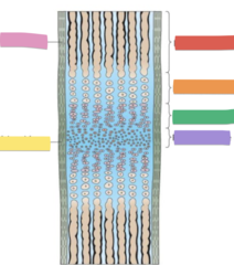
Purple: Progenitor zone
Pink: Periosteum
Yellow: Perichondrium
What are the purple, pink, and yellow sticker covering?
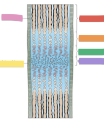
Zones of endochondrial ossification
What is the zones of a synchondrosis similar to?
Purple (progenitor zone)
Which sticker is similar to the resting zone of endochondrial ossification?

1. Progenitor zone has undifferentiated cells
2. Proliferation zones on either side of the progenitor zone where chondrocytes differentiate and divide
3. Hypertropic zone: As the cells divide, they form into columns and deposit cartilage matrix in between. The cells enlarge as they move further along these columns in the hypertropic zone until they degenerate, leaving spaces within the cartilage matrix
4. These spaces are invaded by osteoblasts that deposit bone, which is remolded into compact and cancellous bone.
What are the steps of bone formation in a synchondrosis?
Zone of endochondral ossification (like the growth plate): Has asymmetric, unidirectional growth. Cartilage proliferates on the epiphyseal side and is replaced by bone on the diaphyseal side, pushing the bone in one direction to lengthen it.
Synchondrosis (like the spheno-occipital synchondrosis): Has bilateral or bidirectional growth. Each side has growth zones that mirror each other, producing cartilage that is replaced by bone on both sides, allowing two bones to separate symmetrically as the cranial base expands.
What is the main difference between zone of endochondrial ossification and zones of a synchondrosis?
False. Growth rate is not always the same on both sides of a synchondrosis.
T/F. Growth rate is always the same on both sides of a synchondrosis.
It matches the growth rate, so the synchondrosis maintains a constant thickness.
Once joints are established in a synchondrosis, what does the rate of bone formation depend on?
Spheno-occipital synchondrosis
Which synchondrosis is a major contributor to postnatal growth and influences the overall facial skeletal pattern?
Fuse: 12-15 years old
Ossify: 20 years old
When does the Spheno-occipital synchondrosis fuse and ossify?
It displaces the face down and out
How does the growth of the Spheno-occipital synchondrosis influence the face?
Cranial base, facial skeleton, cranial vault
*The cranial base and facial skeleton are important for dentistry
What are the three major divisions of the skull that are important to measure?
1. Intramembranous ossification
2. Neural growth pattern
Which bone formation pattern does the cranial vault use and what is the growth pattern?
1. Intramembranous ossification
2. Somatic growth curve
Which bone formation pattern does the facial skeleton use, and what is the growth pattern?
1. Endochondral ossification
2. Neural growth curve for several years and then adjusts to the somatic growth pattern later
Which bone formation pattern does the cranial base use, and what is the growth pattern?
cranial vault
What part of the skull does not change much in size as we age?
facial skeleton
What part of the skull grows in size as we age?
Sex differences come from sex hormones and they fully established during adolescence
When are the sex differences in the facial apperance formed and by what?
Yellow: anterior fontanelle
Red: sphenoid fontanelle
Pink: Posterior fontanelle
Blue: Mastoid fontanelle
What are the yellow, red, pink, and blue stickers covering?

Intramembraneous osteogenesis
After birth, what closes the open spaces of the fontanelles?
Phase 1: Newborn-5
What postnatal skull development phase has intensive growth mainly of the posterior part of the skull?
Phase 2: 7 to puberty
What postnatal skull development phase is the phase of relative rest?
Phase 3: 13-16 to 20-23
The postnatal skull development phase has intensive growth mainly in the anterior (facial) part of the skull?
Week 6 to Week 16
When does the maxilla develop?
1. Displacement
2. Sutural growth
3. Surface remoldeling
What are the 3 mechanisms we use for growth in the maxilla postnatally?
Maxillary sinus growth
What is responsible for the displacement of the maxillary anterior and inferiorly?
Functional Matrix concept: The soft tissues continue to grow, the bones become passively carried along (displaced) with the soft tissues attached to the bones by Sharpey fibers
What is the "theory/concept" of why the maxilla is displaced?
The cranial base, particulary the area surrounding the maxilla, actively grows, displacing the maxilla downward and forward
How does the cranial base growth displace the maxilla?
This refers to the displacement caused by the maxilla's own enlargement, particularly at the maxillary tuberosity (a bony prominence)
How does the primary displacement displace the maxilla?
This refers to displacement caused by the growth and enlargement of adjacent bones, like the mandible
How does the secondary displacement displace the maxilla?
The downward and forward movement of the maxilla creates tension at the sutures, which stimulates bone growth and remodeling at those sites
How does the tension and stimuli of maxilla displacement affect growth?
The position and function of the tongue and mandible also play a role in maxillary development, potentially influencing its shape and growth patterns
How do soft tissue factors displace the maxilla?
Purple: tooth germ
Blue: Merkel's cartilage
Pink: Bone of the mandible
*Merkels cartilage tells us mandible
What are the purple, blue, and pink stickers labeling, and which jaw is this histological section taken from?

Begins week 5 and ends week 14
When does the prenatal growth of the mandible occur?
It is transitory because it helps guide the bone and then dies by apoptosis. The cartilage never forms bone
What is the function of Merckel's cartilage?
Development starts week 5 and ends week 16. Continues to grow until 20 years old because it has a growth plate
What is the condylar development timeline?
Early bell stage (Has to develop at the same time the teeth form for the attachment of tooth to bone)
When does the development of the alveolar process begin?
The dental artery enters the tooth as the growth of the alveolar process because the growth of bone needs to leave space for the nerves and arteries
When does the dental artery enter the tooth?
lost teeth = lost muscle = lost bone *function is related to maintainace
- the bone reverts in some measure to the infantile condition as its height is diminished
What happens to the bone if we lose teeth?