giglio- normal abdomen GI tract
 Call Kai
 Learn
 Practice Test
 Spaced Repetition
 Match
 Flashcards
 Knowt Play
1/53

Earn XP
Description and Tags
up through slide 37, not including cecum and equine content
54 Terms
what are indications for an abdominal radiographs?
patient history (GI signs, urinary signs, repro evaluation, weight loss, trauma), physical exam findings (abdominal distention, organomegaly/palpable masses, body wall swelling/hernias)
how many projections are typically taken for abdominal radiographs? what is the difference in large dogs?
minimum of 3 orthogonal views (VD, right and left lateral); large dogs require cranial and caudal views in large dogs

describe how you would position the patient for a lateral view, what are the collimation landmarks?
pull pelvic limbs caudally, center slightly caudal to the last rib; collimation from the diaphragm to the greater trochanter and from the spine to the ventral body wall

describe how you would position the patient for a VD view, what are the collimation landmarks?
use v-trough, center on last rib; collimation from the diaphragm to hips and lateral borders of body wall
why do we typically choose VD over DV?
DV has higher chances of movement; more difficult to position well

describe the effect of right lateral recumbency on the stomach portions and the gas/fluid distribution in each
fundus is up/pylorus is down, gas in fundus/fluid in pylorus

describe the effect of left lateral recumbency on the stomach portions and the gas/fluid distribution in each
fundus is down/pylorus is up, fluid in fundus/gas in pylorus
what is the importance of getting both right and left laterals?
allows for redistribution of gas in the GI tract

these show the redistribution of gas in GI: which one is the DV and has gas in the fundus?
1

these show the redistribution of gas in GI: which one is the VD and has gas that moved to the cardia and pylorus?
2
what is a special abdominal projection that can be used to evaluate for bladder stones, mild uterine enlargement, or hiatal hernia?
abdominal compression

how does the abdominal compression projection work?
using a wooden spoon or a compression bandage you can displace the intesstine off the urinary bladder and get more organ separation

what is the name of this view and why is it used?
caudal abdominal view with legs pulled cranially (‘the butt shot’) used to visualize the urethra
what is the name of the special projection used for looking for free peritoneal gas?
horizontal beam
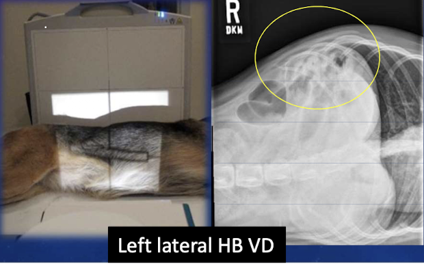
what are the different projections of the horizontal beam?
left lateral recumbency VD, dorsal recumbency right or left lateral
as a measure of quality control when we look at the x-ray penetration of organs. the degree of tissue penetration is a guide for ________ ________.
kilovolt peak (kVp)

which radiograph is underexposed?
1

which radiograph is overexposed?
2

what is the rule that ensure the entire abdomen is included?
from the diaphragm to the level of the coxofemoral joints
quality control: how can you determine if the abdomen is lateral?
superimposition of the rib heads and iliac wings; superimposition of lumbar spine and transverse processes (nike swoosh and horse heads)

what do you look for to determine if the abdomen is straight?
symmetric rib heads and iliac wings, dorsal spinous processes superimposed (tear drop shape), transverse processes symmatric

what abdominal structures are not normally seen?
pancreas, gallbladder, adrenals, lymph nodes, ureters, uterus, ovaries, retained testies, aorta, CVC
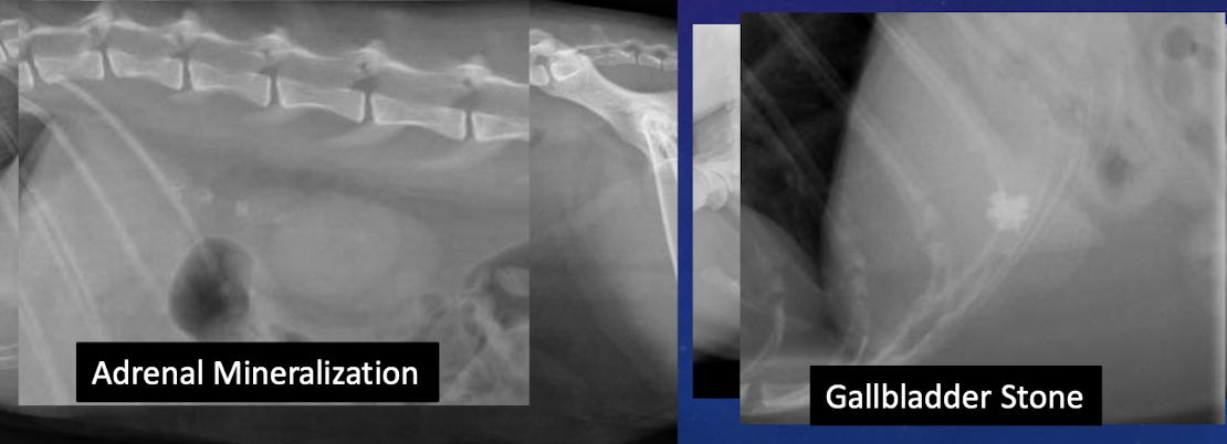
what case would we see the pancreas or aorta in cats?
very fat cats

label the abdominal divisions from A-D
right cranial, left cranial, central, caudal

label the abdominal divisions from A-E
craniodorsal, cranioventral, central, caudodorsal, caudoventral
when it comes to the abdominal divisions, what is the mass effect?
pushing organs out of the way as a mass grows within the abdominal cavity

which image (right or left) shows normal peritoneal and retroperitoneal detail?
left
do we assess peritoneal and retroperitoneal together or individually?
separate spaces assessed individually
what can cause decreased peritoneal and retroperitoneal detail?
abdominal fluid, decreased intra-abdominal fat, young animal (brown fat, small amount of fluid), large masses, carcinomatosis/sarcomatosis, peritonitis

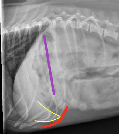
in lateral projection, how do you determine normal liver size?
parallel with gastric axis (purple), caudoventral margin contained behind costochondral arch (red), liver margins taper to a point and have sharp margins (yellow)
in a VD projection, how do you determine normal liver size?
gastric axis perpendicular to spine

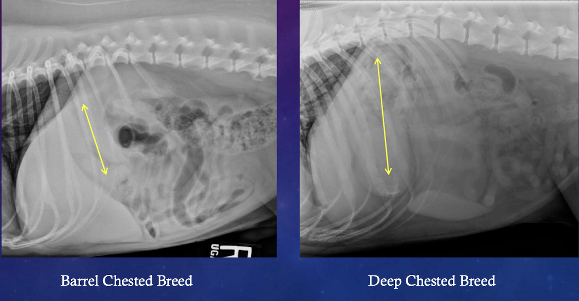
breed differences in gastric axis
deep chested breeds liver appears smaller
what is normal liver size in a puppy?
appears larger relative to the dog

describe the appearance of the gastric axis seen with microhepatia (small liver)
angled more cranially

describe the appearance of the gastric axis seen with hepatomegaly (large liver)
angled more caudally

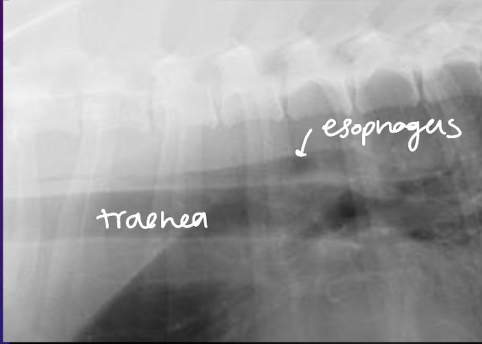
describe the normal appearance of the esophagus
small volume of luminal gas can be seen in left lateral view, can occasionally see caudal thoracic esophagus (fluid-filled)
where is the esophagus located relative to the trachea?
dorsal
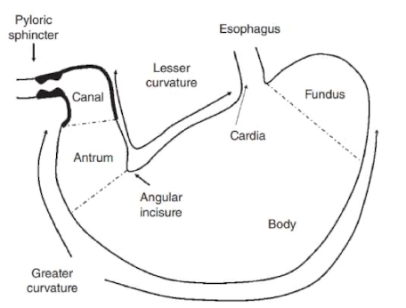
what are the parts of the stomach?
cardia, fundus, body, antrum and pylorus
describe the pylorus in the dog
pylorus right of midline, elongated

describe the pylorus in the cat
pylorus on the midline, “U” shaped

what impacts the appearance of the gastric anatomy?
content, volume, and patient position (when empty: parallel to the ribs/intercostal spaces)

which one (left or right) is the cat stomach?
right
why are there differences in the radiographic lateral projections?
air rises to different portions of the GI tract

what is the “abnormality” shown here?
pyloric pseudo mass- NORMAL fluid fillled pylorus seen on the right lateral projection (take left lateral projection and gas rises)

why do you have to be cautious when radiographically evaluating for wall thickness vs luminal content? how to solve this?
air/fluid interface (border effacement) results in false mucosal thickening; contrast radiology and ultrasonography

small intestinal anatomy: where is the duodenum?
right side to midline

small intestinal anatomy: where is the jejunem?
middle of the abdomen (differences in dog vs cat)

small intestinal anatomy: where is the ileum?
mid-right abdomen
between dogs and cats, which species typically contains more SI gas?
dogs contain 30-60% gas if fasted, cats contain a small amount of SI gas

what normal variations are seen in abdominal radiographs?
soft tissue mixed with small gas bubbles (normal ingesta), mineral/metal (cat litter, osseous fragments, medication)

what normal variation is shown in this cat?
string of pearls (jejunal appearance in cats)— secondary to peristalsis

what normal variation is shown in this dog?
duodenal pseudo-ulcers (peyer patches)— ONLY in duodenum
what is the normal cat SI diameter?
12 mm, serosa to serosa, little amounts of intraluminal gas
overall shape of large intestine
? or shepards crook shape
