Methods
 Call Kai
 Learn
 Practice Test
 Spaced Repetition
 Match
 Flashcards
 Knowt Play
1/22

There's no tags or description
Looks like no tags are added yet.
23 Terms

What kind of imaging is this?
fMRI

What kind of imaging is this?
(anatomical) MRI

What kind of imaging is this?
PET

What kind of imaging is this?
MEG
Methods to study brain function
Electrophysiological recordings
Functional imaging
Lesion studies
Electrical stimulation of the brain
The Wada Test
The Split Brain
Electrophysiological recordings
Electroencephalogram (EEG)
Cap with 20 electrodes put at precise locations
One electrode used behind the ears as a reference to check the potential (voltage), similarly to how an ECG uses the ankle
Average EEG looks different depending on the state of the brain (awake, drowsy, etc.)
Is used to see if one is struggling with one of the senses to see if it is a brain issue, and where that would be.
Great temporal resolution
Determined by the number of electrodes?
Types of waves in EEG
Delta waves
Up to 3 Hz
High amplitude waves
Seen in
adults during slow wave sleep
frontally
awake babies
posteriorly
May occur focally with subcortical lesions
Theta waves
4-7 Hz
Seen in (location unrelated to the task at hand)
Young children
Drowsy adults
During meditation
Alpha waves
8-12 Hz
Basic rhythm in posterior regions, higher amplitudes on dominant side
Found at central sites during rest
Strongly correlated with visual input
Emerges with eye closure and relaxation
Prominent indicator of a coma
Beta waves
12-30 Hz
Normal with low amplitude
Usually seen in symmetrical distribution and most prominent frontally
Associated with active thinking and concentration
Changes in beta waves indicates drug use
Lack of beta waves indicates cortical damage
Gamma waves
30-100 Hz
Thought to represent binding of different populations of neurons together into a network for carrying out a certain cognitive or motor function
Typical in cross-modal sensory processing
Decrease in gamma waves indicates cognitive decline
EEGs are described in terms of
Rhythmic activity
These are subdivided by frequency bands
Transient activity
Event related potentials (ERPs)
Event related potentials (ERPs)
Stimulus-correlated in the EEG
(Transient activity?)
Usually averages 9 lines into 1 line
(Picture):
Information about e.g. a click sound. Takes ~250ms to perceive the sound, and at around ~400ms the brain tries to recognize the sound
P3 becomes flatter as we habituate/adapt/get used to the sound
During adaptation, the entire signal becomes flatter

Epilepsy
Reoccurring abnormal brain activity
Can be traced in the brain to see where it originates
Very prominent changes suggest where the epilepsy may originate
Not heritable
May be caused by e.g. damage in the brain, drug use, lesions, brain tumours causing pressure —> neurons fire impulses.
EEG pros/cons
Great temporal resolution
Can spot it in real time (within ms)
Restricted by number of electrodes
May not see signals deep within the brain (microvolts are not always detectable through the skull) without drilling a hole in the skull to record deeper areas in the brain
Functional imaging methods
fMRI
functional Magnetic Resonance Imaging
MRI
anatomical Magnetic Resonance Imaging
Pick up ALL protons in the brain (not just in the blood) to get an anatomical imagine of the brain more accurate than x-rays
PET
Positron Emission Tomography
MEG
Magnetoencephalography
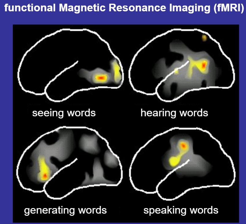
How does fMRI work?
Neural activity is correlated with blood flow
Differences in the blood flow is visualised by the big machine
Head is fixated to stay still
Machine applies alternating magnetic fields to test the hemoglobin
distinguishes it from oxygenated (diamagnetic) and deoxygenated (paramagnetic)
Diamagnetic blood is repulsed by the magnetic field
Paramagnetic blood is attracted by the magnetic field
Proton has a spin and acts as a compass needle
When the magnetic field turns on, the proton points north
When the magnetic field is turned off, the proton returns to its original position
This is recorded by the machine
As a result, we can reconstruct the regions of the brain in 3D
Shows us which part of the brain is active when exposed to different things
How does PET work?
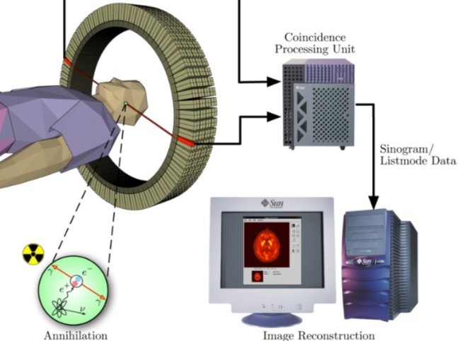
Blood flow is seen by a radioactive labelled glucose inserted in your blood
Accumulates in the area that the brain is active
Beta decay: Radioactive tracer emits positrons
When an electron and positron meet, they annihilate and produce 2 photons that fly away in opposite directions
If e.g. there is more blood flow on the left side of the brain, the left side of the machine receives the photon signal a little bit faster than the right side, and we can produce a picture of the brain
Half life of the isotope is 4 minutes, but don’t go around doing this often in your life (maybe once or twice at most)
How does MEG work?
Records tiny magnetic fields in the brain
With a “HUGE HAIRDRYER” / mask from hell
High temporal resolution
Neurons are basically cables and also produce magnetic fields
PROBLEMS WITH MEG:
Very low (microvolts) so produce very small magnetic fields
To pick up on these very small magnetic fields, the hospital must be very isolated form other magnetic fields (Faraday’s cage)
Range only 1-2 mm (cannot see deeper tissues)
Lesion studies
Right/Left hemisphere damage does different things
(E.g.)
strokes
meningioma
tumour of meninges
leads to loss of speech
tumours
leads to loss of touch, temperature etc
cysts
membrane-like filled with liquid due to uncontrolled growth, puts pressure on surrounding tissue
leads to issues with problem-solving, personality change etc
What is a stroke?
Death of nerves due to poor blood supply
Strokes can be
Ischemic strokes
Lack of blood flow due to clotted arteries
Hemorrhagic strokes
Lack of blood flow due to bursting of an artery (and thus due to bleeding)
Electrical stimulation of the brain
Instead of passively listening to the brain (EEG), we add electricity
TMS
Transcranial Magnetic Stimulation
Apparatus produces strong magnetic pulse
Can be directed at a certain brain area
Induces electrical activity in the neurons
e.g. a magnetic pulse in the visual centre causes a bright flash
How does electrical stimulation of the brain work?
Open the skull (about 1 cm) and place electrodes on the surface of the brain and apply tiny amounts of electricity and see how the patient responds
Patient is awake and aware during the procedure
Does not hurt as the brain lacks pain receptors
The Wada Test
Takes advantage of the fact that the right and left hemisphere have separate blood supplies
Inject anaesthesia into one brain half to put the hemisphere at sleep
Lasts 3-4 minutes before it wakes up again
E.g. gives the person a spoon in the left hand, the right hemisphere recognizes the touch of it but does not know the word as that is the left hemisphere’s job
When the patient is asked when the left hemisphere is awake again, they do not know the name of the object, but the left hand can point at the correct option when given choices.
The Split Brain
Cut the corpus callosum so the hemispheres cannot communicate
This process is called callosotomy
Historically done to alleviate life-threatening epilepsy that did not respond to medication
When seeing things with the right eye, the patient knows the word for the object (left hemisphere sees it and knows it)
When seeing things with the left eye, the patient does not see anything, but can pick it out with their right hand from different options
Corpus callosum
Bunch of neurons that cross information over between the hemispheres
Left brain / Right brain abilities
Left brain
Sequential
Analytical
Verbal
Logical
Grammar
Words
Right brain
Simultaneous
Holistic
Imaginistic
Intuitive
Intonation (languague)
Context