Ap Psych Unit 3
 Call Kai
 Learn
 Practice Test
 Spaced Repetition
 Match
 Flashcards
 Knowt Play
1/119

Earn XP
Description and Tags
idk
120 Terms
Nervous System
Made up from the Peripheral and central system.
The nervous system is responsible for transmitting signals between different parts of the body, coordinating actions and responses to stimuli.
Central Nervous System
Contains the Brain and the Spinal cord.
It is responsible for processing information and coordinating the body's activities. The Central Nervous System integrates sensory information and sends signals to the Peripheral Nervous System.
The brain
Decision maker of the Nervous system.
Allows for humanity, our thinking, actions and feleings.
Takes in information from sensory neurons and processes it.
Spinal cord
Takes in information from sensory neurons and processes it.
A two-way highway that allows for information to connect with the peripheral nervous system and the brain.
Spinal cord reflexes
The neutral pathways that the spinal cord has, controls our reflexes.
Reflex arc
Made up of a single sensory neuron and a single motor neuron.
Each communicates through the spinal cord which has interneurons.
Somatic NS
Allows for the enabling of voluntary control of the skeletal muscles we have,
Autonomic NS
Controls our internal organ muscles and also our glands.
Autonomic means “self regulating.”
ANS influence functions like heartbeat, digestion and glandular activity.
Sympathetic NS
Either expands or arouses energy, like “fight or flight."
Accelerates one’s heartbeat, blood pressure, but in contrast slows down digestion
Also raises blood sugar
Cools people down when sweaty
IT DOES NOT ENERGIZE EVERYTHING
IT ONLY ENGERGIZES WHAT IS NEEDED FOR “FIGHT OR FLIGHT”
IT ALSO INHIBITS UNNECESSARY SYSTEMS
It is Catabolic : Meaning it releases energy
Parasympathetic NS
Does the opposite effects .
RETURNS BODY TO HOMEOSTASIS
DOESN'T JUST TURN Sympatheitc “off”, it reverses
It is Anabolic: saves energy
Saves/conserves energy (believes in “rest and digest.”)
Glial Cells
Non-neural cells
Provide: Structural support, Metabolic support, Insulation, Make up the myelin sheath.
Multiple Sclerosis
DEFINITION: Central nervous system disorder that affects the brain, spinal cord, and optic nerves. A disease of less control and slower reaction time. Muscles and brain regions have communication that slows down. Autoimmune disease, & Progressive disorder.
CAUSES: Genetics, an Autoimmune response where the body targets its own myelin/nerve fibers, age. Inflammation destroys the myelin sheath of internours (CNS-brain,eyes, & spinal chord) leaving multiple areas of scar tissue (sclerosis).
SYMPTOMS: Less muscle control, lack of correlation, impairment of vision & speech, can lead to impaired cognition, double vision. Severity and symptoms vary widely. Onset usually early adulthood.
PROGNOSIS/TREATMENT/CURE: There is no cure, people still live a normal life but might need help with things. Often episodes of crisis & remission. ITS PROGRESSIVE.
Guillain-Barré Syndrome
Another demyelinating illness
Attacks the myelin sheaths in the peripheral nervous system
PROGNOSIS: Good
ITS TEMPORARY
Degree of severity, like paralysis
Myasthenia Gravis:
DEFINITION: pericardial system is attacked. When ACh which is usually released to the muscle cell receptors, (so certain targeted muscles contract) is blocked and muscles can't contract. Basically the body’s immune system attacks its own healthy tissues by mistake, breaking down the communication between nerves and voluntary movement of skeletal muscles.
CAUSES: Antibody attacks the acetylcholine receptors. Triggers like stress, & the thymus gland could have a tumor or is enlarged.
SYMPTOMS: The early caught, better chances of less severity. Double vision, dropping eye lids, trouble speaking, chewing, swallowing, weak arms, legs, neck fatigue (heavy head, hard to hold up), muscle weakness overall (also when it gets so bad you need medical care because muscles are too weak). MOST COMMON IN WOMEN IN EARLY 30’s, AND MEN OVER 50.
PROGNOSIS/TREATMENT/CURE: CHRONIC. Leading to hardships with muscle control, weakness and even paralysis. There is no permanent cure, there is a surgery that removes the thymus gland (thymectomy) to lead to remission. Patients live a normal lifespan with proper management. Drugs used to suppress the immune system, blood can be taken out, get the antibodies taken out from the blood, and put back in the body too.
ACh (acetylcholine)
Plays a role in learning & memory
Allows for muscle action/voluntary movement.
Acts like a messenger for every connection between motor neurons and skeletal muscles/ (It carries information from the brain, to spinal cord and body tissues)
Types of neuron
Sensory, Motor, & Interneurons.
Sensory neurons (Afferent)
They send messages from the body’s tissues & sensory receptors to the brain & spinal cord for processing. / Carry in information from sense receptors to the brain & spinal cord. Estimated to have a few million.
Motor neurons (Efferent)
In the peripheral nervous system. They carry the instructions from the Central Nervous System, out to the body’s muscles & its glands. / Carry out information from brain & spinal cord to muscles & glands. Estimated to have a few million. Whether voluntary or involuntary movement is a result of motor neurons.
Interneurons
Between the Sensory inputs & the Motor outputs, processing the information transported. (billions are in our nervous system, and only millions of sensory or motor neurons.) / Do the communication between the brain & spinal cord, processing information between inputs from Sensory neurons & outputs from Motor neurons. ONLY FOUND in three areas which are BRAIN, SPINAL CORD & EYE. estimated to have 1-100 billion.
Dendrite
Often bushy fibers that receive & integrate information, directing it to the cell body.
cell body/soma
Has branching fibers, a long axon fiber and in a neuron. Receives signals from the bushy dendrites, sending signals back out through its axons.
axon and myelin sheath
Lengthy, sending messages through its own terminal branches (away from cell body), to other neurons or muscles or glands. Myelin sheaths are a layer of fatty tissue that keep axons insulated, and help speed their impulses. Once myelin sheaths start to deteriorate, it leads to less control and slower reaction time. Implied by neurons (Action potential), it travels through the axon (axon must be recharged and returned to a resting state).
synaptic gap
Separation of axon terminal from the receiving neuron. Like a “protoplasmic kiss.”
SYNAPSE: interference, brief, during the transmission of neurons, and thus the meeting point of neurons is a synapse. (British physiologist Sir Charles Sherrington)
neurotransmitter
The release of chemical messengers from the action potential reaching terminals at an axon’s end. Neurotransmitters cross the synaptic gap and bind to receptor sites which receive neurons, unlocking small channels at the sites, then electrically letting charged atoms in to excite receiving neurons for preparation to fire.
Can be serotonin or dopamine
Can affect, (depending where) behavior and emotions
Resting Potential:
In neuron chemistry-to-electricity process, ions are exchanged. The fluid outside of an axonś memberans is mainly positively charged ions, so a resting axonś fluid interior is mainly negative charge. This is the postive-outside/negative-inside state.
Depolarization
Neurons, when they fire, cause the first section of an axon to open its “gates.” Positively charged ions (which attract to the negative interior) “flood” through the open slides. Depolarization is the difference of the loss on the inside/outside.
Action Potential
Caused by depolarization, since the next section of an axon’s channels to open, and so on and so forth, like “falling dominos.” The short term inflow of the positive ions is the neutral impulse/action potential.
Myelin sheath
Insulates some axons (encases them in fatty tissue), to speed their impulses.
Neurotransmitters
Triggered from the action potential and when it reaches the terminals at an axon’s end. Neurotransmitters cross the synaptic gap in 1/10,000 a second, binding the receptor sites on the receiving neuron. Very quickly, neurotransmitters open tiny channels at the receiving site and electrically charged atoms go in, either exciting or inhibiting the receiving neurons and their readiness to fire.
Reuptake
The excess of neurotransmitters, and how they drift away to either be broken down by enzymes or reabsorbed by a sending neuron.
Degradation
Receiving neurons degrades what it doesn't use.
Refractory Period
When neurons need a short break, so quick, it's a small fraction of an eyeblink. The resting pause is called Refractory period, and after action potentials cannot occur until the axon is recharged and returns to its resting state, then the neuron can once again fire.
The “all-or-none” principle:
Once a neuron/ muscle fiber responds entirely to a stimulus, or it doesn't respond at all. If a stimulus falls short upon reaching the threshold, there is no action potential fired. If a stimulus does reach/exceed the threshold, the neuron is fired at full-strength action potential. A neuron either fires or it doent…IT CAN NOT FIRE HARDER OR SOFTER! THEY ONLY FIRE FASTER.
Nerves
Like a bunch of cable bundles of fibers that send chemical & electrical signals between the brain, spinal cord & rest of the body. The bridge between the gaps between the physical nervous system & human thoughts,feelings & our behavior.
Drugs
Can be agonists
Excite
Meaning drug molecule is similar enough to neurotransmitter to mimic its effect or block/slow reuptake
Prozac, Zoloft, paxil, etc are examples of SSRIs which are agonists
Agonists
Excite
Meaning drug molecule is similar enough to neurotransmitter to mimic its effect or block/slow reuptake
Antagonistis
Inhibit
Drug molecules inhibit the release of the neurotransmitter is similar enough to block the receptor site but not close enough to stimulate
FYI: Direct antagonist= receptor blocker
The effects of neurotransmitters depend on the receptors - a given neurotransmitter can trigger different types of receptors with different results
Direct antagonist
receptor blocker
Neurotransmitters
Plays role in arousal,attention,anger,aggression,sexuality, thirst
Nicotine stimulates ACh receptors
Poison of Black Widow Spider-violent muscle contractions conclusion, can be fatal
Acetylcholine
Allows for muscle action, memory, & learning.
Excitatory or Inhibitory: Both-depending on receptor
Particularly prevalent in hippocampus-deterioration of ACh-producing neurons mark of early Alzheimer's (if too little) disease
Plays role in arousal,attention,anger,aggression,sexuality, thirst
Nicotine stimulates ACh receptors
Poison of Black Widow Spider-violent muscle contractions conclusion, can be fatal
Norepinephrine (NE)/Epinephrine
Helps for controlling arousal & alertness.
Excitatory or Inhibitory: Both-depending on receptor
Involved in stress response (Especially fight/flight response), i.e alertness & arousal
Also mood changes, sleep & learning
In conjunction with serotonin, appears to play important role in depression
In conjunction with dopamine, appears to play role in focus & attention, i.e ADHD meds increase levels of both
Appears to play role in bipolar disorder (Lithium is used to treat manic episodes because it lowers NE
Cocaine(takes quick effect but wears off fast) as well as amphetamines (slower affect/control, but lasts longer) prolong the effects of NE by blocking reuptake (as well as blocking reuptake of serotonin & dopamine)
Gamma - aminobutyric Acid (GABA)
An inhibitory neurotransmitter, major.
Excitatory or Inhibitory: Both-depending on receptor
Major inhibitory transmitter
May be involved in eating & sleep disorders
Inhibits CNS (FYI: excitatory in early development / inhibitory with maturity)
Regulates anxiety & movement
Associated with anxiety disorders & Huntington´s disease (few disorders with 1 genetic disorder, aka if you have a gene, you get it.)
Valium as well as other anti-anxiety drugs & depressants alcohol work at GABA sites
Antagonist if GABA is blocked and not allowed to do what it does
Dopamine
Guides/influences movement, attention, learning, movement, & emotion.
Excitatory or Inhibitory: Wide ranging effects on nervous system
Influences posture & movement (whole body movement)
Also plays role in motivation reward & socialization
Too little = Association with Parkinson's disease
Too much = Association with Schizophrenia (Tardive dyskinesia)
Many drugs ranging from marijuana to heroin increase the impact of dopamine
Cocanine & amphetamines mimic dopamine
Serotonin (5-HT)
Has affects on hunger,sleep, mood & arousal.
Excitatory or Inhibitory: Inhibitory (In most sites)
Regulates mood, emotion, especially in inhibiting aggression, sleep, appetite, pain.
Alone with NE, appears to contribute to depression & OCD
Prozac & other anti-depressants enhance effects (SSRIs)
LSD & Ecstasy are similar in chemical structure
¨Comfort foods¨ increase serotonin
Endorphins
Neurotransmitters that have a guide/influence on the perception of pleasure or pain.
Discovered in 1975
Excitatory or Inhibitory: Appears mostly inhibitory (inhibits pain)
Released in response to pain & vigorous activity
Reduces pain, appears to allow brain to produce/experience feeling of pleasure
Found in large amounts in people with chronic pain disorders, during childbirth, etc.
¨Runners high¨
Opium, morphine &similar narcotics resemble endorphins & operate on its receptor sites
Glutamate (Glu)
A neurotransmitter that has major excitatory, and is usually involved with memory.
Excitatory or Inhibitory: Major excitatory transmitter in CNS
Important in learning & memory
Associated with memory loss in Alzheimer´s
Negative reactions to MSG
Substance p
Involved with pain perception & the immune response.
Excitatory or Inhibitory: Generally excitatory
Transmit pain signals & inflammation signaling - plays role in pain perception
Oversupply in chronic pain
FYI: Substance p is thought to play a role in asthma, psoriasis & inflammatory bowel disease & in the CNS, migrans, schizophrenia, depression & anxiety.
Methods for studying the brain
Ablation, lesioning, direct stimulation/ single cell recording.
Ablation
Animal research / psycho surgery I.E frontal lobotomy
Lesioning
The intentional or accidental destruction / removal / damage to a specific cluster of brain cells whether normal or defective in the brain / spinal cord tissue
accident victims / lesions
Phinease gage
Direct stimulation
Applying a mild electrical current / shock directly to a specific area in the brain, observing the resulting behavior. Researchers trigger the brian region to see what it controls.
Proving causation rather than correlation itself.
Single cell recording
Inserting a thin microelectrode into or right next to a single neuron, measuring its electrical activity (action potentials ) while an organism does something.
Specialization on the cellular level. Allows researchers to see the exact stimulus which makes a neuron fire.
Brain Images
BRAIN IMAGES ARE CORRELATIONAL STUDIES, NOT CAUSE AND EFFECT.

EEG (electroencephalograms)
An amplified readout of the electrical activity waves in the brain from billions of neurons that sweep across its surface.
Researchers record these brain waves though a shower cap-hat with electrodes covered in conductive gel.
Researchers are limited to direct access to the brain, but they can present a stimulus multiple times and let a computer filter out brain activity that doesn't deal with the stimulus.
How people certify brain dead, if you have no electrical charge in the brain.
Neurons use energy which can light up a light bulb.
Mainly used in sleep studies / research.
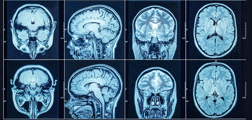
CAT (computerized axial tomography)
Allows us to see inside the living brain.
Examines the brain with X-ray photographs that reveal the damage to the brain.
They show structure only
Good: Relatively cheap
Bad: uses radiation
Unethical for research as you expose someone to radiation

PET (positron emission tomography)
Portrays brain acting by shows each brain area’s consumption of the chemical fuel it has, the sugar glucose.
Neurons that are active eat/gobble glucose.
Measures how much the brain is being used.
The brain consumes 20% of our calorie intake.
A precision revives a temporary radioactive (isotope) glucose, and the gamma rays which are released are tracked by the PET scan, as a task is performed.
Areas of the brain that are lightened up, are the areas being used.
Hot spots show the most active brain areas as a person does a task. (daydream, look at images or do mathematical calculations.)
Show function only
Ethical issues for research as it exposes people to radioactivity
There is a chemical that sticks to the gummy plaques that expose where Alzheimer's is killing the brain (you die from complications like forgetting to swallow / breathe, not just Alzheimer's.)
Though now we know, there's not much people can do to prevent Alzheimer's.
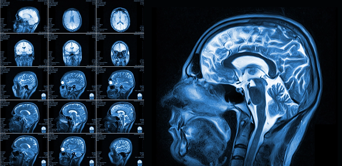
MRI / NMR (nucleo magnetic resonance imaging)
A person’s head is put into a strong magnetic field, which aligns with the spinning atoms in brain molecules. Radio-wave pulse for a moment disorientates the atoms.
When an atom returns to its normal spin, it emits signals that give detailed pictures of soft tissues, including the brain.
Structure only
Good: no radiation and very detailed
Bad: Very expensive

fMRI (functional nucleo magnetic resonance imaging)
Measures the blood flow to brain regions by comparing multiple MRI scans.
Combines PET scan and MRI in real time
Prior to surgery various languages and speech tasks were employed to chart functional language areas in the vicinity of the tumor.
Divisions of the brain
Forebrain , Midbrain , Hindbrain.

Forebrain
Includes: Telencephalon (Cerebrum / Endbrain), Cerebral cortex, Corpus callosum, Limbic system, Basal ganglia, Diencephalon, Thalamus, Hypothalamus.
The largest / most complex division of the brain. Split into 2 main subdivisions during development (telencephalon & diencephalon.)
Location : Most anterior (front) division of the brain.

Midbrain
Indcludes: Mesencephalon, Tectum, Tegmentum, Upper RAS.
Smallest region of the brain, sitting at the top of the brainstem between the forebrain and hindbrain.

Hindbrain
Includes: Metencephalon, Cerebellum, Pons, Mylencephalon (medulla), Lower RAS.
Located at the lower back of the skull. Connects the brain to the spinal cord. Dived into metencephalon and myelencephalon.

Telencephalon (Cerebrum / Endbrain)
Location: The most anterior & superior region of the brain. Wraps around and sits above (superior) to the diencephalon.
SHAPE: Two hemispheres, wrinkled surface.
Forms the largest part of the human brain. (85%)
MAIN FUNCTION: Controls conscious thought / memory /language / voluntary movements.

Cerebral cortex
Location: The wrinkled outer layer of the cerebrum.
SHAPE: Thin, wrinkled, folded inward.
Thin sheet of gray matter, divided into left and right hemispheres.
Its folds and grooves have massive surface area in the skull.
MAIN FUNCTION: Control conscious thought / memory /language / voluntary movements / sensory perception.
DAMAGE: Depends on the lobe which is injured. Paralysis can occur, or personality changes (frontal), sensory loss (parietal), blindness (occipital), or speech . memory loss (temporal).

Corpus callosum
Location:Deep in the center of the brain.
SHAPE: C-shaped band.
Thick of a band of white matter nerve fibers that connect the brain's 2 hemispheres.
MAIN FUNCTION: Allow left and right hemispheres of the brain to communicate / share information.
“Split brain procedure” (cutting it) is done to treat severe epilepsy, yet stopping cross-hemisphere communication.
DAMAGE: Leads to “split brain” syndrome (the 2 hemispheres cant share sensory or motor information.
People can struggle to name things seen in their left visual field since the information isn't reaching the right hemisphere, and the information can't go back to the left hemisphere which allows for language.

Limic System
Location: Under the Cerebral Cortex, wrapped around the top of the brainstem.
SHAPE: Ring-shaped network of deep structures.
Includes hippocampus and amygdala.
The amygdala (almond shaped) acts as the brain's threat detector for fear/ aggression.
The hippocampus (seahorse shaped) converts short-term memories into lasting ones.
MAIN FUNCTION: To control emotion / drives of behavior / memory foundation.
DAMAGE: Hippocampal damage leads to antegrade amnesia (inability to form new memories), and damage to the Amygdala leads to less fear response and less abilities to recognize emotional cues from others.

Basal ganglia
Location: Deep in the white matter of the cerebral hemispheres. Cluster of gray matter nuclei that work with the motor cortex.
SHAPE: Cluster of gray matter.
In constant loop with the cerebral cortex and thalamus to filter out unwanted movements. (while allowing wanted movements.)
Process memories through the Basal ganglia and become explicit in the cerebellum.
MAIN FUNCTION: Initiate smooth / coordinate voluntary movement / restricts (suppress) involuntary movements.
DAMAGE: To the Basal Ganglia or its dopamine supply leads to movement disorders.
(underactivity) Parkinson's disease / (Overactivity) Huntington's disease.

Diencephalon
Location: Deep in the brain, between the cerebral hemispheres and midbrain.
SHAPE: Size of an apricot, roughly symmetrical.
MAIN FUNCTION: Act as primary relay / processing century for sensory and motor signals to the cerebral cortex. Also manages the autonomic nervous system.
DAMAGE: Neurological and psychological disruptions / sensory and motor deficits / long lasting amnesia.

The hippocampus (seahorse shaped)
Converts short-term memories into lasting ones.

The amygdala (almond shaped)
Acts as the brain's threat detector for fear/ aggression.

Thalamus
Location: Paired, egg shaped, located centrally-directly superior to the brainstem, and deeply under the cerebral cortex.
SHAPE: Two egg shaped masses of gray matter.
MAIN FUNCTION: Act as the brain's sensory relay station, filters & routes incoming sensory signals (sight / sound / touch / taste, all except SMELL) to cortical regions.
DAMAGE: Sensory loss on the opposite side of the body, burning pain, movement issues, even a coma.

Hypothalamus
Location: Directly below the Thalamus, and a bit above the Pituitary gland.
SHAPE: Tiny, cone shaped. (Size of pearl / almond).
Controls the entire autonomic nervous system.
MAIN FUNCTION: Maintains homeostasis (internal balance).
Regulates body temperature /hunger / thirst / sleep cycles / hormone release ( by way of pituitary gland).
DAMAGE: Messes up basic survival drives. Leads to heavy weight gain or loss / uncountable thirst / body temperature is not stable / hormone imbalance.

Mesencephalon
Location: Most superior (cranial) part of the brainstem, between the pons and forebrain.
SHAPE: SHort bridge.
MAIN FUNCTION: Governs visual and auditory reflexes / coordinate motor control / relay nerve impulses between the spinal cord and higher regions.
DAMAGE: Lead to movement disorders / eye movement abnormalities.

Tectum
Location: The dorsal (back) surface of the midbrain.
SHAPE: Has 4 small rounded bumps, (Colliculi).
The 2 top bumps (Superior colliculi) control visual reflexes, and the bottom 2 (inferior colliculi) handle auditory reflexes.
MAIN FUNCTION: Process auditory tracking, reflexive visual.
EX: Turning one’s head to a sudden loud noise.
DAMAGE: Impairment to eye movement tracking, and the diminishment of the automatic “startle” reflexes to sudden sounds / sight.

Tegmentum
Location: Ventral (front)core of the midbrain, under the tectum.
MAIN FUNCTION: Regulate movement / suppression to pain /reward pathways. Houses key dopamine centers.
DAMAGE: Destruction of dopamine neurons leads to Parkinson’s symptoms, and nerve damage causes double vision.

Upper RAS
Location: Core of midbrain, into thalamus.
SHAPE: Net like mesh of neurons.
Anesthesia works as it suppresses activity in this network.
MAIN FUNCTION: Be the brain’s alertness / sends signals to the cortex to maintain wakefulness / focus / consciousness.
DAMAGE: Immediate loss of consciousness / irreversible coma.
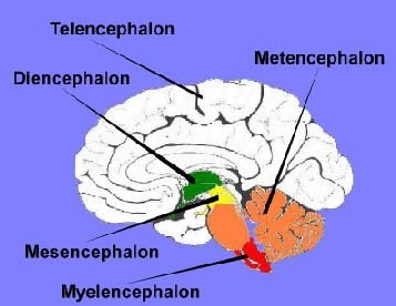
Metencephalon
Location: Upper division of the hindbrain, in the lower back of the skull, directly below the midbrain, and above the medulla.
MAIN FUNCTION: Cranial nerve control / signal relay / motor coordination / balance.
DAMAGE: Paralysis, movement / balance disorders.

Cerebellum
Location: Under the back of the cerebrum, behind the brainstem.
SHAPE: Densely folded, cauliflower shaped.
It only takes up 10% of the brain's volumes; yet it uses more than 50% of the neurons in the brain.
MAIN FUNCTION: Coordinate voluntary movements / balance/ equilibrium / posture / fine motor skills.
DAMAGE: Leads to clumsy / uncoordinated movements / slurred speech / tremors.

Pons
Location: In the middle of the brainstem, above the medulla and in front of the cerebellum.
SHAPE: Rounded, horse shoe shaped, bulging band of nerve fibers.
MAIN FUNCTION: Relay signals between the Cerebrum and cerebellum /help regulate breathing rhythm . control facial expressions / helps with chewing / eye movements.
DAMAGE: “Locked-in syndrome (Body is paralyzed yet one is fully conscious),” facial paralysis / sleep and breathing disruptions.

Mylencephalon (medulla)
Location: The lowest part of the brainstem, smaller as it goes into the spinal cord.
SHAPE: Cone shaped stalk.
Nerve trucks cross over the medulla. (The reasons are that each side of the brain controls the opposite side of the body).
MAIN FUNCTION: Automatically let life supporting functions run, like heart rate / blood pressure / breathing / swallowing.
DAMAGE: Fatal since it leads to immediate respiratory / cardiac arrest.
Smaller injuries lead to hardships with swallowing/ gag reflex gone / paralysis.

Lower RAS
Location: Extends through the pons and medulla, lower portion.
SHAPE: Net like.
Helps transition the brain between deep sleep / REM sleep cycles.
MAIN FUNCTION: Filter out repetitive background sensory noises during sleep / regulates muscle tone/ helps the cardiovascular and respiratory reflexes.
DAMAGE: Disruption in sleep-wake cycles / weakens muscle tone reflex / impaired automatic breathing and blood pressure responses.
Cells at rest
cell polarized i.e slightly more negative inside than out
Resting potential
Neurons just sit there, polarized & resting. The inside of the axon has a negative charge unlike the outside.
STEP 1
Dendrite stimulated by neurotransmitter from adjacent neuron (STEP 1)
Receiving signals at the Dendrites
Neighboring neurons/ sensory receptors send chemical / physical messages to branching extensions of the neuron.
STEP 2
Depolarization(STEP 2): when the cell is stimulated.
Depolarization / domino effect
STEP 3
the permeability of the cell wall changed - the ion pumps allow Na+ & Cl- (sodium chloride & STEP 3) in; pushing K+ out - creates an electrical charge
Tiny gates on the axon open up, NA+ flood in the negatively-charged amazon, flipping the charge to positive as it moves down. (like line of falling dominoes)
STEP 4
Action Potential: the electrical charge (which is created & STEP 4) travels down the axon
STEP 5
(action potential travels & STEP 5) travels down the axon away from the cell body to the synaptic terminal.
Traveling down the axon
Electrical impulse goes down a tail of the neuron to the end, if the axon has a myelin sheath wrapping the signal skips faster down gaps.
STEP 6
when the electrical charge reaches synaptic terminal (STEP 6)
STEP 7
it induces the release of the neurotransmitter (STEP 7)
STEP 8
the neurotransmitter diffuse across the synaptic gap (STEP 8)
STEP 9
stimulates the dendrite of the adjacent neuron (STEP 9) binding in a lock in key fit.
Chemical messengers float across the synaptic gap, fitting like a key into special locks on the receiving neurons' dendrites, telling if the next neuron to fire or not.
(STEP 10)
Original neuron enters refractory period
Reuptake
Leftover chemicals in the gap get sucked back into the sending neuron to be recycled, Firing neuron takes a break before firing again.
Degradation
Chemical messengers in the brain are broken down or cleared from the synapse after transmitting a signal, stopping their action and keeping the nervous system balanced.

Brain Stem
Connects the spinal cord to higher brain regions.
Cerebrum and cerebellum to spinal cord.
Medulla & pons, they both together control foundational life functions EX: Heart rate.
Midbrain is in it too.

Medulla (often considered part of the brain stem)
Location: Part of the Brain stem, at the base (lowest), directly above the spinal cord.
Main function: To control breathing / heart rate
Important for automatic functions

Pons
Location: Above the Medulla, in the middle of the brainstem, and below midbrain.
Connects the two hemispheres in the brain / cerebellum.
Latin for bridge,
Main function: To help regulate sleep / facial expressions
They act as a bridge, relaying movement
and sensory signals between the cerebrum and cerebellum.

Reticular Formation aka Reticular Activan System (RAS)
Net-like network of neurons that move
vertically though the core of the brainstem in the thalamus.
Filters incoming sensory stimuli.
Measures electrical activity.
Main function: Regulate arousal / alertness / attention
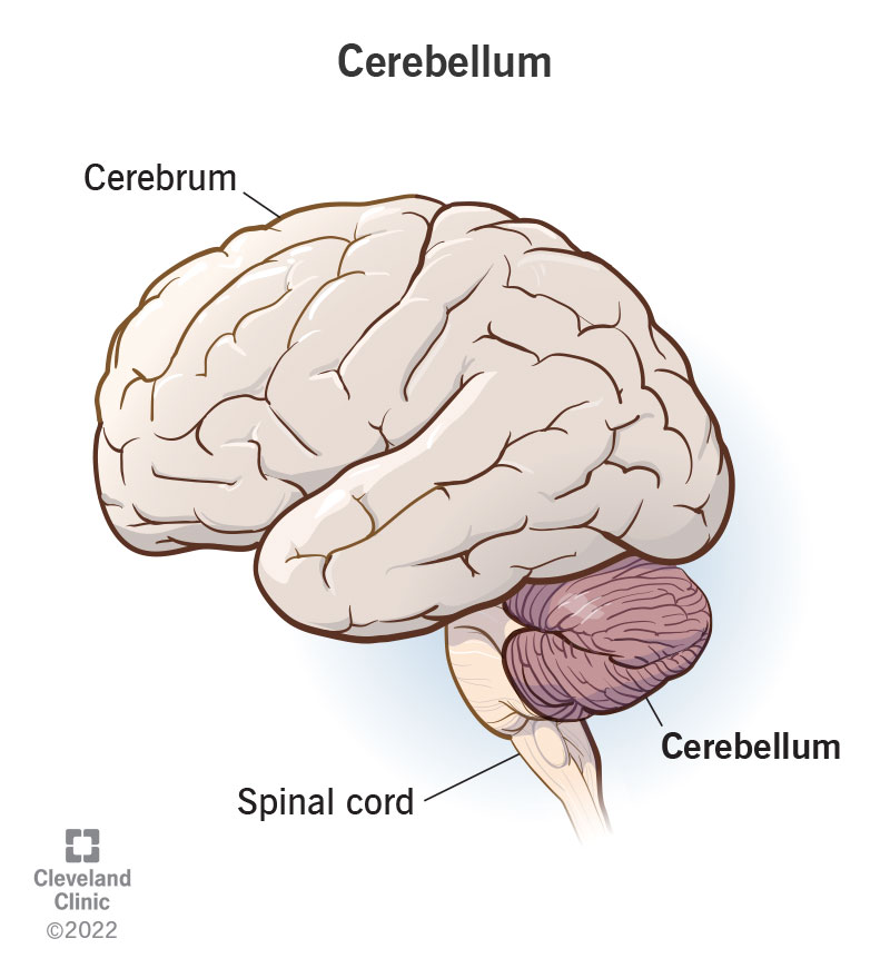
Cerebellum
Location: back of the skull, underneath the occipital
& temporal lobes behind the brainstem.
Called “little brain”
Main function: coordinate voluntary muscle movements /
Posture / balance / equilibrium / motor learning
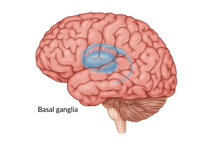
Basal Ganglia
Groups of gray matter nuclei
Location: Within the cerebral hemispheres, surrounding
the thalamus.
Groups work with the motor cortex to inhibit
voluntary movements.
Main function: smooth out voluntary movement
and procedural learning.