Chapter 10 Mitosis review

0.0(0)
Studied by 0 people
call kaiCall Kai
learnLearn
examPractice Test
spaced repetitionSpaced Repetition
heart puzzleMatch
flashcardsFlashcards
GameKnowt Play
Card Sorting

1/131

flashcard set

Earn XP

Description and Tags

Cell cycle and Mitosis

132 Terms

1
New cards

Functions of cell division

Multicellular organisms use cell division for:

  • Growth

  • Maintenance

  • Repair injured cells and tissues

  • Development

  • Single-celled organisms use cell division to reproduce


2
New cards

Cell division: cell growth

Cell growth via cell division refers to the cell cycle—an ordered, regulated sequence of events where a parent cell increases in size, duplicates its genetic material (DNA), and divides to produce two genetically identical daughter cells

  • the coordinated process in which a cell increases in size and mass, duplicates its genetic material, and splits into two genetically identical daughter cells

  • cells can use cell division to grow in size and number


<p><span><strong>Cell growth via cell division</strong></span> refers to the <strong>cell cycle</strong>—an ordered, regulated sequence of events where a parent cell increases in size, duplicates its genetic material (DNA), and divides to produce two genetically identical daughter cells</p><ul><li><p>the coordinated process in which a cell increases in size and mass, duplicates its genetic material, and splits into two genetically identical daughter cells</p></li></ul><ul><li><p>cells can use cell division to grow in size and number</p></li></ul><p></p>
3
New cards

Cell division: maintenance

The process by which eukaryotic cells divide (via mitosis and cytokinesis) to replace damaged, dead, or worn-out cells and preserve normal tissue structure and function

  • replace old cells

  • the process by which somatic (body) cells undergo mitosis and cytokinesis to replace damaged, dead, or worn-out cells and preserve tissue integrity and homeostasis


<p>The process by which eukaryotic cells divide (via mitosis and cytokinesis) to <strong>replace damaged, dead, or worn-out cells</strong> and preserve normal tissue structure and function</p><ul><li><p>replace old cells </p></li><li><p>the process by which somatic (body) cells undergo mitosis and cytokinesis to replace damaged, dead, or worn-out cells and preserve tissue integrity and homeostasis</p></li></ul><p></p>
4
New cards

Cell division: development

The process by which a less specialized cell becomes a specialized cell type with a distinct structure and function

  • the process by which a less specialized cell (such as a stem cell or zygote) develops into a specialized cell type with a distinct structure and function (such as a neuron, muscle cell, or red blood cell)

  • development (new function)


<p>The process by which a less specialized cell becomes a specialized cell type with a distinct structure and function</p><ul><li><p>the process by which a less specialized cell (such as a stem cell or zygote) develops into a specialized cell type with a distinct structure and function (such as a neuron, muscle cell, or red blood cell)</p></li><li><p>development (new function)</p></li></ul><p></p>
5
New cards

Functions of cell division

Multicellular organisms use cell division for:

  • Growth

  • Maintenance

  • Repair injured cells and tissues

  • Development

  • Single-celled organisms use cell division to reproduce


6
New cards

Purpose of cell division:

  • Cell division

  • Growth

  • Development

  • Repair

  • Cancer


7
New cards

Purpose of cell division:

  • Cell division

  • Growth

  • Development

  • Repair

  • Cancer


8
New cards

What do single-celled organisms use cell division for?

Single-celled organisms use cell division to reproduce

  • Single-celled organisms use cell division (such as binary fission or mitotic division) as their sole method of asexual reproduction, creating two new, genetically identical individual organisms from a single parent cell


<p><span>Single-celled organisms use cell division to reproduce</span></p><ul><li><p><span>Single-celled organisms use cell division (such as binary fission or mitotic division) as their </span><strong>sole method of asexual reproduction</strong>, creating two new, genetically identical individual organisms from a single parent cell</p></li></ul><p></p>
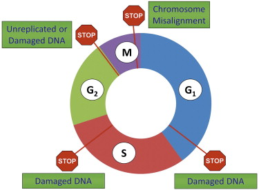
9
New cards

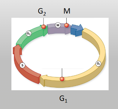
Genome

The complete set of genetic material (DNA or RNA) in an organism, including all of its genes and noncoding regulatory sequences

  • the complete set of genetic material in an organism. It contains all the instructions needed for an organism to develop, grow, function, and reproduce

  • What is the genome? - all the cell’s DNA


<p>The complete set of genetic material (DNA or RNA) in an organism, including all of its genes and noncoding regulatory sequences</p><ul><li><p>the complete set of genetic material in an organism. It contains all the instructions needed for an organism to develop, grow, function, and reproduce</p></li></ul><ul><li><p>What is the genome? - all the cell’s DNA </p></li></ul><p></p>
10
New cards

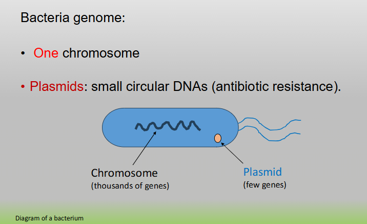
Bacteria genome

The complete set of DNA within a single bacterial cell, consisting of its main chromosomal DNA and any extra-chromosomal plasmid DNA

  • One chromosome (containing thousands of genes)

  • Plasmids: small circular DNAs (antibiotic resistance)

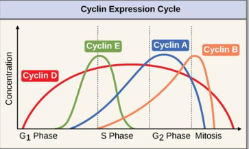
  • Main chromosome: usually a single, circular double-stranded DNA molecule located in an unenclosed region of the cytoplasm called the nucleoid


<p>The complete set of DNA within a single bacterial cell, consisting of its main chromosomal DNA and any extra-chromosomal plasmid DNA</p><ul><li><p>One chromosome (containing thousands of genes)</p></li><li><p><span>Plasmids: small circular DNAs (antibiotic resistance)</span></p></li><li><p>Main chromosome: usually a single, circular double-stranded DNA molecule located in an unenclosed region of the cytoplasm called the nucleoid</p></li></ul><p></p>
11
New cards

Plasmid

Small, circular pieces of DNA that exist separately from an organism's main chromosome. Found in prokaryotes and some eukaryotes, they carry extra genes (like antibiotic resistance) and can move between cells

  • A small, circular, double-stranded DNA molecule that is physically separate from a cell's main chromosomal DNA and can replicate independently

  • They typically carry "bonus" or non-essential genes that provide a survival advantage, such as antibiotic resistance genes

  • Plasmids: small circular DNAs (antibiotic resistance)

<p><span>Small, circular pieces of DNA that exist separately from an organism's main chromosome. Found in prokaryotes and some eukaryotes, they carry extra genes (like antibiotic resistance) and can move between cells</span></p><ul><li><p>A small, circular, double-stranded DNA molecule that is physically separate from a cell's main chromosomal DNA and can replicate independently</p></li><li><p>They typically carry "bonus" or non-essential genes that provide a survival advantage, such as <strong>antibiotic resistance</strong> genes</p></li><li><p>Plasmids: small circular DNAs (antibiotic resistance)</p></li></ul><p></p>
12
New cards

The eukaryotic genome: animals

The eukaryotic genome of animals is defined as the total genetic material contained within an animal cell, characterized by multiple linear chromosomes housed inside a membrane-bound nucleus, as well as circular DNA within the mitochondria

Made of:

  • Chromosomes

  • Mitochondria


<p>The eukaryotic genome of animals is defined as the total genetic material contained within an animal cell, characterized by multiple linear chromosomes housed inside a membrane-bound nucleus, as well as circular DNA within the mitochondria</p><p><span>Made of:</span></p><ul><li><p><span>Chromosomes</span></p></li><li><p><span>Mitochondria</span></p></li></ul><p></p>
13
New cards

The eukaryotic genome: plants

The eukaryotic plant genome is defined as the entire set of genetic material (DNA instructions) distributed across three distinct compartmentalized structures within a plant cell: the nucleus, the mitochondria, and the chloroplasts

Made of:

  • Chromosomes

  • Mitochondria

  • Chloroplasts


<p>The eukaryotic plant genome is defined as the entire set of genetic material (DNA instructions) distributed across three distinct compartmentalized structures within a plant cell: the nucleus, the mitochondria, and the chloroplasts</p><p><span>Made of:</span></p><ul><li><p><span>Chromosomes</span></p></li><li><p><span>Mitochondria</span></p></li><li><p><span>Chloroplasts</span></p></li></ul><p></p>
14
New cards

The eukaryotic genome

The entire complement of DNA contained within a eukaryotic cell, which is uniquely characterized by being partitioned into multiple linear chromosomes enclosed within a membrane-bound nucleus, along with non-nuclear DNA found in organelles like mitochondria and chloroplasts

Animals:

  • Chromosomes

  • Mitochondria

Plants:

  • Chromosomes

  • Mitochondria

  • Chloroplasts


15
New cards

Cancer

A disease of uncontrolled cell division that occurs when cell cycle checkpoints and regulatory proteins fail to halt the cycle, usually due to accumulated genetic mutations

  • uncontrolled, unrestrained cell division that results from a breakdown in the normal regulatory mechanisms of the cell cycle

  • cell division: cancer, unchecked cell growth


<p>A disease of <strong>uncontrolled cell division</strong> that occurs when cell cycle checkpoints and regulatory proteins fail to halt the cycle, usually due to accumulated genetic mutations</p><ul><li><p><strong>uncontrolled, unrestrained cell division</strong> that results from a breakdown in the normal regulatory mechanisms of the cell cycle</p></li><li><p>cell division: cancer, unchecked cell growth</p></li></ul><p></p>
16
New cards

Metastasis

The process by which cancer cells spread from their original (primary) site to distant parts of the body through the bloodstream or lymphatic system

  • the process where cancer cells break away from a primary tumor, travel through the blood or lymphatic system, and form new secondary tumors in other parts of the body


<p>The process by which cancer cells spread from their original (primary) site to distant parts of the body through the bloodstream or lymphatic system</p><ul><li><p>the process where cancer cells break away from a primary tumor, travel through the blood or lymphatic system, and form new secondary tumors in other parts of the body</p></li></ul><p></p>
17
New cards

Eukaryotic chromosomes

A eukaryotic chromosome is a linear, double-stranded DNA molecule tightly packaged with histone proteins inside a membrane-bound nucleus

  • The number of chromosomes varies among species

  • In the same species, chromosome number is identical

  • Example: humans have 46 chromosomes


<p><span>A </span>eukaryotic chromosome is a linear, double-stranded DNA molecule tightly packaged with histone proteins inside a membrane-bound nucleus</p><ul><li><p><span>The number of chromosomes varies among species</span></p></li><li><p><span>In the same species, chromosome number is identical</span></p></li><li><p><span>Example: humans have 46 chromosomes</span></p></li></ul><p></p>
18
New cards

Chromosome number varies among species in Eukaryotes

Every eukaryotic species has a characteristic and fixed number of chromosomes in its cell nuclei, but this number varies widely across different species and does not correlate with organism complexity

  • Each eukaryotic species has a distinct chromosome number in somatic (body) cells

  • Chromosome number varies among species in eukaryotes


<p><span>Every eukaryotic species has a characteristic and fixed number of chromosomes in its cell nuclei</span>, but this number varies widely across different species and does <strong>not</strong> correlate with organism complexity</p><ul><li><p>Each eukaryotic species has a distinct chromosome number in somatic (body) cells</p></li><li><p><span>Chromosome number varies among species in eukaryotes</span></p></li></ul><p></p>
19
New cards

Types of cells

  • Somatic: 2 sets of chromosomes: diploid (2n)

  • Gametes: (eggs and sperm cells) half the number of chromosomes: haploid (1n)


20
New cards

Somatic cells

Any body cells in a multicellular organism except for reproductive cells (gametes like sperm and eggs)

  • 2 sets of chromosomes: diploid (2n)

  • In humans and most eukaryotic species, somatic cells are diploid (2n), meaning they contain two full sets of chromosomes—one set inherited from each parent (46 total chromosomes in humans, made up of 23 pairs)

  • Somatic cells divide strictly by mitosis, producing two genetically identical daughter cells to support organismal growth, tissue repair, and maintenance

  • Mutations that occur in somatic cells can affect the individual organism (e.g., leading to cancer), but they cannot be passed on to offspring


<p>Any body cells in a multicellular organism except for reproductive cells (gametes like sperm and eggs)</p><ul><li><p>2 sets of chromosomes: diploid (2n)</p></li><li><p>In humans and most eukaryotic species, somatic cells are diploid (2n), meaning they contain two full sets of chromosomes—one set inherited from each parent (46 total chromosomes in humans, made up of 23 pairs)</p></li><li><p>Somatic cells divide strictly by mitosis, producing two genetically identical daughter cells to support organismal growth, tissue repair, and maintenance</p></li><li><p>Mutations that occur in somatic cells can affect the individual organism (e.g., leading to cancer), but they cannot be passed on to offspring</p></li></ul><p></p>
21
New cards

Diploid (2n)

A diploid cell or organism is one that contains two complete sets of chromosomes (abbreviated as 2n)

  • In a diploid cell, one complete set of chromosomes is inherited from the mother (maternal set) and the other set is inherited from the father (paternal set)

  • A diploid cell is a cell that contains two complete sets of chromosomes (2n), with one set inherited from each biological parent

  • Humans have a diploid number of 2n = 46 chromosomes (23 pairs of homologous chromosomes)


<p><span>A </span>diploid cell or organism is one that contains two complete sets of chromosomes (abbreviated as 2n)</p><ul><li><p>In a diploid cell, one complete set of chromosomes is inherited from the mother (maternal set) and the other set is inherited from the father (paternal set)</p></li><li><p><span>A </span><strong>diploid cell</strong> is a cell that contains <strong>two complete sets of chromosomes</strong> (2n), with one set inherited from each biological parent</p></li><li><p>Humans have a diploid number of <strong>2n = 46 chromosomes</strong> (23 pairs of homologous chromosomes)</p></li></ul><p></p>
22
New cards

Gametes (haploid)

A haploid (1n) reproductive or sex cell (such as a sperm or egg) produced by meiosis that fuses with another gamete during fertilization to form a diploid (2n) zygote

  • Haploid Status (1n): gametes contain only one set of chromosomes (half the number of somatic/body cells). In humans, this means each gamete has 23 chromosomes rather than the full set of 46

  • Form via meiosis

  • haploid (1n) reproductive or sex cell produced by meiosis in sexually reproducing organisms


<p>A haploid (1n) reproductive or sex cell (such as a sperm or egg) produced by meiosis that fuses with another gamete during fertilization to form a diploid (2n) zygote</p><ul><li><p>Haploid Status (1n): gametes contain only one set of chromosomes (half the number of somatic/body cells). In humans, this means each gamete has 23 chromosomes rather than the full set of 46</p></li><li><p>Form via meiosis</p></li><li><p>haploid (1n) reproductive or sex cell produced by meiosis in sexually reproducing organisms</p></li></ul><p></p>
23
New cards

Haploid (1n)

A cell that contains a single set of unpaired chromosomes, denoted by the symbol n

  • A cell that contains one complete set of unpaired chromosomes (n), meaning it has only a single copy of each chromosome type rather than homologous pairs

  • In humans, haploid cells have 23 chromosomes (n = 23). This includes gametes (sperm and egg cells)

  • Form via meiosis


<p>A cell that contains a single set of unpaired chromosomes, denoted by the symbol<em> n</em></p><ul><li><p><span>A cell that contains one complete set of unpaired chromosomes (</span>n<span>), meaning it has only a single copy of each chromosome type rather than homologous pairs</span></p></li><li><p>In humans, haploid cells have 23 chromosomes (n = 23). This includes gametes (sperm and egg cells)</p></li><li><p>Form via meiosis</p></li></ul><p></p>
24
New cards

Karyotype

The number and visual appearance of an individual’s complete set of chromosomes

  • a visual profile or diagram that displays an individual’s complete set of condensed chromosomes, organized and numbered into homologous pairs

  • chromosomes arranged by size


<p>The number and visual appearance of an individual’s complete set of chromosomes</p><ul><li><p>a visual profile or diagram that displays an individual’s complete set of condensed chromosomes, organized and numbered into homologous pairs</p></li><li><p>chromosomes arranged by size</p></li></ul><p></p>
25
New cards

Human karyotype

A karyotype is a visual representation or organized profile of an individual’s complete set of chromosomes, isolated from a single cell and arranged in homologous pairs by size, centromere position, and banding pattern

  • A normal human somatic (body) cell is diploid (2n = 46), containing 46 total chromosomes organized into 23 pairs

  • Autosomes: 22 pairs (44 total) of non-sex chromosomes, numbered 1 through 22 from largest to smallest

  • Sex Chromosomes: 1 pair (2 total) that determines biological sex


<p>A karyotype is a visual representation or organized profile of an individual’s complete set of chromosomes, isolated from a single cell and arranged in homologous pairs by size, centromere position, and banding pattern</p><ul><li><p><span>A normal human somatic (body) cell is diploid (</span>2n = 46<span>), containing <strong>46 total chromosomes</strong> organized into <strong>23 pairs</strong></span></p></li><li><p><strong>Autosomes:</strong> 22 pairs (44 total) of non-sex chromosomes, numbered 1 through 22 from largest to smallest</p></li><li><p><strong>Sex Chromosomes:</strong> 1 pair (2 total) that determines biological sex</p></li></ul><p></p>
26
New cards

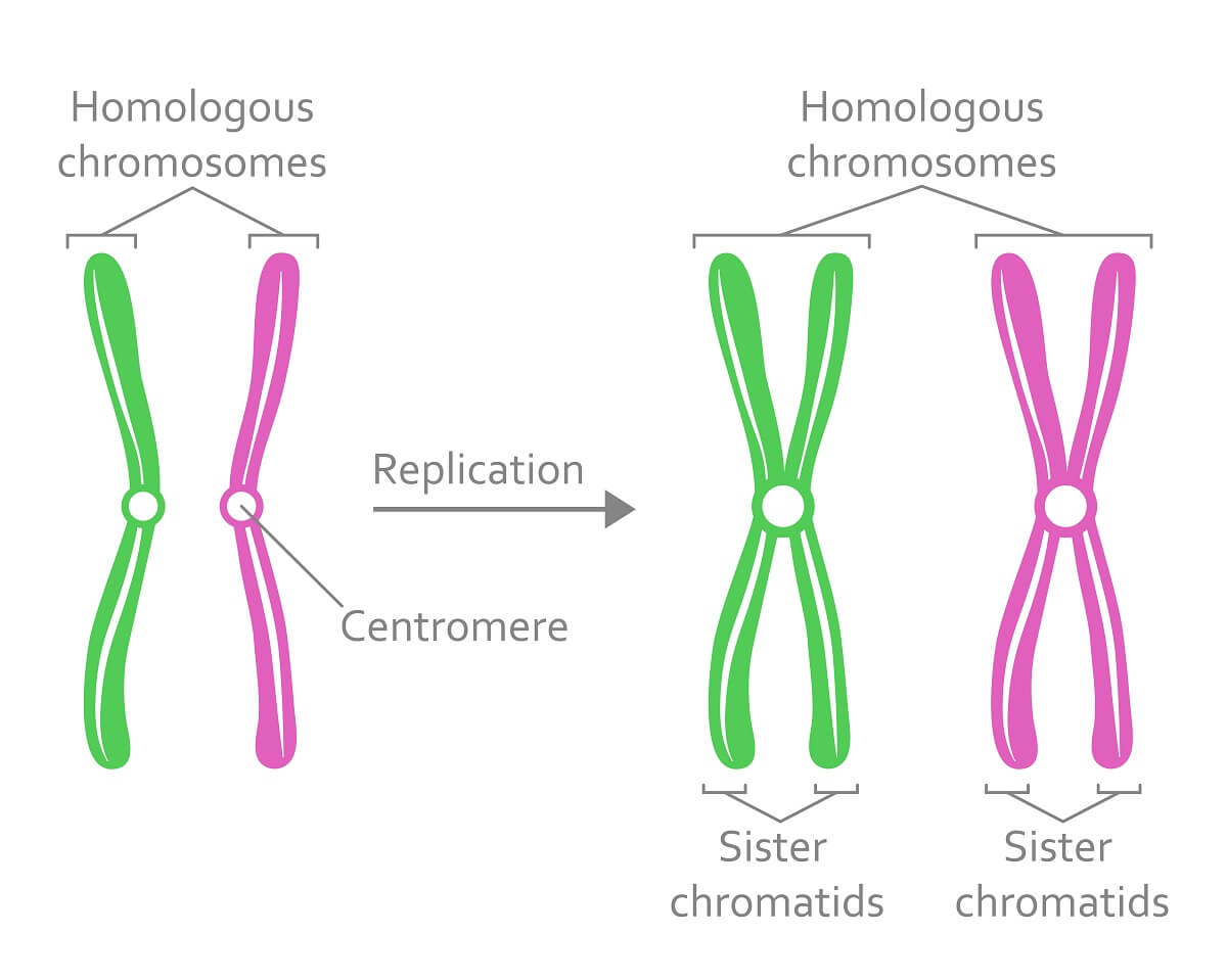
Homologous chromosomes

A matched pair of chromosomes in diploid cells—one inherited from the maternal parent (egg) and one from the paternal parent (sperm)

  • a matched pair of chromosomes—one inherited from the mother and one from the father—that share the same length, centromere position, and gene sequence, though they may carry different alleles

  • Homologous chromosomes are separate chromosomes from different parents with the same genes (different alleles). Sister chromatids are exact, replicated copies of a single chromosome joined at the centromere


<p>A matched pair of chromosomes in diploid cells—one inherited from the maternal parent (egg) and one from the paternal parent (sperm)</p><ul><li><p>a matched pair of chromosomes—one inherited from the mother and one from the father—that share the same length, centromere position, and gene sequence, though they may carry different alleles</p></li><li><p>Homologous chromosomes are separate chromosomes from different parents with the same genes (different alleles). Sister chromatids are exact, replicated copies of a single chromosome joined at the centromere</p></li></ul><p></p>
27
New cards

Homologous chromosomes

A matched pair of chromosomes in diploid cells—one inherited from the maternal parent (egg) and one from the paternal parent (sperm)

  • a matched pair of chromosomes—one inherited from the mother and one from the father—that share the same length, centromere position, and gene sequence, though they may carry different alleles

  • Homologous chromosomes are separate chromosomes from different parents with the same genes (different alleles). Sister chromatids are exact, replicated copies of a single chromosome joined at the centromere

  • similar in size, types of genes


<p>A matched pair of chromosomes in diploid cells—one inherited from the maternal parent (egg) and one from the paternal parent (sperm)</p><ul><li><p>a matched pair of chromosomes—one inherited from the mother and one from the father—that share the same length, centromere position, and gene sequence, though they may carry different alleles</p></li><li><p>Homologous chromosomes are separate chromosomes from different parents with the same genes (different alleles). Sister chromatids are exact, replicated copies of a single chromosome joined at the centromere</p></li><li><p><span> similar in size, types of genes</span></p></li></ul><p></p>
28
New cards

Homologous chromosomes vs. Sister chromatids

  • Homologous chromosomes are a pair of matching chromosomes—one inherited from your mother and one from your father—that carry the same genes in the same locations but may have different alleles (versions)

  • Sister chromatids are the two identical, replicated copies of a single chromosome that are joined together at the centromere


<ul><li><p><span>Homologous chromosomes are a pair of matching chromosomes—one inherited from your mother and one from your father—that carry the same genes in the same locations but may have different alleles (versions)</span></p></li><li><p>Sister chromatids are the two identical, replicated copies of a single chromosome that are joined together at the centromere</p></li></ul><p></p>
29
New cards

Sister chromatids

Two identical copies of a single replicated chromosome that are connected to each other at a specialized region called the centromere. They contain the exact same genes and the same alleles

  • two identical copies of a single chromosome, made during S phase and held together at the centromere

  • they separate during anaphase of mitosis and anaphase II of meiosis, ensuring each new cell gets a complete chromosome copy


<p>Two identical copies of a single replicated chromosome that are connected to each other at a specialized region called the centromere. They contain the exact same genes and the same alleles</p><ul><li><p><span>two identical copies of a single chromosome, made during S phase and held together at the centromere</span></p></li><li><p><span>they separate during anaphase of mitosis and anaphase II of meiosis, ensuring each new cell gets a complete chromosome copy</span></p></li></ul><p></p>
30
New cards

Heterologous chromosomes

Often called non-homologous chromosomes - are chromosomes that belong to different pairs and contain different gene sequences, sizes, shapes, and centromere positions

  • chromosomes that belong to different pairs and carry completely different sets of genes, lengths, and centromere positions

  • In humans X and Y chromosomes are not homologous (these are heterologous )


<p>Often called non-homologous chromosomes - are chromosomes that belong to different pairs and contain different gene sequences, sizes, shapes, and centromere positions</p><ul><li><p>chromosomes that belong to different pairs and carry completely different sets of genes, lengths, and centromere positions</p></li><li><p><span>In humans X and Y chromosomes are not homologous (these are heterologous )</span></p></li></ul><p></p>
31
New cards

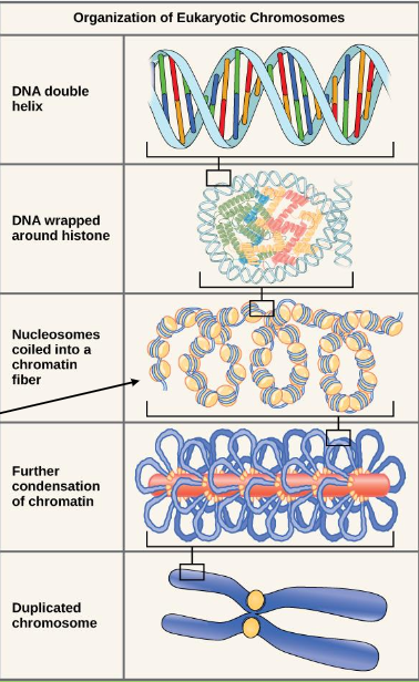
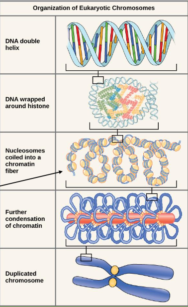
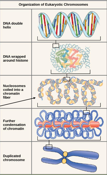
Organization of Eukaryotic chromosomes

Eukaryotic DNA is tightly condensed into compact chromosomes to fit into the nucleus

  • DNA double helix

  • DNA wrapped around histone

  • Nucleosomes coiled into a chromatin fiber

  • Further condensation of chromatin

  • Duplicated chromosome

  • Chromatin: nucleosomes coiled into a fiber


<p><span>Eukaryotic DNA is tightly condensed into compact chromosomes to fit into the nucleus</span></p><ul><li><p>DNA double helix</p></li><li><p>DNA wrapped around histone</p></li><li><p>Nucleosomes coiled into a chromatin fiber</p></li><li><p>Further condensation of chromatin</p></li><li><p>Duplicated chromosome </p></li><li><p>Chromatin: nucleosomes coiled into a fiber</p></li></ul><p></p>
32
New cards

Organization of eukaryotic chromosomes

The organization of eukaryotic chromosomes refers to the multi-level hierarchical packaging of multiple linear double-stranded DNA molecules with histone proteins to form condensed chromatin within a membrane-bound nucleus

  • Eukaryotic DNA is tightly condensed into compact chromosomes to fit into the nucleus

  • Chromatin: nucleosomes coiled into a fiber


<p>The organization of eukaryotic chromosomes refers to the multi-level hierarchical packaging of multiple linear double-stranded DNA molecules with histone proteins to form condensed chromatin within a membrane-bound nucleus</p><ul><li><p>Eukaryotic DNA is tightly condensed into compact chromosomes to fit into the nucleus</p></li><li><p>Chromatin: nucleosomes coiled into a fiber</p></li></ul><p></p>
33
New cards

Packaging of DNA

  • DNA wraps around histone proteins (like a string of beads)

  • The histone-DNA complex is called nucleosome

  • This structure then coils to form a chromatin fiber

  • The chromatin further packs into a chromosome

<ul><li><p><span>DNA wraps around histone proteins (like a string of beads)</span></p></li><li><p><span>The histone-DNA complex is called nucleosome </span></p></li><li><p><span>This structure then coils to form a chromatin fiber</span></p></li><li><p><span>The chromatin further packs into a chromosome</span></p></li></ul><p></p>
34
New cards

Nucleosome

The fundamental structural unit of chromatin in eukaryotic cells, consisting of a segment of DNA wrapped around a core of histone proteins

  • the basic repeating structural and functional unit of eukaryotic chromatin, consisting of a segment of DNA wrapped around a core of histone proteins

  • nucleosomes are the basic structural and repeating building blocks of chromatin

  • DNA wraps around histone proteins (like a string of beads)

  • The histone-DNA complex is called nucleosome

  • This structure then coils to form a chromatin fiber


<p>The fundamental structural unit of chromatin in eukaryotic cells, consisting of a segment of DNA wrapped around a core of histone proteins</p><ul><li><p>the basic repeating structural and functional unit of eukaryotic chromatin, consisting of a segment of DNA wrapped around a core of histone proteins</p></li><li><p>nucleosomes are the basic structural and repeating building blocks of chromatin</p></li><li><p><span>DNA wraps around histone proteins (like a string of beads)</span></p></li><li><p>The histone-DNA complex is called nucleosome</p></li><li><p>This structure then coils to form a chromatin fiber</p></li></ul><p></p>
35
New cards

Chromatin

The complex of DNA and structural proteins (primarily histones) that makes up eukaryotic chromosomes

  • The complex of DNA and proteins (primarily histones) that makes up eukaryotic chromosomes. When a cell is not actively dividing (during interphase), chromatin exists in a relaxed, uncoiled, diffuse mass of long, thin fibers within the nucleus

  • Nucleosomes coil into a chromatin fiber

  • The chromatin further packs (condenses) into a chromosome

<p>The complex of DNA and structural proteins (primarily histones) that makes up eukaryotic chromosomes</p><ul><li><p><span>The complex of DNA and proteins (primarily histones) that makes up eukaryotic chromosomes. When a cell is not actively dividing (during interphase), chromatin exists in a relaxed, uncoiled, diffuse mass of long, thin fibers within the nucleus</span></p></li><li><p>Nucleosomes coil into a chromatin fiber</p></li><li><p><span>The chromatin further packs (condenses) into a chromosome</span></p></li></ul><p></p>
36
New cards

Histones

A basic, positively charged protein found in eukaryotic cell nuclei that DNA wraps around to condense, pack, and organize into structural units called nucleosomes

  • A protein that DNA wraps around to condense and package it into the linear chromosomes of eukaryotic cells

  • A protein that DNA coils around so it can fit inside a cell

  • DNA wraps around histone proteins (like a string of beads)

  • The histone-DNA complex is called nucleosome


<p>A basic, positively charged protein found in eukaryotic cell nuclei that DNA wraps around to condense, pack, and organize into structural units called <strong>nucleosomes</strong></p><ul><li><p><span>A protein that DNA wraps around to condense and package it into the linear chromosomes of eukaryotic cells</span></p></li><li><p><span>A </span>protein<span> that DNA coils around so it can fit inside a cell</span></p></li><li><p><span>DNA wraps around histone proteins (like a string of beads)</span></p></li><li><p><span>The histone-DNA complex is called nucleosome</span></p></li></ul><p></p>
37
New cards

Chromosomes

A thread-like, condensed cellular structure made of a single long molecule of DNA tightly wrapped around protein complexes (histones). They serve as the primary organizational and inheritance units of genetic information in cells

  • a thread-like, gene-carrying structure made of a single long molecule of DNA tightly wrapped around associated histone proteins

  • chromatin further packs (condenses) into a chromosome


<p>A thread-like, condensed cellular structure made of a single long molecule of DNA tightly wrapped around protein complexes (histones). They serve as the primary organizational and inheritance units of genetic information in cells</p><ul><li><p>a thread-like, gene-carrying structure made of a single long molecule of DNA tightly wrapped around associated histone proteins</p></li><li><p>chromatin further packs (condenses) into a chromosome</p></li></ul><p></p>
38
New cards

Packaging of DNA:

  • DNA wraps around histone proteins (like a string of beads)

  • The histone-DNA complex is called nucleosome

  • This structure then coils to form a chromatin fiber

  • The chromatin further packs into a chromosome

<ul><li><p><span>DNA wraps around histone proteins (like a string of beads)</span></p></li><li><p><span>The histone-DNA complex is called nucleosome </span></p></li><li><p><span>This structure then coils to form a chromatin fiber</span></p></li><li><p><span>The chromatin further packs into a chromosome</span></p></li></ul><p></p>
39
New cards

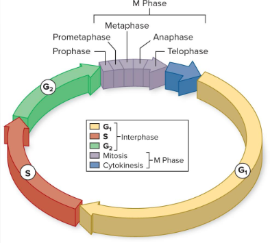
Cell cycle

The ordered sequence of growth, DNA replication, and division that a eukaryotic cell goes through from its origin until it divides into two genetically identical daughter cells

  • A series of events in the life of every cell

<p>The ordered sequence of growth, DNA replication, and division that a eukaryotic cell goes through from its origin until it divides into two genetically identical daughter cells</p><ul><li><p>A series of events in the life of every cell</p></li></ul><p></p>
40
New cards

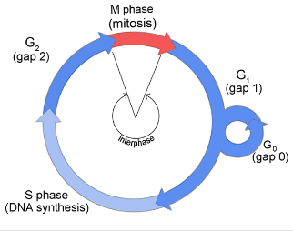
The Cell Cycle has 2 major phases

  • Interphase (blue) is the cell’s normal growth and preparation for cell division

  • Mitotic phase (red): the DNA and cytoplasm are split and the cell divides

<ul><li><p><span>Interphase (blue) is the cell’s normal growth and preparation for cell division</span></p></li><li><p><span>Mitotic phase (red): the DNA and cytoplasm are split and the cell divides</span></p></li></ul><p></p>
41
New cards

Interphase

The longest preparatory stage of the cell cycle (making up about 90% of a cell's life) where a eukaryotic cell grows, replicates its DNA, and prepares for cell division (mitosis or meiosis)

  • the cell’s normal growth and preparation for cell division

  • the longest portion of the cell cycle (making up about 90% of a cell's life) during which the cell grows, carries out normal metabolic functions, duplicates its organelles, and replicates its DNA in preparation for cell division

  • Interphase occurs in 3 stages


<p>The longest preparatory stage of the cell cycle (making up about 90% of a cell's life) where a eukaryotic cell grows, replicates its DNA, and prepares for cell division (mitosis or meiosis)</p><ul><li><p><span>the cell’s normal growth and preparation for cell division</span></p></li><li><p>the longest portion of the cell cycle (making up about 90% of a cell's life) during which the cell grows, carries out normal metabolic functions, duplicates its organelles, and replicates its DNA in preparation for cell division</p></li><li><p>Interphase occurs in 3 stages </p></li></ul><p></p>
42
New cards

Interphase occurs in 3 stages

  • G1 Phase (first gap): cell grows and performs normal functions

  • S Phase: dna replication/synthesis occurs

  • G2 Phase (second gap): cell gets ready for mitosis


<ul><li><p>G1 Phase (first gap): cell grows and performs normal functions</p></li></ul><ul><li><p>S Phase: dna replication/synthesis occurs</p></li><li><p>G2 Phase (second gap): cell gets ready for mitosis</p></li></ul><p></p>
43
New cards

Interphase occurs in 3 stages

  • G1 Phase (first gap): cell grows and performs normal functions

  • S Phase: dna replication/synthesis occurs

  • G2 Phase (second gap): cell gets ready for mitosis


<ul><li><p>G1 Phase (first gap): cell grows and performs normal functions</p></li><li><p>S Phase: dna replication/synthesis occurs</p></li><li><p>G2 Phase (second gap): cell gets ready for mitosis</p></li></ul><p></p>
44
New cards

Apoptosis

Programmed cell death—an orderly, controlled "cellular suicide" where a cell intentionally destroys itself for the good of the organism

  • a controlled, self-destruction process that the body uses to cleanly eliminate old, unneeded, or damaged cells without causing inflammation


45
New cards

G0 phase

A non-dividing, quiescent (resting) state of the cell cycle where a cell exits the active cycle and stops preparing to divide

  • Cells typically enter G0 from the G1 phase when they do not receive the signal ("go-ahead" checkpoint) to continue toward the S phase (DNA replication)

  • Even though the cell is not dividing, it remains metabolically active and continues carrying out its specialized, normal everyday functions

  • a resting or quiescent state in the cell cycle where a cell has temporarily or permanently exited active division


<p>A non-dividing, quiescent (resting) state of the cell cycle where a cell exits the active cycle and stops preparing to divide</p><ul><li><p>Cells typically enter G0 from the <strong>G1 phase</strong> when they do not receive the signal ("go-ahead" checkpoint) to continue toward the <strong>S phase</strong> (DNA replication)</p></li><li><p>Even though the cell is not dividing, it remains <strong>metabolically active</strong> and continues carrying out its specialized, normal everyday functions</p></li><li><p>a resting or quiescent state in the cell cycle where a cell has temporarily or permanently exited active division</p></li></ul><p></p>
46
New cards

G1 (gap 1) phase

The first growth stage of interphase in the eukaryotic cell cycle, occurring just after cell division (M phase) and right before DNA replication (S phase)

  • Cell grows and performs normal functions

  • Cell Growth: the cell increases in physical size and volume

  • Protein & Organelle Synthesis: the cell produces new proteins, RNA, and duplicates non-chromosomal organelles (such as mitochondria and ribosomes)

  • Normal Metabolic Activity: the cell carries out its specialized everyday functions

  • Preperation for DNA replication


<p>The first growth stage of <strong>interphase</strong> in the eukaryotic cell cycle, occurring just after cell division (M phase) and right before DNA replication (S phase)</p><ul><li><p><span>Cell grows and performs normal functions</span></p></li></ul><ul><li><p><span><strong>Cell Growth:</strong> the cell increases in physical size and volume</span></p></li><li><p><span><strong>Protein &amp; Organelle Synthesis:</strong> the cell produces new proteins, RNA, and duplicates non-chromosomal organelles (such as mitochondria and ribosomes)</span></p></li><li><p><span><strong>Normal Metabolic Activity:</strong> the cell carries out its specialized everyday functions</span></p></li><li><p>Preperation for DNA replication</p></li></ul><p></p>
47
New cards

S Phase

The middle stage of Interphase in the eukaryotic cell cycle where the cell replicates its entire genome (nuclear DNA) and duplicates its centrosomes

  • Sister Chromatids Form: each single-chromatid chromosome is duplicated into two identical copies called sister chromatids, which remain firmly joined at the centromere by cohesin proteins

  • The period during interphase of the cell cycle where the cell replicates its nuclear DNA and duplicates its centrosomes before entering cell division


<p>The middle stage of Interphase in the eukaryotic cell cycle where the cell replicates its entire genome (nuclear DNA) and duplicates its centrosomes</p><ul><li><p><strong>Sister Chromatids Form:</strong> each single-chromatid chromosome is duplicated into two identical copies called <strong>sister chromatids</strong>, which remain firmly joined at the centromere by cohesin proteins</p></li><li><p>The period during <strong>interphase</strong> of the cell cycle where the cell replicates its nuclear DNA and duplicates its centrosomes before entering cell division</p></li></ul><p></p>
48
New cards

S Phase

The middle stage of Interphase in the eukaryotic cell cycle where the cell replicates its entire genome (nuclear DNA) and duplicates its centrosomes

  • Sister Chromatids Form: each single-chromatid chromosome is duplicated into two identical copies called sister chromatids, which remain firmly joined at the centromere by cohesin proteins

  • The period during interphase of the cell cycle where the cell replicates its nuclear DNA and duplicates its centrosomes before entering cell division


<p>The middle stage of Interphase in the eukaryotic cell cycle where the cell replicates its entire genome (nuclear DNA) and duplicates its centrosomes</p><ul><li><p><strong>Sister Chromatids Form:</strong> each single-chromatid chromosome is duplicated into two identical copies called <strong>sister chromatids</strong>, which remain firmly joined at the centromere by cohesin proteins</p></li><li><p>The period during <strong>interphase</strong> of the cell cycle where the cell replicates its nuclear DNA and duplicates its centrosomes before entering cell division</p></li></ul><p></p>
49
New cards

G2 phase

The final subphase of interphase in the eukaryotic cell cycle, occurring right after DNA replication (S phase) and just before mitosis (M phase)

Cell gets ready for mitosis

  • Makes proteins needed for mitosis

  • Produces new organelles

  • Checks for DNA damage

  • The G2 Checkpoint: a major regulatory checkpoint verifies that DNA replication is completely finished and checks for any DNA damage before giving the green light to enter mitosis. If severe damage is detected, the cell pauses for repair or triggers apoptosis (programmed cell death)


<p>The final subphase of <strong>interphase</strong> in the eukaryotic cell cycle, occurring right after DNA replication (S phase) and just before mitosis (M phase)</p><p>Cell gets ready for mitosis</p><ul><li><p><span>Makes proteins needed for mitosis</span></p></li><li><p><span>Produces new organelles</span></p></li><li><p><span>Checks for DNA damage</span></p></li><li><p><strong>The G2 Checkpoint: </strong>a major regulatory checkpoint verifies that DNA replication is completely finished and checks for any DNA damage before giving the green light to enter mitosis. If severe damage is detected, the cell pauses for repair or triggers apoptosis (programmed cell death)</p></li></ul><p></p>
50
New cards

G2 phase

The final subphase of interphase in the eukaryotic cell cycle, occurring right after DNA replication (S phase) and just before mitosis (M phase)

Cell gets ready for mitosis

  • Makes proteins needed for mitosis

  • Produces new organelles

  • Checks for DNA damage

  • The G2 Checkpoint: a major regulatory checkpoint verifies that DNA replication is completely finished and checks for any DNA damage before giving the green light to enter mitosis. If severe damage is detected, the cell pauses for repair or triggers apoptosis (programmed cell death)


<p>The final subphase of <strong>interphase</strong> in the eukaryotic cell cycle, occurring right after DNA replication (S phase) and just before mitosis (M phase)</p><p>Cell gets ready for mitosis</p><ul><li><p>Makes proteins needed for mitosis</p></li><li><p>Produces new organelles</p></li><li><p>Checks for DNA damage</p></li><li><p><strong>The G2 Checkpoint: </strong>a major regulatory checkpoint verifies that DNA replication is completely finished and checks for any DNA damage before giving the green light to enter mitosis. If severe damage is detected, the cell pauses for repair or triggers apoptosis (programmed cell death)</p></li></ul><p></p>
51
New cards

The Mitotic Phase involves several steps

  • Mitosis is the first step

  • The second step is called cytokinesis, when the cytoplasm and the organelles are divided into two new cells

  • Mitotic phase: the DNA and cytoplasm are split and the cell divides


<ul><li><p>Mitosis is the first step</p></li><li><p>The second step is called cytokinesis, when the cytoplasm and the organelles are divided into two new cells</p></li><li><p>Mitotic phase: the DNA and cytoplasm are split and the cell divides</p></li></ul><p></p>
52
New cards

Mitosis

The process in eukaryotic cell division where the nucleus of a single cell divides to produce two genetically identical diploid (2n) daughter cells

  • a process of nuclear division in eukaryotic somatic cells where a single parent cell replicates its DNA once and divides once to produce two genetically identical daughter cells, maintaining the original diploid (2N) chromosome number


<p>The process in eukaryotic cell division where the nucleus of a single cell divides to produce <strong>two genetically identical diploid (2n) daughter cells</strong></p><ul><li><p>a process of nuclear division in eukaryotic somatic cells where a single parent cell replicates its DNA once and divides once to produce <strong>two genetically identical daughter cells</strong>, maintaining the original diploid (2N) chromosome number</p></li></ul><p></p>
53
New cards

Prophase

The first official phase of mitosis (the division of the nucleus) in eukaryotic cells, following the completion of Interphase (G1, S, and G2)

  • Chromosomes condense and become visible

  • Spindle fibers emerge from the centrosomes

  • Nuclear envelope breaks down

  • Nucleolus disappears


<p>The first official phase of mitosis (the division of the nucleus) in eukaryotic cells, following the completion of <span><strong>Interphase</strong></span> (G1, S, and G2)</p><ul><li><p>Chromosomes condense and become visible</p></li><li><p>Spindle fibers emerge from the centrosomes</p></li><li><p>Nuclear envelope breaks down</p></li><li><p>Nucleolus disappears</p></li></ul><p></p>
54
New cards

Prometaphase

The transitional phase of mitosis that follows prophase and precedes metaphase. During prometaphase, the nuclear envelope breaks down completely, and the mitotic spindle microtubules interact directly with the chromosomes

  • Chromosomes continue to condense

  • Kinetochores appear at the centromeres

  • Mitotic spindle microtubules attach to kinetochores

  • Centrosomes move toward opposite poles


<p>The transitional phase of mitosis that follows prophase and precedes metaphase. During prometaphase, the nuclear envelope breaks down completely, and the mitotic spindle microtubules interact directly with the chromosomes</p><ul><li><p>Chromosomes continue to condense</p></li><li><p>Kinetochores appear at the centromeres</p></li><li><p>Mitotic spindle microtubules attach to kinetochores </p></li><li><p>Centrosomes move toward opposite poles </p></li></ul><p></p>
55
New cards

Metaphase

The stage of mitosis where the cell's condensed chromosomes align single-file along the metaphase plate (the equatorial plane or center of the cell)

  • Mitotic spindle is fully developed, centrosomes are at opposite poles of the cell

  • Chromosomes are lined up at the metaphase plate

  • Each sister chromatid is attached to a spindle fiber originating from opposite poles


<p>The stage of mitosis where the cell's condensed chromosomes align single-file along the <strong>metaphase plate</strong> (the equatorial plane or center of the cell)</p><ul><li><p>Mitotic spindle is fully developed, centrosomes are at opposite poles of the cell</p></li><li><p>Chromosomes are lined up at the metaphase plate</p></li><li><p>Each sister chromatid is attached to a spindle fiber originating from opposite poles</p></li></ul><p></p>
56
New cards

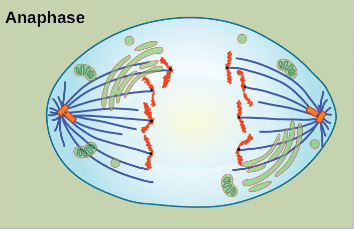
Anaphase

The stage of mitosis where the protein glue holding sister chromatids together breaks down, allowing them to separate and move toward opposite poles of the cell

  • Cohesin proteins binding the sister chromtids together breaks down

  • Sister chromatids (now called chromosomes) are pulled toward opposite poles

  • Non-kinetochore spindle fibers lengthen, elongating the cell

<p>The stage of mitosis where the protein glue holding sister chromatids together breaks down, allowing them to separate and move toward opposite poles of the cell</p><ul><li><p>Cohesin proteins binding the sister chromtids together breaks down</p></li><li><p>Sister chromatids (now called chromosomes) are pulled toward opposite poles</p></li><li><p>Non-kinetochore spindle fibers lengthen, elongating the cell</p></li></ul><p></p>
57
New cards

Telophase

The final stage of mitosis during which the genetic material arrives at opposite poles of the cell and the nucleus reconstructs itself to prepare for cell division

  • Chromosomes arrive at opposite poles and begin to decondense

  • Nuclear envelope material surrounds each set of chromosomes

  • The mitotic spindle breaks down


<p>The final stage of mitosis during which the genetic material arrives at opposite poles of the cell and the nucleus reconstructs itself to prepare for cell division</p><ul><li><p>Chromosomes arrive at opposite poles and begin to decondense </p></li><li><p>Nuclear envelope material surrounds each set of chromosomes </p></li><li><p>The mitotic spindle breaks down</p></li></ul><p></p>
58
New cards

Cytokinesis

The physical process in the cell cycle that divides the cytoplasm, organelles, and cell membrane of a single eukaryotic cell into two separate, genetically identical daughter cells

  • Animal cells: a cleavage furrow seperates the daughter cells (via contractile ring)

  • Plant cells: a cell plate seperates the daughter cells


<p>The physical process in the cell cycle that divides the cytoplasm, organelles, and cell membrane of a single eukaryotic cell into <strong>two separate, genetically identical daughter cells</strong></p><ul><li><p>Animal cells: a cleavage furrow seperates the daughter cells (via contractile ring)</p></li><li><p>Plant cells: a cell plate seperates the daughter cells </p></li></ul><p></p>
59
New cards

Mitosis begins with prophase

The first official phase of mitosis in eukaryotic cells, during which the loose chromatin condenses into distinct, visible sister chromatids and the mitotic spindle begins to form

  • Chromosomes condense

  • The nucleus membrane breaks down

  • The organelles move toward edges of the cell

  • Centrosomes move to the poles of the cell

  • The spindle microtubules form


<p>The first official phase of mitosis in eukaryotic cells, during which the loose chromatin condenses into distinct, visible sister chromatids and the mitotic spindle begins to form</p><ul><li><p>Chromosomes condense</p></li><li><p>The nucleus membrane breaks down</p></li><li><p>The organelles move toward edges of the cell</p></li><li><p>Centrosomes move to the poles of the cell</p></li><li><p>The spindle microtubules form</p></li></ul><p></p>
60
New cards

Prophase: spindle microtubules form

Spindle microtubules begin to form during prophase (and continue organizing during early/prometaphase) of mitosis and meiosis

  • Spindle microtubules are made of tubulin protein subunits

  • Tubulin Dimers: each microtubule is a hollow, straw-like cylinder built from polymerized dimers

  • Prophase: spindle microtubules form


<p>Spindle microtubules begin to form during <strong>prophase</strong> (and continue organizing during early/prometaphase) of mitosis and meiosis</p><ul><li><p>Spindle microtubules are made of <strong>tubulin protein subunits</strong></p></li><li><p><strong>Tubulin Dimers:</strong> each microtubule is a hollow, straw-like cylinder built from polymerized dimers</p></li><li><p>Prophase: spindle microtubules form</p></li></ul><p></p>
61
New cards

Kinetochore

A specialized protein structure that assembles on the centromere of each sister chromatid during cell division. Its primary purpose is to act as the physical anchor site where mitotic spindle fibers (microtubules) attach to pull the chromatids apart

  • protein structure that builds itself on top of that centromere region to hook onto the spindle fibers

  • begin forming during prophase and finish assembling in prometaphase of mitosis

  • sister chromatids form a protein called kinetochore in the centromere region which attaches the chromatids to the spindle fibers


<p>A specialized protein structure that assembles on the centromere of each sister chromatid during cell division. Its primary purpose is to act as the physical anchor site where mitotic spindle fibers (microtubules) attach to pull the chromatids apart</p><ul><li><p>protein structure that builds itself on top of that centromere region to hook onto the spindle fibers</p></li><li><p>begin forming during <strong>prophase</strong> and finish assembling in <strong>prometaphase</strong> of mitosis</p></li><li><p>sister chromatids form a protein called kinetochore in the centromere region which attaches the chromatids to the spindle fibers</p></li></ul><p></p>
62
New cards

Cohesin

A ring-shaped protein complex that holds sister chromatids together after DNA replication (during S phase) until they separate during mitosis or meiosis

  • a multi-subunit protein complex that holds sister chromatids together from the time they are replicated during the S phase of interphase until they are separated during mitosis


<p>A ring-shaped protein complex that holds <strong>sister chromatids</strong> together after DNA replication (during S phase) until they separate during mitosis or meiosis</p><ul><li><p>a multi-subunit protein complex that <strong>holds sister chromatids together</strong> from the time they are replicated during the S phase of interphase until they are separated during mitosis</p></li></ul><p></p>
63
New cards

Metaphase

The stage of mitosis where condensed chromosomes align in a single file along the cell's equatorial plane, known as the metaphase plate

  • Chromosomes line up in the middle (metaphase plate)

  • Sister chromatids remain attached


<p>The stage of mitosis where condensed chromosomes align in a single file along the cell's equatorial plane, known as the <strong>metaphase plate</strong></p><ul><li><p><span>Chromosomes line up in the middle (metaphase plate)</span></p></li><li><p><span>Sister chromatids remain attached</span></p></li></ul><p></p>
64
New cards

Anaphase

The stage of mitosis where the protein "glue" (cohesin) holding sister chromatids together breaks down, allowing the sister chromatids to separate and move toward opposite poles of the cell

  • Cohesin proteins break down allowing the chromatids to separate

  • Sister chromatids move in opposite directions toward the centrosomes to which their microtubules are attached

  • The cell elongates


<p>The stage of mitosis where the protein "glue" (cohesin) holding sister chromatids together breaks down, allowing the sister chromatids to separate and move toward opposite poles of the cell</p><ul><li><p>Cohesin proteins break down allowing the chromatids to separate</p></li><li><p>Sister chromatids move in opposite directions toward the centrosomes to which their microtubules are attached</p></li><li><p>The cell elongates</p></li></ul><p></p>
65
New cards

Telophase

The final stage of mitosis, during which the genetic material has arrived at opposite poles of the cell and nuclear organization is rebuilt

  • Chromosomes reach poles and become less dense

  • Spindles break into tubulin monomers

  • Nuclear membrane forms around chromosomes

  • Nuclear Envelope Re-formation: a new nuclear membrane and nucleolus form around each set of separated chromosomes at opposite poles, creating two distinct nuclei within the single cell

  • Chromosomes Decondense: the tightly coiled chromosomes loosen and uncoil back into a diffuse, less-condensed state (chromatin)

  • Spindle Disassembly: the mitotic spindle apparatus and its microtubules break down and disappear


<p>The final stage of mitosis, during which the genetic material has arrived at opposite poles of the cell and nuclear organization is rebuilt</p><ul><li><p><span>Chromosomes reach poles and become less dense</span></p></li><li><p><span>Spindles break into tubulin monomers</span></p></li><li><p><span>Nuclear membrane forms around chromosomes</span></p></li><li><p><span><strong>Nuclear Envelope Re-formation:</strong> a new nuclear membrane and nucleolus form around each set of separated chromosomes at opposite poles, creating two distinct nuclei within the single cell</span></p></li><li><p><span><strong>Chromosomes Decondense:</strong> the tightly coiled chromosomes loosen and uncoil back into a diffuse, less-condensed state (chromatin)</span></p></li><li><p><span><strong>Spindle Disassembly:</strong> the mitotic spindle apparatus and its microtubules break down and disappear</span></p></li></ul><p></p>
66
New cards

Cytokinesis

The physical division of the cytoplasm that splits a single eukaryotic parent cell into two separate, genetically identical daughter cells at the end of mitosis

  • The final stage of cell division when the cytoplasm physically splits into two daughter cells, completing the process started by mitosis

  • Cytokinesis is different between plants and animals

  • Animal cells: cleavage furrow forms (via contractile ring)

  • Plant cells: cell plate forms (eventually becomes a new cell wall)


67
New cards

Cytokinesis in animal cells

The physical division of the cytoplasm that splits one parent cell into two genetically identical daughter cells, occurring via contractile cytokinesis

  • The contractile ring: a band of microfilaments made of actin filaments interacting with myosin proteins forms just beneath the plasma membrane

  • Cleavage furrow: as the actin-myosin ring tightens (like a drawstring or a coin purse), it pinches the plasma membrane inward from the outside, creating a visible indentation called the cleavage furrow

  • Animals: forms a cleavage furrow


<p>The physical division of the cytoplasm that splits one parent cell into two genetically identical daughter cells, occurring via contractile cytokinesis</p><ul><li><p><span>The contractile ring: a band of microfilaments made of actin filaments interacting with myosin proteins forms just beneath the plasma membrane</span></p></li><li><p><span>Cleavage furrow: as the actin-myosin ring tightens (like a drawstring or a coin purse), it pinches the plasma membrane inward from the outside, creating a visible indentation called the cleavage furrow</span></p></li><li><p>Animals: forms a cleavage furrow</p></li></ul><p></p>
68
New cards

Cytokinesis in plant cells

The physical division of the cytoplasm and plasma membrane that follows mitosis (during telophase), resulting in two separate daughter cells

  • Cell Plate Formation: Golgi-derived vesicles fuse together at the equatorial plane. This flat, membrane-bound structure is called the cell plate

  • Outward Growth: the cell plate grows outward toward the perimeter until it fuses with the existing plasma membrane and cell wall

  • Plants in cytokinesis form a cell plate


<p>The physical division of the cytoplasm and plasma membrane that follows mitosis (during telophase), resulting in two separate daughter cells</p><ul><li><p><span>Cell Plate Formation: Golgi-derived vesicles fuse together at the equatorial plane. This flat, membrane-bound structure is called the cell plate</span></p></li></ul><ul><li><p><span>Outward Growth: the cell plate grows outward toward the perimeter until it fuses with the existing plasma membrane and cell wall</span></p></li><li><p>Plants in cytokinesis form a cell plate </p></li></ul><p></p>
69
New cards

Mitosis in cells (labeled)

A. Interphase

B. Prophase

C. Metaphase

D. Anaphase

E. Telophase

F. Cytokinesis

<p>A. Interphase</p><p>B. Prophase</p><p>C. Metaphase</p><p>D. Anaphase</p><p>E. Telophase</p><p>F. Cytokinesis</p>
70
New cards

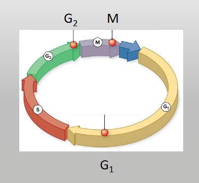
Cells internal checkpoints regulate their growth

Quality-control stopping points that evaluate a cell’s internal and external conditions before allowing it to advance to the next phase of division

  • G1 checkpoint

  • G2 checkpoint

  • M checkpoint

  • New cell must be identical to the original: fix errors

<p>Quality-control stopping points that evaluate a cell’s internal and external conditions before allowing it to advance to the next phase of division</p><ul><li><p>G1 checkpoint</p></li><li><p>G2 checkpoint</p></li><li><p>M checkpoint</p></li><li><p>New cell must be identical to the original: fix errors</p></li></ul><p></p>
71
New cards

Regulation at internal checkpoints

Mistakes that affect cell function (wrong number or mutated chromosomes) are regulated at 3 checkpoints:

  1. At end of G1

  2. At G2 to Mitosis transition

  3. In metaphase of mitosis


<p>Mistakes that affect cell function (wrong number or mutated chromosomes) are regulated at 3 checkpoints:</p><ol><li><p>At end of G1</p></li><li><p>At G2 to Mitosis transition</p></li><li><p>In metaphase of mitosis</p></li></ol><p></p>
72
New cards

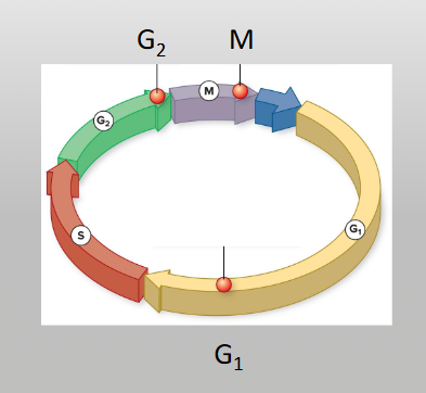
Control of the cell cycle

External signals can stimulate or inhibit the cell cycle

  • Death of nearby cells (such as a wound or cut in skin)

  • Release of growth hormones

  • Cell crowding

  • Internal factors also regulate progress of the cell cycle


<p>External signals can stimulate or inhibit the cell cycle</p><ul><li><p>Death of nearby cells (such as a wound or cut in skin)</p></li><li><p>Release of growth hormones</p></li><li><p>Cell crowding</p></li><li><p>Internal factors also regulate progress of the cell cycle</p></li></ul><p></p>
73
New cards

Control of the cell cycle

External signals can stimulate or inhibit the cell cycle

  • Death of nearby cells (such as a wound or cut in skin)

  • Release of growth hormones

  • Cell crowding

  • Internal factors also regulate progress of the cell cycle

<p>External signals can stimulate or inhibit the cell cycle</p><ul><li><p>Death of nearby cells (such as a wound or cut in skin)</p></li><li><p>Release of growth hormones</p></li><li><p>Cell crowding</p></li><li><p>Internal factors also regulate progress of the cell cycle</p></li></ul><p></p>
74
New cards

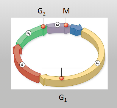
G1 checkpoint

Determines whether all conditions are favorable for cell division

  • External influences, adequate cell reserves and size are important

  • There is a check for DNA damage

  • A cell that does not meet all the requirements does not enter the S phase

<p>Determines whether all conditions are favorable for cell division</p><ul><li><p>External influences, adequate cell reserves and size are important</p></li><li><p>There is a check for DNA damage</p></li><li><p>A cell that does not meet all the requirements does not enter the S phase</p></li></ul><p></p>
75
New cards

G1 checkpoint

Determines whether all conditions are favorable for cell division

  • External influences, adequate cell reserves and size are important

  • There is a check for DNA damage

  • A cell that does not meet all the requirements does not enter the S phase


<p>Determines whether all conditions are favorable for cell division</p><ul><li><p>External influences, adequate cell reserves and size are important</p></li><li><p>There is a check for DNA damage</p></li><li><p>A cell that does not meet all the requirements does not enter the S phase</p></li></ul><p></p>
76
New cards

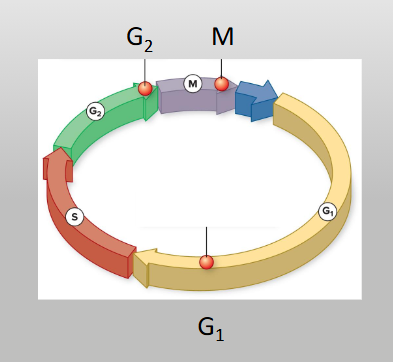
G1 checkpoint options

G1 Checkpoint determines whether all conditions are favorable for cell division

The cell has 2 options if conditions aren’t met:

  • Stop the cycle and try to fix the problem

  • Enter G0 (becomes dormant) and wait for signs that conditions are better

<p>G1 Checkpoint determines whether all conditions are favorable for cell division</p><p>The cell has 2 options if conditions aren’t met:</p><ul><li><p>Stop the cycle and try to fix the problem</p></li><li><p>Enter G0 (becomes dormant) and wait for signs that conditions are better</p></li></ul><p></p>
77
New cards

G1 checkpoint options

G1 Checkpoint determines whether all conditions are favorable for cell division

The cell has 2 options if conditions aren’t met:

  • Stop the cycle and try to fix theproblem

  • Enter G0 (becomes dormant) and wait for signs that
    conditions are better


<p>G1 Checkpoint determines whether all conditions are favorable for cell division</p><p>The cell has 2 options if conditions aren’t met:</p><ul><li><p>Stop the cycle and try to fix theproblem</p></li><li><p>Enter G0 (becomes dormant) and wait for signs that<br>conditions are better</p></li></ul><p></p>
78
New cards

The G2 checkpoint

  • Cell size and protein reserves are checked

  • G2 checkpoint makes sure that all chromosomes have been replicated and that the DNA is not damaged

  • If problems detected, the cell cycle stops while the cell completes DNA replication or repairs the damaged DNA


<ul><li><p>Cell size and protein reserves are checked</p></li><li><p>G2 checkpoint makes sure that all chromosomes have been replicated and that the DNA is not damaged</p></li><li><p>If problems detected, the cell cycle stops while the cell completes DNA replication or repairs the damaged DNA</p></li></ul><p></p>
79
New cards

The G2 checkpoint

  • Cell size and protein reserves are checked

  • G2 checkpoint makes sure that all chromosomes have been replicated and that the DNA is not damaged

  • If problems detected, the cell cycle stops while the cell completes DNA replication or repairs the damaged DNA

<ul><li><p>Cell size and protein reserves are checked</p></li><li><p>G2 checkpoint makes sure that all chromosomes have been replicated and that the DNA is not damaged</p></li><li><p>If problems detected, the cell cycle stops while the cell completes DNA replication or repairs the damaged DNA</p></li></ul><p></p>
80
New cards

The M checkpoint

At the end of metaphase

  • Determines whether all sister chromatids are attached to the spindle

  • The cycle will not proceed until kinetochores of sister
    chromatids are firmly attached to two spindle fibers

  • Failure to correct this, can lead to new cells getting wrong number of chromatids

<p>At the end of metaphase</p><ul><li><p>Determines whether all sister chromatids are attached to the spindle</p></li><li><p>The cycle will not proceed until kinetochores of sister<br>chromatids are firmly attached to two spindle fibers</p></li><li><p>Failure to correct this, can lead to new cells getting wrong number of chromatids</p></li></ul><p></p>
81
New cards

The M checkpoint

At the end of metaphase

  • Determines whether all sister chromatids are attached to the spindle

  • The cycle will not proceed until kinetochores of sister
    chromatids are firmly attached to two spindle fibers

  • Failure to correct this, can lead to new cells getting wrong number of chromatids


<p>At the end of metaphase</p><ul><li><p>Determines whether all sister chromatids are attached to the spindle</p></li><li><p>The cycle will not proceed until kinetochores of sister<br>chromatids are firmly attached to two spindle fibers</p></li><li><p>Failure to correct this, can lead to new cells getting wrong number of chromatids</p></li></ul><p></p>
82
New cards

G1 Checkpoint (restriction point) Summary

Checkpoint name/phase

  • G₁ Checkpoint (Restriction Point )

  • Late G₁ phase

Key questions asked by the cell

  • Is the cell large enough?

  • Are nutrient sufficient?

  • Is there growth factor signaling?

  • Is the DNA damaged?

Primary Cyclin-CDK complex involved

  • Cyclin D/ CDK4

  • Cyclin E/ CDK2


<p>Checkpoint name/phase</p><ul><li><p>G₁ Checkpoint (Restriction Point )</p></li></ul><ul><li><p>Late G₁ phase</p></li></ul><p>Key questions asked by the cell</p><ul><li><p>Is the cell large enough?</p></li><li><p>Are nutrient sufficient?</p></li><li><p>Is there growth factor signaling?</p></li><li><p>Is the DNA damaged?</p></li></ul><p>Primary Cyclin-CDK complex involved</p><ul><li><p>Cyclin D/ CDK4</p></li><li><p>Cyclin E/ CDK2</p></li></ul><p></p>
83
New cards

G2 Checkpoint (G2/M transition) - summary

Phase

  • Boundary of G2 and M phase

Key questions asked by the cell

  • Did the DNA replicate completely in S phase?

  • Is the replicated DNA free of damage or errors?

Primary Cyclin-CDK complex involved

  • Cyclin B / CDK1


<p><strong>Phase</strong></p><ul><li><p>Boundary of G2 and M phase</p></li></ul><p><strong>Key questions asked by the cell</strong></p><ul><li><p>Did the DNA replicate completely in S phase?</p></li><li><p>Is the replicated DNA free of damage or errors?</p></li></ul><p><strong>Primary Cyclin-CDK complex involved</strong></p><ul><li><p>Cyclin B / CDK1</p></li></ul><p></p>
84
New cards

M Checkpoint (Spindle checkpoint) - summary

Phase

  • During metaphase of mitosis

Key questions asked by the cell

  • Are all sister chromatids properly attached to spindle fibers?

  • Is there equal tension across the kinetochores?


<p><strong>Phase</strong></p><ul><li><p>During metaphase of mitosis</p></li></ul><p><strong>Key questions asked by the cell </strong></p><ul><li><p>Are all sister chromatids properly attached to spindle fibers?</p></li><li><p>Is there equal tension across the kinetochores?</p></li></ul><p></p>
85
New cards

Two groups of intracellular molecules regulate the cell cycle

  • Positive Regulators promote moving to next step of the cell cycle

  • Negative Regulators inhibit/stop moving to the next cell cycle


86
New cards

Positive Regulators

Molecules that promote, accelerate, or allow the cell cycle to progress to the next stage

  • promote moving to next step of the cell cycle

  • molecules—primarily proteins—that drive the cell cycle forward, promoting progression from one phase to the next or triggering events like chromosome condensation, nuclear envelope breakdown, and mitotic exit

  • proteins that drive a cell forward through its growth and division stages, primarily consisting of cyclins and cyclin-dependent kinases (CDKs)


87
New cards

Positive regulators

Specific proteins that promote or advance the cell cycle through its various checkpoints toward cell division

  • Promote moving to next step of the cell cycle

  • Proteins that drive a cell forward through its growth and division stages, primarily consisting of cyclins and cyclin-dependent kinases (CDKs)

  • Cyclins: proteins whose levels fluctuate (cycle) throughout the cell cycle. They bind to and activate CDKs

  • Cyclin-Dependent Kinases (CDKs): enzymes that remain at a constant concentration but are inactive until bound by a specific cyclin. Once activated, they phosphorylate target proteins to advance the cell cycle


88
New cards

Negative regulators

Inhibit/stop moving to the next cell cycle

  • molecules that halt, slow down, or inhibit cell progression through mitosis. They act as structural brakes at specific cell cycle checkpoints to prevent the cell from dividing when conditions are unfavorable (such as when DNA is damaged or the cell is too small)

  • proteins that halt or slow down cell division when the cell has DNA damage, lacks proper size, or faces adverse environmental conditions. Acting primarily as tumor suppressors, these molecules prevent damaged or abnormal cells from replicating

  • negative Regulators p53 and p21


89
New cards

Negative Regulators

Molecules that halt, delay, or inhibit the cell cycle to prevent the cell from dividing when conditions are unfavorable

  • Inhibit/stop moving to the next cell cycle

  • proteins that halt or slow down cell division when the cell has DNA damage, lacks proper size, or faces adverse environmental conditions. Acting primarily as tumor suppressors, these molecules prevent damaged or abnormal cells from replicating

  • negative Regulators p53 and p21


90
New cards

Cylins and cyclin-dependent kinases (Cdks)

  • Levels of these proteins fluctuate over the cell cycle

  • Internal and external signals can trigger increases in cyclin protein levels

  • core regulatory proteins that drive the cell cycle forward


<ul><li><p>Levels of these proteins fluctuate over the cell cycle</p></li><li><p>Internal and external signals can trigger increases in cyclin protein levels</p></li><li><p>core regulatory proteins that drive the cell cycle forward</p></li></ul><p></p>
91
New cards

Cyclins

A group of regulatory proteins whose concentrations rise and fall in a predictable, cyclical pattern throughout the cell cycle

  • Internal and external signals can trigger increases in
    cyclin protein levels

  • Function: they control the progression of a cell through the different phases of the cell cycle by partnering with enzymes

  • Interaction with CDKs: cyclins bind to and activate CDKs (cyclin-dependent kinases). CDKs are always present in the cell in a steady concentration, but they remain inactive until the right cyclin binds to them

  • Phosphorylation: once the cyclin-CDK complex forms, it adds a phosphate group (phosphorylates) to target proteins. This phosphorylation acts as a green light to drive the cell into the next phase (such as DNA replication or mitosis)


<p>A group of regulatory proteins whose concentrations rise and fall in a predictable, cyclical pattern throughout the cell cycle</p><ul><li><p><span> Internal and external signals can trigger increases in</span><br><span>cyclin protein levels</span></p></li></ul><ul><li><p><span><strong>Function:</strong> they control the progression of a cell through the different phases of the cell cycle by partnering with enzymes</span></p></li><li><p><span><strong>Interaction with CDKs:</strong> cyclins bind to and activate <strong>CDKs</strong> (cyclin-dependent kinases). CDKs are always present in the cell in a steady concentration, but they remain inactive until the right cyclin binds to them</span></p></li><li><p><span><strong>Phosphorylation:</strong> once the cyclin-CDK complex forms, it adds a phosphate group (phosphorylates) to target proteins. This phosphorylation acts as a green light to drive the cell into the next phase (such as DNA replication or mitosis)</span></p></li></ul><p></p>
92
New cards

Cylin-dependent kinases (CDKs)

A type of enzyme (kinase) that adds phosphate groups to target proteins (phosphorylation) to drive a cell forward through the cell cycle

  • Dependency on Cyclin: a CDK is inactive on its own. It only turns on when it binds to a regulatory protein called a cyclin to form an active CDK-cyclin complex

  • Constant Levels vs. Fluctuating Activity:

    • CDK levels stay relatively constant (present all the time) in the cell

    • Cyclin levels rise and fall predictably depending on the phase of the cell cycle

  • Checkpoints and Transitions: active CDK-cyclin complexes phosphorylate specific target proteins to pass cell cycle checkpoints (such as moving from G₁ to S phase, or triggering M phase/mitosis)


93
New cards

Cylins vs. Cyclin-dependent kinases (Cdks)

Levels of these proteins fluctuate over the cell cycle

  • Internal and external signals can trigger increases in
    cyclin protein levels

Cyclins

  • A group of regulatory proteins whose concentrations rise and fall (fluctuate cyclically) in a predictable pattern throughout the cell cycle

  • They bind to and activate Cdks, and they also help direct Cdks to specific target proteins needed during that particular phase of the cell cycle

Cyclin-Dependent Kinases (Cdks)

  • A family of enzyme kinases (specifically protein kinases) that are present in relatively constant concentrations throughout the cell cycle, but remain inactive on their own

  • Function: They become active only when they bind to a specific cyclin partner. Once activated, the Cdk-cyclin complex phosphorylates (adds a phosphate group to) target proteins. This phosphorylation acts as an on/off switch that drives the cell past checkpoints (such as G1, G2, and M checkpoints) and into the next phase of division

<p><span>Levels of these proteins fluctuate over the cell cycle</span></p><ul><li><p><span>Internal and external signals can trigger increases in<br>cyclin protein levels</span></p></li></ul><p><strong>Cyclins</strong></p><ul><li><p><span>A group of regulatory proteins whose concentrations <strong>rise and fall (fluctuate cyclically)</strong> in a predictable pattern throughout the cell cycle</span></p></li><li><p><span>They bind to and activate Cdks, and they also help direct Cdks to specific target proteins needed during that particular phase of the cell cycle</span></p></li></ul><p><strong>Cyclin-Dependent Kinases (Cdks)</strong></p><ul><li><p><span>A family of <strong>enzyme kinases</strong> (specifically protein kinases) that are present in relatively <strong>constant concentrations</strong> throughout the cell cycle, but remain <strong>inactive</strong> on their own</span></p></li><li><p><span><strong>Function:</strong> They become active only when they bind to a specific cyclin partner. Once activated, the <strong>Cdk-cyclin complex</strong> phosphorylates (adds a phosphate group to) target proteins. This phosphorylation acts as an on/off switch that drives the cell past checkpoints (such as G1, G2, and M checkpoints) and into the next phase of division</span></p></li></ul><p></p>
94
New cards

How positive regulators (CDKs) work

  1. Binding & Activation: as a specific phase of the cell cycle approaches, the matching cyclin is synthesized and binds to its partner CDK

  2. Target Phosphorylation: once activated, the CDK acts as a kinase, adding a phosphate group (PO₄³⁻) to specific target proteins inside the cell

  3. Triggering Transitions: this phosphorylation acts like an on/off switch—either activating proteins needed for the next step (such as DNA replication or mitosis) or inactivating proteins that block progression


<ol><li><p><span><strong>Binding &amp; Activation:</strong> as a specific phase of the cell cycle approaches, the matching cyclin is synthesized and binds to its partner CDK</span></p></li><li><p><span><strong>Target Phosphorylation:</strong> once activated, the CDK acts as a <strong>kinase</strong>, adding a phosphate group (PO₄³⁻) to specific target proteins inside the cell</span></p></li><li><p><span><strong>Triggering Transitions:</strong> this phosphorylation acts like an on/off switch—either activating proteins needed for the next step (such as DNA replication or mitosis) or inactivating proteins that block progression</span></p></li></ol><p></p>
95
New cards

How positive regulators (CDKs) work

  • As a cell prepares to divide, specific regulatory proteins called cyclins accumulate and rise in concentration in response to external or internal signals

  • The cyclin binds to a catalytic enzyme called a cyclin-dependent kinase (CDK). Without its specific cyclin partner, the kinase remains completely inactive

  • One the cyclin-CDK complex forms, an activating kinase adds a phosphate group (P) to the CDK. This triggers a structural change that opens up the kinases active site

  • The fully activated complex then transfers a phosphate group onto specific target proteins downstream. This phosphorylation alters the function of those target proteins- either activating or inhibiting them- to advance the cell cycle
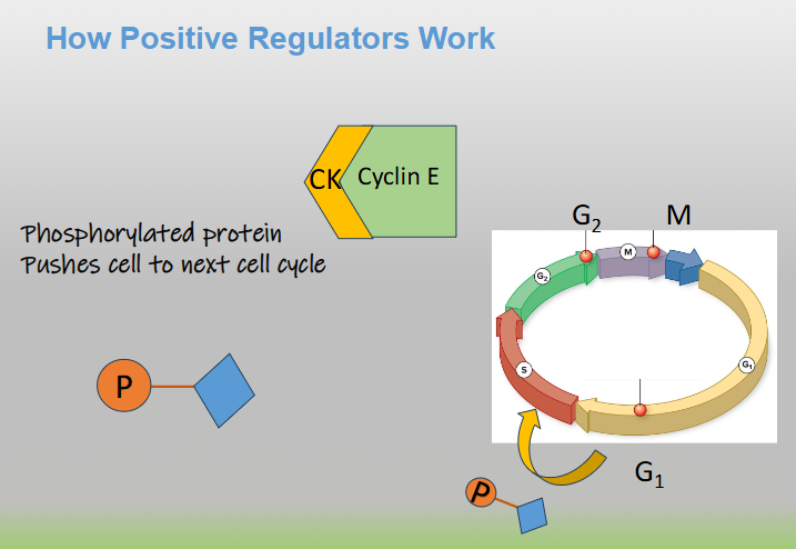

<ul><li><p>As a cell prepares to divide, specific regulatory proteins called cyclins accumulate and rise in concentration in response to external or internal signals</p></li><li><p>The cyclin binds to a catalytic enzyme called a cyclin-dependent kinase (CDK). Without its specific cyclin partner, the kinase remains completely inactive</p></li><li><p>One the cyclin-CDK complex forms, an activating kinase adds a phosphate group (P) to the CDK. This triggers a structural change that opens up the kinases active site</p></li><li><p>The fully activated complex then transfers a phosphate group onto specific target proteins downstream. This phosphorylation alters the function of those target proteins- either activating or inhibiting them- to advance the cell cycle</p></li></ul><p></p>
96
New cards

How positive regulators (CDKs) work

  1. Cyclin binds to cyclin kinase (CK)

  2. CK becomes phosphorylated (phosphate group is added) - phosphorylated proteins can activate target cells

  3. Target protein receives phosphate

  4. Phosphorylated protein pushes cell to next cell cycle


<ol><li><p>Cyclin binds to cyclin kinase (CK)</p></li><li><p>CK becomes phosphorylated (phosphate group is added) - phosphorylated proteins can activate target cells </p></li><li><p>Target protein receives phosphate </p></li><li><p>Phosphorylated protein pushes cell to next cell cycle </p></li></ol><p></p>
97
New cards

How positive regulators (CDKs) work

  1. Cyclin binds to cyclin kinase (CK)

  2. CK becomes phosphorylated (phosphate group is added) - phosphorylated proteins can activate target cells

  3. Target protein receives phosphate

  4. Phosphorylated protein pushes cell to next cell cycle


<ol><li><p>Cyclin binds to cyclin kinase (CK)</p></li><li><p>CK becomes phosphorylated (phosphate group is added) - phosphorylated proteins can activate target cells </p></li><li><p>Target protein receives phosphate </p></li><li><p>Phosphorylated protein pushes cell to next cell cycle </p></li></ol><p></p>
98
New cards

How positive regulators (CDKs) work

  1. Cyclin binds to cyclin kinase (CK)

  2. CK becomes phosphorylated (phosphate group is added) - phosphorylated proteins can activate target cells

  3. Target protein receives phosphate

  4. Phosphorylated protein pushes cell to next cell cycle


<ol><li><p>Cyclin binds to cyclin kinase (CK)</p></li><li><p>CK becomes phosphorylated (phosphate group is added) - phosphorylated proteins can activate target cells </p></li><li><p>Target protein receives phosphate </p></li><li><p>Phosphorylated protein pushes cell to next cell cycle </p></li></ol><p></p>
99
New cards

Negative Regulators p53 and p21

Work at the G1 checkpoint

  • Molecules that halt, delay, or stop the cell cycle to prevent a cell from dividing abnormally or uncontrollably

  • Unlike positive regulators (like cyclins and CDKs) that push the cell cycle forward, negative regulators act as critical biological “brakes” at checkpoints- specifically the G1 checkpoint- to ensure the cell has intact DNA before replicating its genome


100
New cards

Negative Regulators p53 and p21

Work at the G1 checkpoint

  • p53 and p21 are critical tumor suppressor proteins that act as negative regulators of the cell cycle, halting cell division at the G1 checkpoint in response to DNA damage. They prevent uncontrolled cell growth, working like a cellular brake system

  • p53 and p21 are critical tumor suppressor proteins that act as negative regulators of the cell cycle, functioning to halt cell division when DNA damage is detected. While positive regulators (like cyclins and Cdks) drive the cell cycle forward, negative regulators act like vehicle brakes to stop progression, ensuring genetic errors are not passed on to daughter cells