Chest and Abdomen- Exam 3- Bowl Gas Patterns
 Call Kai
 Learn
Learn Practice Test
 Spaced Repetition
 Match
Match Flashcards
 Knowt Play
1/79

There's no tags or description
Looks like no tags are added yet.
Name | Mastery | Learn | Test | Matching | Spaced | Call with Kai | Chat |
|---|
No analytics yet
Send a link to your students to track their progress
80 Terms
What type of gas pattern is normal but sometimes can be abnormal
Intraluminal
Where is it normal to see intraluminal gas pattern
Stomach and bowel.
Where is intraluminal gas pattern abnormal to be seen
Gallbladder, portal vein, urinary bladder
You should recognize what with intraluminal gas pattern
Displacement of normal bowel gas distribution due pathology
-ascites or masses
What gas pattern is always abnormal and most commonly found within the peritoneal cavity (intraperitoneal air)
Extraluminal
What gas patter is normal but when in excess it's abnormal
Intramural air (wall, always abnormal)
What is the last type of gas pattern that is always abdnormal
Abscess
Intestinal obstruction accounts for what percentage of emergency department visits for acute abdominal pain
15%
What is the most common obstruction for intraluminal
Small bowel obstruction
What is the AKA for ileus intraluminal obstruction
Paralytic ileus or adynamic ileus
Types of intraluminal large bowel obstructions
Volvulus: sigmoid and fecal
Colitis
Gastric outlet obstruction
Biliary tree intraluminal obstruction is called
Gallstone ileus
Extraluminal obstructive types
Perforation
Intramural gas
Types of perforation for Extraluminal
Intarperiotneal
Extraperitoneal
Explain intraperitoneal perforation
Within peritoneum covering of the bowels
Explain extraperitoneal perforation
Within the walls themselves
Type of intramural gas locations for Extraluminal
Pneumatosis coli
Ischemic bowel
Gas forming infections
Normal gas pattern

What is special with small bowel obstruction and radiographic findings
May be apparent 6-12 hours before clincal symptoms
Symptoms of small bowel obstruction
Abdominal pain, vomiting and abdominal distention
Important values for the dilates loops of small bowel to be considered small bowel obstruction
> 30 mm (3 cm) proximal (jejunum)
>25 mm (2.5 cm) distal (ileum)
Air fluid levels for small bowel obstruction
>25 mm (2.5 cm) in length
Must be at different positions in the same loop (step ladder appearance)
What is it called in small bowel obstruction when small bubbles are trapped between bowel loops
String of pearls
What is a sign that is less commonly found in small bowel obstruction
Fluid-filled loops= gas less abdomen
-fluid displaces the air and you can't see it anymore
-VERY ADVANCED OBSTRUCTION
What is the most common cause of all intestinal obstructions
Mechanical small bowel obstruction
About 80% of all bowel obstructions are
Small bowel obstruction
Who is most commonly having small bowel obstruction
Western countries: post surgical adhesion
Incarcerated hernias
Gallstone ileus
Foreign bodies
Intussusception
Intraluminal masses
Malignancy
What must you do on x ray in order to see incarcerated hernias that can cause small bowel obstruction
Include inguinal regions of abdominal and limbo pelvic x rays
Gallstones that worked out of bile duct in to GI tract and probably perforated out of it
Gallstone ileus
It is often difficult to distinguish between an ileus and ______ on plain film so
Mechanical bowel obstruction
-so CT is often sued
Lack of forward movement cause a….
Paralytic ileus (aka adynamic ileus)
What can cause paralytic ileus (aka adynamic ileus)
Stasis of bowel contents WITHOUT mechanical obstruction
Postoperative, drugs, metabolic states, intra-abdominal inflammation
Small bowel should be
Less than 3 cm
Large bowel should be
Less than 6 cm
Appendix should be
Less than 6 mm
Cecum should be
Less than 9 cm
Example g the 3 6 9 diameter rule for gas pattern findings
Small bowel should be less than 3 cm
Large bowel should be less than 6 cm
Appendix should be less than 6 mm
Cecum should be less than 9 cm
Small bowel obstruction has what type of location
Central location
What does a small bowel obstruction look like
Stacked coins
Why does a small bowel obstruction look like stacked coins
Valvulae conniventes extending from wall to wall (folds extend from wall to wall)
Accumulations in small bowel obstruction suggest
Adhesions secondary to intra-abdominal surgery, hernias, tumors and Crohn's disease
With large bowel obstruction, the air is normally in
Rectum and sigmoid
Describe location of large bowel obstruction
Peripheral location
What does large bowel obstruction look like
Haustral markings don't extend form wall to wall (folds don`t go all the way across)
Small bowel obstruction vs large bowel obstruction

Example of a patient in a standing position and how can you tell
Because of the positioning of the air fluid levels

Example of another small bowel obstruction with air fluid levels, what position are the patients in
Standing

Example of the air fluid levels on side lying

When changing positions for x ray, do the air fluid levels in bowel obstruction move within or outside of the bowels
Move within the bowels
What happens as more fluid fills up in bowel obstruction
It pushes the air up and out until its passed out
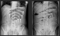
Distended small bowel, showing stacked coins and having a large bowel obstruction

Showing the difference between air fluid levels in standing and supine positioning

Example of string of pearls/beads sign with small bowel obstruction

String of beads sign is seen with
Small bowels obstruction
Example of a large bowel obstruction that is too wide of a diameter

Do you get air fluid levels in both small and large bowel obstruction
Yes
What is more common large or small bowel obstruction
Small bowel obstruction
Example of someone having bowel obstruction and ulcerative colitis, how can you tell?
The smooth appearance of the descending colon

One more example comparing small bowel obstruction and large bowel obstruction

Example of both large and small bowel obstruction showing air fluid levels

Air in the peritoneum is called
Pneumoperitoneum
How can you see if someone has pneumoperitoneum
You can see the top of the liver, normally this should not be seen

What is something that you see with pneumoperitoneum
Riggers sign (aka double wall sign)

Explain out riglers sign (aka double wall sign) works
Normally you cant see the walls cause of the water density of the walls being in contact with the water density of the peritoneum but with air in between the two structures it allows the different layers to be seen
Good example of a normal wall vs a riglers sign (double wall sign)
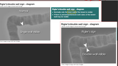
Riglers sign seen with pneumoperitoneum, showing both the luminal surface and the peritoneal surface

Falciform ligament sign can be made visible by
Large amount of free air in the peritoneal cavity

Example of a massive pneumoperitoneum (you can tell its massive because the whole liver and spleen are visible)

Pneumoretroperitoneum is most commonly
Trauma related
How can we tell something is pneumoretroperitoneum
When you can see the whole kidney, normally you should not be able to see the top of it as clear as we do in the x ray

Pneumoretroperitoneum seen on CT

Example of air through the fibers of the psoas muscle
I think this is seen with pneumoretroperitoneum

Another example of pneumoretroperitoneum plus a colostomy bag

Good example of pneumoperitoneum

Football sign example

Normal gastric air bubble showing an air fluid levels

It's okay to have some air where and not where
Okay to see some air under the diaphragm on the left (gastric air bubble)
But when air is on the right side of the patient then its a problem
Patients who have small livers (cirrhosis) or flattened diaphragm due to lung hyperinflation (emphysema) a void is created within the upper abdomen above the liver. This space may be filled by bowel and this bowel is air filled then it may mimic free gas What is this called
Chilaiditi`s phenomenon

How do you get a false rigler/double wall sign
One big loop of bowel that doubled back on itself

Sigmoid volvulus showing coffee bean sign
