CH 27: opportunistic molds
 Call Kai
 Learn
 Practice Test
 Spaced Repetition
 Match
 Flashcards
 Knowt Play
1/29

There's no tags or description
Looks like no tags are added yet.
30 Terms
Opportunistic fungi
rarely cause disease in healthy individuals
medical conditions in pts who are immunocompromised
found in high numbers in environment
inhalation
respiratory infx
pneumonia, sinusitis, bronchitis, eye infx, ear infx, and allergies
Aspergillus spp.
rapidly growing environmental mold
usually blue to green to black color macroscopically
identifiable by swollen vesicle at end of conidiophores

Aspergillus fumigatus
conidia in single rows that only cover 2/3 top of vesicle

Aspergillus flavus
circumferential conidia

Aspergillus niger
black macroscopic mold
2 rows of circumferential, dark conidia

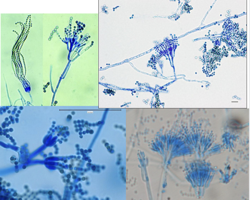
Penicillum spp.
branching conidiophores w/ elongated phialides and chains of microconidia
‘brush-like’
Fusarium spp.
canoe-shaped, multi-cellular, macroconidia

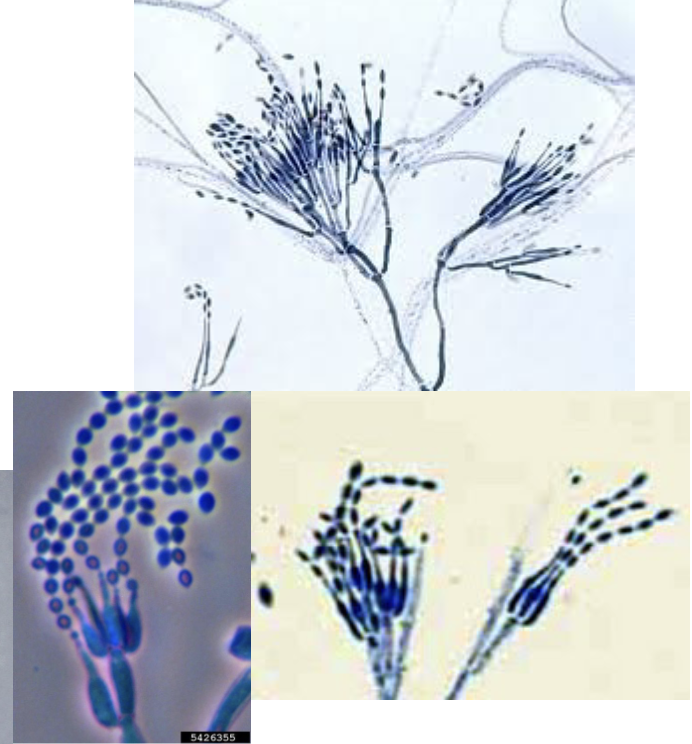
Paecilomyces spp.
branching conidiophores w/ elongated, flask-shaped phialides
long chains of oval conidia
Zygomycetes
group of opportunistic molds
very serious and fatal infx
hyphae are usually broad, no septation, and little to no branching
principal agents
Rhizppus spp.
mucor spp.
Rhizopus
large sporangia that arises directly from rhizoids

Mucor spp.
large spherical sporangia
NO rhizoids

Cutaneous mycoses
fungal diseases of keratinized tissues
collectively called ‘dermatophytes’
cause infx of hair, skin, and nails
Trichophyton
Microsporum
Epidermophyton
Cutaneous Mycoses infections
tinea pedis - athlete’s foot
tinea corporis - ringworm
tinea unguium - onychomycosis
tinea capitis - scalp infx
tinea barbae - barber’s itch
tinea cruris - jock itch
Trichophyton mentagrophytes
macroconidia: thin-walled, smooth, and cigar-shaped
microconidia: numerous , grape-like clusters

Trichophyton rubrum
macroconidia: thin-walled, smooth, and cylindrical to club-shaped
microconidia: sparse, club-shaped, and in singles

Epidermophyton floccosum
macroconidia: thin-walled, smooth, and club-shaped; ‘beaver tail’ clusters
microconidia: none

Microsporum canis
macroconidia: thick-walled, spindle-shaped, tapered ends
microconidia: unremarkable

Microsporum gypseum
macroconidia: moderately thick walls, fusiform shape w/ rounded ends
microconidia: numerous, but unremarkable

Subcutaneous mycoses
fungal infx of subcutaneous tissues
lesions are often dark in color, hard, or crusted
organisms are collectively called ‘dematiaceous molds’
‘dematiaceous’ refers to dark color
Mycetoma
subcutaneous infx that cause granulomatous, pus-filled lesions, and draining tracts
can be bacterial (actinomyces) or fungal
conidia/spores introduced to subcutaneous tissue by skin trauma
most common on feet/ankles
infx most common in Africa
most common causes
Pseudallescheria boydii
Exophiala
Acremonium
Madurella

Pseudallescheria boydii
asexual phase: rounded microconidia w/ truncated base
sexual phase: large cleistochecia filled w/ spores

Exophiala
short conidiophore w/ clusters of elongated conidia w/ rounded ends

Chromoblastomycosis
chronic infx characterized by hard, scaly, or tumor-like lesions
introduced via skin trauma
most common on feet/legs
tropical areas
principal agents:
Fonsecaea pedrosoi
Phialophora verrucosa
Cladophialophora carrionii

Fonsecaea pedrosoi
clusters of short chains of oval conidia from a single conidiophore

Phialophora verrucosa
vase-like conidiophore w/ cup shaped top

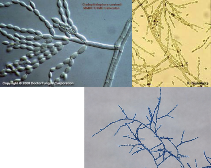
Cladophialophora carrionii
long, branching chains of oval conidia
Phaeohyphomycosis
subcutaneous infx that can become systemic
brain abscesses, endocarditis, etc
principal agents:
Alternaria
Bipolaris
Curvularia
Exophiala
Phialophora
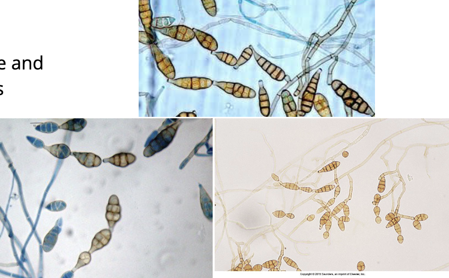
Bipolaris
oval, clustered, septate macroconidia from genticulate (bent) conidiophore
each conidia has 3-5 septations

Alternaria
chains of club-shaped conidia w/ transverse and longitudinal septations
golden brown color
Curvularia
rounded, septate conidia w/ 4 cells
distinct swelling in cells when mature
