Microbiology Lab Practical Study set
 Call Kai
 Learn
 Practice Test
 Spaced Repetition
 Match
 Flashcards
 Knowt Play
1/129

There's no tags or description
Looks like no tags are added yet.
130 Terms
What type of Lab are we?
BSL-2 Safety
Enter lab
wash hand --> lab coat --> gloves --> wipe desk
Leave Lab
wipe desk --> toss gloves --> lab coat --> wash hand
Turret with Objective Lenses
Magnifies the image for viewing by the eye. Our microscopes have four objective lenses -4x, 10x, 40x, and 100x.

Stage
Holds your slide

Mechanical Stage Control Knobs
Moves the stage around so you can look at different parts of your slide.

Eyepiece/Ocular Lens
Magnifies the image for viewing by the eye. In our microscopes, the magnification of this lens is 10x

Head of the Microscope
Holds lenses and optical components in place

Knob for adjusting Microscope Head
Holds the head to the rest of the microscope

Power Cord Storage
Keeps the power cord organized and tangle-free

Power Switch
Turns the microscope on or off

Brightness Adjustment
Controls light intensity for better image visibility

Condenser
Focuses light onto the specimen for clearer viewing
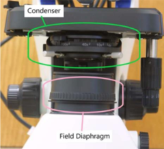
Field Diaphragm
Regulates the amount of light reaching the specimen. Closing/opening this aperture widens/narrows the light beam reaching your slide

Iris Diaphragm Control
Adjusts light aperture for contrast and clarity. A way to control the brightness of the light

Condenser Centering Knobs
Aligns condenser for optimal light focus
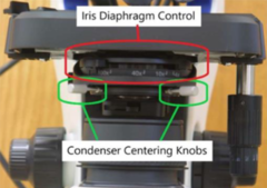
Condenser Focusing Knob
Fine-tunes the focus of the light source

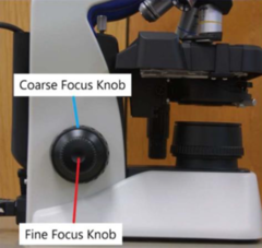
Coarse Focus Knob
Moves the stage or objective lens for rough focusing

Fine Focus Knob
Fine-tunes focus for clearer image details
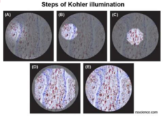
"Kohler" Illumination
Provides the best sample illumination
Evenly illuminated field
Bright image without glare
Minimum heating
Total Magnification = ocular power x objective lens power
Oil Immersion
Oil is required for the 100x Lens
● Decreases diffraction of light rays
● Fills the air gap between the specimen and the lens. The 100x Lens
● The only power you should use when determining size, shape, and staining characteristics of organisms
Always clean oil off with lens paper and lens cleaner
Smear Preparations (we are pros at this)
From a broth medium- Ensure bacteria are in suspension, transfer a drop or two onto a slide using a loop
From a solid medium- Add a small drop of water onto the slide, then use a loop to pick up a colony from your solid medium, and inoculate the water on the slide
Spread out your sample in the etched circle
A thin smear is a better smear- More likely to stain correctly and see individual cells
Air Drying ≠ Heat Fixation- Air Drying: occurs on the heat block, near body temp, to remove moisture- Heat Fixation: After air drying, the shelf above the incinerator is very hot, making the surface proteins of bacteria stick to the glass
Simple Staining
Almost any stain can be used, including- Crystal Violet, Safranin, Malachite Green, Carbol-Fuchsin, Methylene Blue, etc.
All cells are stained the same color
Good for determining size, shape, and arrangement of cells (for some organisms, daughter cells remain stuck together after division occurs)

Gram Stain
The most common differential stain - Used to distinguish between Gram-Positive and Gram-Negative organisms
Gram-Positive Organisms
Thick peptidoglycan layer, retains crystal violet after decolonization, end color is purple
Gram-Negative Organisms
Thin peptidoglycan layer, crystal violet washes out after decolorization and safranin counterstains, end color is pink
Gram Stain Steps (also good at this)
1. Flood the prepared slide with crystal violet for 1 min. This is the primary stain. Rinse off with DI water after 1 min
2. Flood prepared slide with Gram's Iodine for 2 min. This is the mordant, which fixes the crystal violet to the cell wall. Rinse off with DI water after 1 min
3. Decolorize using acetone-alcohol. The most important step rinse off with DI water almost instantly after application
4. Flood the prepared slide with safranin for 1 min. This is the counterstain. Rinse off with DI water after 1 min
Acid Fast Stains
Acid-fast organisms contain a large amount of mycolic acid in their cell walls
Mycolic acids make these organisms resistant to ordinary staining methods
Mycolic acid is a type of lipid
Ziehl-Neelsen Stain
Ziehl-Neelsen is a type of acid-fast stain
1. Carbol fuchsin stains every cell red
- Heating is used to help this primary
stain penetrate the mycolic acid layer
of acid-fast organisms
1. Acid alcohol decolorizes
- Only non acid-fast bacteria will be
decolorized, acid-fast bacteria remains
red
1. Methylene blue counterstains
- Non acid-fast organisms become blue

Kinyoun Stain
Kinyoun stain is a different type of acid-fast
staining
Same procedure as Ziehl-Neelsen EXCEPT
- No heat is required
- Tergitol, a type of detergent, is added
to the carbol fuchsin stain
- Tergitol softens they mycolic acid layer
allowing carbol fuchsin to stain the
acid-fast bacteria
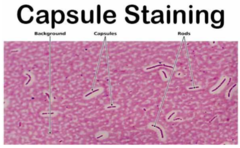
Capsule Stain
This method of staining only stains the
cells and the capsule remains colorless
Despite the name, the capsule does not
stain (a 'negative' stain)
Endospore Staining
Endospore appear green against the rest of the bacteria
- Malachite Green and heat are used to stain the endospore
- Safranin is used to stain the surrounding bacteria (vegetative cells)

Liquid Media
Contain no agar
Solid Media
Broth mixed with agar to solidify the
medium
Loop
Best for broth cultures,
streaking for isolation,
inoculation of small agar slants
Less likely to tear up the media
Needle
Best for placing organisms
deep into media, anaerobic
environment
- Stab and streak
Used for TSIA, LIA, and motility
Aseptic Technique (easy)
Start with sterilizing the loop or needle in the incinerator
Pick up you culture from the plate to tube with a loop or needle
- If tube media, hold the cap with your pinky finger
- Pass the opening of the tube in front of the incinerator opening
- Recap after obtaining your loopful of bacteria
Insert the loop or needle into whatever media you wish to innoculate
Re-sterilize your inoculating instrument
Streak for Isolation
Best way to obtain a pure culture and isolated
colonies and observe hemolysis
Flame the loop between each quadrant of the streak
- This ensures a dilution of bacteria between each
streak quadrant
Selective Medium
Contains at least one type of inhibitor
Inhibits the growth of certain types of organisms, while allowing the growth of other types of organisms
Differential Medium
Contains at least one potential substrate and indicator
Color change is observed
Does not inhibit the growth of any organism
Selective/Differential Medium
Contains at least one inhibitor AND at least one potential substrate and indicator
Inhibits certain organisms, allows for other types of organisms, AND indicates if the organism(s) that are capable of growth used the substrate or not
Controls
Controls allow for test results to be more clearly defined in the lab and ensure tests/reagents are working properly.
Review all of the tests that require the use of controls to be successful
● Catalase, Oxidase, Streptocard, ect.
What is the pH indicator used in carbohydrate broths?
Phenol red
What substrates can be found in carbohydrate broths in our lab?
Glucose, sucrose, or lactose
What indicates a positive reaction when a bacteria uses a carbohydrate?
Phenol red turns yellow due to acid by-products and a drop in pH
What indicates a negative reaction when a bacteria does not use a carbohydrate?
Phenol red becomes darker due to increased pH from protein utilization
What is the purpose of a Durham tube in phenol red glucose broths?
To determine if CO2 gas is produced
What should be compared to phenol red broth results?
TSIA results
What indicates a positive reaction in carbohydrate broth?
Yellow, Acidic
What indicates a negative reaction in carbohydrate broth?
Red, Alkaline
What does a Durham tube in phenol red glucose broths determine?
If CO2 gas is produced
TSIA determines
- Carbohydrate utilization and gas production
- Hydrogen sulfide production
- Atmospheric requirements
TSIA contains
- pH indicator: phenol red
- Hydrogen sulfide indicator: ferrous sulfate
- Substrates: 0.1% Glucose, 1% Lactose, 1% Sucrose, sodium thiosulfate
TSIA K/A
Bacteria is facultative anaerobe and uses glucose sugar only
Glucose is quickly used up on the slant, aerobic environment
- Bacteria resort to using peptones to produce energy
- Peptone usage is aerobic, requires oxygen, and occurs only
In the slant
- Creates alkaline by-products, K, red
The butt of the tube still has glucose
- Utilization of this carbohydrate creates acid by products, A, yellow
TSIA A/A or A/A+G
Bacteria is facultative anaerobe and uses glucose,
lactose and/or sucrose
- Bacteria uses the glucose first and continues to use
lactose and/or sucrose later
A large amount of acid by-products are generated
- A, yellow
Gas, CO2, will show as cracking in the media
- + G
TSIA K/N
Bacteria is an obligate aerobe due to no change *********** of the tube
- N
Some organisms cannot ferment glucose
The organism will use peptones in the slant to grow, peptones create the alkaline by-products
- K, red
Nitrate Test
The medium is rich in potassium nitrate
There are three results an organism can provide
1. Does NOT produce nitrate reductase
2. Produces ONLY nitrate reductase
3. Produces BOTH nitrate and nitrite reductase
After incubation Nitrate Reagents are added, these
reagents only react with nitrite
- Reagent A: Sulfanilic acid
- Reagent B: alpha-Naphthylamine
Durham tube collects N2 gas if it is produced

NO Nitrate Reductase
There is still nitrate in the medium
After adding reagents A&B there will be no color change
Zn dust will be added
If the medium turns red with Zn, the organism is nitrate reductase negative
Zn reduced nitrate to nitrite, then reagents A&B will react with nitrite turning red
ONLY Nitrate Reductase
The organism produces only nitrate reductase
Nitrate will be reduced to nitrite
Reagents A&B will react with nitrite turning red
The organism is nitrate reductase positive
BOTH Nitrate & Nitrite Reductase
Produces both nitrate and nitrite reductase
Nitrate will be reduced to nitrite, then further reduced to NO and N2
Reagents A&B only react with nitrite and will not change color
Zn dust is added, to react with nitrate, but no change will occur because it has been fully reduced
The organism is nitrate reductase positive and nitrite reductase positive
Nitrogen gas may collect in the Durham tube
Urea Test
Determine the ability of an organism to split urea by the enzymatic action of urease
- Substrate: Urea
- pH indicator: Phenol Red
- Contains glucose as a food source
Products of urea hydrolysis includes 2 ammonia molecules, 1 CO2 molecule, and 1 water molecule
Bacteria that produce urease will make the medium highly alkaline, turns hot pink
Bacteria that do not produce urease will use glucose if saccharolytic, turns yellow
Asaccharolytic bacteria that do not produce urease will use peptones, turns very light pink

Motility Test
Semi-solid medium used to determine if an organism is motile or non-motile
- Motile organisms have flagella
Tetrazolium salt creates a reddish pink color where the bacteria grows
Needle is used to stab the media and innoculate
Obligate aerobes will only grow where oxygen is present, motile organisms may not grow in the stab, but across the top of the media

Catalase Test (gram +)
Catalase enzyme is produced by some bacteria to detoxify peroxide anion
Add 1-2 drops of 3% hydrogen peroxide
Positive results: bubbles of oxygen gas
- Staphylococcus spp. or Micrococcus spp.
Negative results: no bubbles
- Streptococcus spp. or Enterococcus spp.
(Among medically important bacteria, the most common Gram + cocci are in the genera Staphylococcus, Micrococcus, Streptococcus, and Enterococcus.)

Coagulase Test (gram +)
Coagulase is not an enzyme, it is an extracellular protein
Coagulase test uses Rabbit Plasma to test the pathogenicity of Staphylococcus spp.
Positive test: rabbit plasma gels up, coagulates
- Potential pathogen
Negative test: rabbit plasma remains liquid
- Typically non-pathogenic
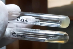
Mannitol Salt Agar (gram +)
Selective/Differential Medium
- Inhibitor: high salt concentration, prevents the growth of non-halophilic organisms
- Substrate: Mannitol
- pH Indicator: Phenol Red
Bacteria that grow & use mannitol substrate will turn the medium yellow, lowering the pH
Bacteria that grow & do not use the mannitol substrate will turn the medium bright pink, raising the pH

Antibiotic Susceptibility Testing:
Novobiocin (gram +)
Presumptive test for Staphylococcus saprophyticus
- Staphylococcus saprophyticus is the only Staphylococcus sp. resistant to Novobiocin
Zone of Inhibition is present for any Staphylococcus sp. other than Staphylococcus saprophyticus
- Staphylococcus saprophyticus will not have a zone of inhibition

Antibiotic Susceptibility Testing:
Bacitracin (gram +)
Presumptive test for Streptococcus pyogenes
- Streptococcus pyogenes is the only Streptococcus sp. susceptible to Bacitracin
Zone of Inhibition is not present for any Streptococcus sp. other than Streptococcus pyogenes
- Streptococcus pyogenes will have a zone of inhibition

Antibiotic Susceptibility Testing:
Optochin (gram +)
Presumptive test for Streptococcus pneumoniae
- Streptococcus pneumoniae is the only Streptococcus sp. susceptible to Optochin
Zone of Inhibition is not present for any Streptococcus sp. other than Streptococcus pneumoniae
- Streptococcus pneumoniae will have a zone of inhibition

Bile Esculin Agar (gram +)
Selective/Differential Medium
- Inhibitor: Bile Salts
- Substrate: Esculin
- pH Indicator: Iron
Used to differentiate Streptococcus sp. from Enterococcus sp. and Streptococcus bovis
Growth on surface: an organism can grow in the presence of bile salts
Growth on surface & black color: the organism can break down esculin to esculetin, esculetin reacts with iron to make a black precipitate.
All Enterococcus sp. hydrolyze esculin and turn the medium black.
Enterococcus sp. Grow very slowly, so growth may not be visible on the surface of the medium.

CAMP Test (gram +)
Determines if an organism can make CAMP factor
Presumptive test for Streptococcus agalactiae
Middle line of Staphylococcus aureus produces double zone hemolysis, both complete and incomplete hemolysis
- CAMP factor from S. agalactiae will interact with the incomplete hemolysis to produce complete hemolysis in an arrowhead shape

Streptocard Enzyme Test
Carbohydrates found in the cell wall of most Streptococcus sp. can be used to classify the bacteria into Lancefield Groups
Agglutination will occur when the appropriate antiserum is mixed with a sample containing antigens extracted from a Streptococcus sp. Of the matching group

RapID ONE
Rapid identification of unknown bacteria
- Advantages: easy, fast, saves incubator space, less waste, large database
- Disadvantages: very expensive RapID One strip is designed for the identification of oxidase-negative
Enterobacteriaceae (new name of the
group is Enterobacteriales)

Scoring a RapID card
Each + indicates that the points listed above it are added
Each box in the red row should reflect the sum of the +
boxes above it
This results in a 7 digit code that is entered into the database
- The code for the RapID test to the right is 4415551

Relative Size
Smallest
Viruses
Bacteria
Fungi
Parasites
Largest
Hemolysis
Some organisms produce hemolysins that lyse RBCs
○ Most hemolysins are phospholipase enzymes that break
down phospholipids of RBC cytoplasmic membranes
○ Other hemolysins are porins, which create channels in the RBC cytoplasmic membrane, allowing contents to leak out
Goal?
○ Nutrients from RBC, specifically iron from hemoglobin
No Hemolysis
Most organisms are non-hemolytic and get nutrients from something other than RBC
Sometimes referred to as Gamma reaction

Complete Hemolysis
Complete degradation of hemoglobin and lysis of RBC
Clearing of red color from the media
Sometimes referred to as Beta hemolysis

Incomplete Hemolysis
RBC are lysed, but NO enzymatic effect on hemoglobin
Hemoglobin degrades to methemoglobin naturally, which
has a green color
Sometimes referred to as Alpha hemolysis
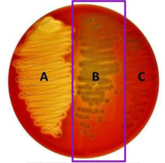
Double Zone Hemolysis
Very few organisms will show two zones of hemolysis
- One enzyme diffuses through the agar rapidly and lyses RBC (produces incomplete hemolysis)
- Another enzyme diffuses slowly and breaks down hemoglobin (produces complete hemolysis)

Oxidase Test (Gram -)
Used to test for Cytochrome c Oxidase
Used for differentiation of Enterobacteriales (formerly called Enterobacteriaceae) from Pseudomonadaceae
Timing is important, so controls are needed!

MacConkey
Selective/differential
- Selective inhibitory chemicals: bile salts and crystal violet that inhibit growth of gram + organisms and most non-enteric organisms.
- Substrate: lactose
- pH indicator: Neutral red
Results
- Did it produce isolated colonies?
- Lactose Fermenters: lowers pH and turns pink
- Non-lactose Fermenters: raises pH and turns yellow/translucent

Eosin Methylene Blue
Selective/differential
- Eosin and methylene blue serve as pH indicators and inhibits growth of gram + bacteria and most non-enteric bacteria
- Substrate: Lactose
Results
- Did it produce isolated colonies?
- Lactose fermenters: blue black or pink colonies with dark centers
- E. coli (sometimes other fecal coliforms): green metallic sheen
- Non-lactose fermenter: colorless/translucent colonies
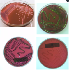
Salmonella-Shigella (SS)
Selective/differential
- Inhibitors: brilliant green dye and bile salts that inhibit the growth of gram + organisms, and most non-enteric organisms
- Substrates: lactose, sodium thiosulfate
- pH indicator: Neutral red
- H2S indicator: ferric citrate
Results
- Did it produce isolated colonies?
- Lactose Fermenters: lowers pH and turns pink/red colonies
- Non-lactose fermenters: raises pH and turns colorless/yellow
- H2S producing bacteria: colonies will have black center

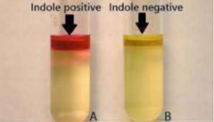
IMViC: Indole
Goal: Determine if an organism can split indole from tryptophan
- Substrate: tryptophan
We add Kovac's reagent to this test
- If the reagent turns red within one minute, indole is present
IMViC: Methyl Red Test and Voges Proskauer
These tests use the same broth, which has NO pH indicator

What is the procedure for the Methyl Red Test?
Add 1 or 2 drops of methyl red.
What indicates a positive result in the Methyl Red Test?
pH less than 4.5, remains red.
What does a negative result in the Methyl Red Test indicate?
Orange or yellow; does not use mixed acid pathway.
What is the procedure for the Voges Proskauer Test?
Add VP reagent I and II.
What indicates a positive result in the Voges Proskauer Test?
Acetoin is being produced, turns pink/red.
What does a negative result in the Voges Proskauer Test indicate?
No Acetoin, turns yellow/brown.
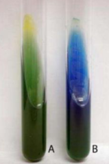
IMViC: Citrate
Citrate is a carbohydrate
- If the organism can use citrate as its sole source of carbon for energy and metabolism, it will make acid by-products and CO2, turning the medium blue
Despite the use of carbohydrate, the pH does increase because of a secondary reaction between the CO2, Na, and water
CO₂ + H₂O + Na⁺ → NaHCO₃ (sodium bicarbonate)
Lysine Iron Agar
LIA determines
- Identification of bacteria in Enterobacteriales (formerly called Enterobacteriaceae)
LIA contains
- pH indicator: Bromocresol purple
- Hydrogen sulfide indicator: Ferric ammonium
citrate
- Substrates: Lysine, sodium thiosulfate, small
amount of glucose
NO organism will produce both LDA and LDC
LIA Purple/Purple [LDA - / LDC+]
Saccharolytic, facultative anaerobe
- During fermentation of glucose, acidic byproducts activate LDC, which removes a carboxyl group from lysine to form cadaverine
- Cadaverine is alkaline and will increase the pH of the butt
- Bacteria on the slant will quickly use up the glucose then resort to peptones for energy, making the final slant pH alkaline

LIA Purple/Yellow [LDA - / LDC -]
Saccharolytic, facultative anaerobe
- Bacteria do NOT produce LDC and will NOT
be activated by acidic conditions to produce
adaverine
- The butt will remain acidic
- Bacteria on the slant will quickly use up the glucose, then resort to peptones for energy, making the final slant pH alkaline

LIA Port Wine/Yellow [LDA + / LDC -]
Saccharolytic, facultative anaerobe
- Bacteria do NOT produce LDC and will NOT be activated by acidic conditions to produce cadaverine
- The butt will remain acidic
- Bacteria on the slant will quickly use up the glucose then resort to peptones for energy, producing alkaline by-products, but see below
Some bacteria produce LDA
- LDA deamination process creates only slightly acidic byproducts
- The combination of peptone usage on the slant AND LDA slight acid makes the portwine color of the slant

LIA Summary

Ornithine Decarboxylase
ODC Broth
- Substrate: Ornithine, some glucose
- pH indicator: Bromocresol purple
Decarboxylase enzymes are activated by acidic, anaerobic conditions
ODC Positive: glucose is initially used and makes medium acidic pH which activates ODC
- This removes the carboxyl group from ornithine to form putrescine, an alkaline compound
- pH is increased, medium turns purple
ODC Negative: glucose is initially used and makes medium acidic pH, but there is no ODC to activate
- pH will remain low, medium will stay yellow

Virus Characteristics
Not the same as bacteria
Genome of DNA or RNA(not both)
Can only replicate inside a host cell
Some are surrounded by an envelope
Cytopathic viruses destroy the host cell when new virions are released
Most are very small
Bacteriophage
A virus that infects bacteria
Required for our Plaque Assay Experiment
- Plaque assay allows us to determine a virus titer, which is a measure of the concentration of the virus
- Allows us to demonstrate the process of viral replication
What is a plaque?
- An area where the host cells have been ruptured
- More bacteria are destroyed than are created by division of healthy bacterial cells
- We observed this plaque-forming unit, PFUs, which represent the number of virions in this sample