Histology and Layers
 Call Kai
 Learn
Learn Practice Test
 Spaced Repetition
 Match
Match Flashcards
 Knowt Play
1/113

There's no tags or description
Looks like no tags are added yet.
Name | Mastery | Learn | Test | Matching | Spaced | Call with Kai | Chat |
|---|
No analytics yet
Send a link to your students to track their progress
114 Terms
Tissue
An aggregation of cells and cell products of similar structure and embryonic origin that perform a common function
Histology
The Study of tissue, especially their structure and arrangement
Four Primary Types
Epithelium, Connective, Muscle, Nervous
Epithelium
Protects body from microbes and gives Hydration Ex: Skin
Connective
Protects, supports and binds other tissue together bones, cartilage, tendons, fat and blood
Muscle
Contracts to produce movement and exert force, Ex Bones, Cardiac Muscle
Nervous
Transmits electrical impulses for fast communication
Epithelial Tissues are found where?
Body Coverings
Body Linings
Glandular tissue
Charicteristics and Functions of epithelial tissue
Cells fit closely together(Sheet like cover)
Tissue layer always has one free surface(apical surface)
The lower surface is always bound by a basement membrane
Avascular(Meaning no blood supply)/ Can regenerate easily if well nourished
Functions are to protect (Through diffusion)
Absorbtion
Filtration
Secretion
Epithelium Characteristics
Epithelium lines pathways that are open to the external environment ( GI Tract, Respiratory tract, etc)
Endothelium
(Lines the inside) Epithelium that lines the blood vessels, heart and lymphatic vessels
Mesothelium
Lines the cavities. Epithelium that lines peritoneum (abdomen cavity, pleura, lung cavity) and paricardium(heart cavity)
Layers
Simple- one layer
Stratified- more than one
Shape of cells
Squamous- flattened ( squeezed nucleus)
Cuboidal- cube shaped ( normal nucleus)
Columnar- cloumn like ( oval shape towards basement membrane)
Epithelial types
Simple Squamous- inside blood vessels
Simple cuboidal- lines ducts ex. kidney, tubules and mucous glands
Simple columnar- lining - of small intestine
Pseudostratified- columnar- trachea- bronchi

Simple Squamous
Single flat layer of cells
Usually forms membranes
lines lungs and capilaries

Squamous definition
The epithelium is a single layer of flat, or squamous cells
The peripheral cytoplasm of each cell is so attenuated that it is usually not resolvable; the nucleus is usually the only cellular structure available
Nucleus usually has a flattened appearance due to the width of the cells
No speciallized structures at the top like cilia or microvilli
Simple Cuboidal
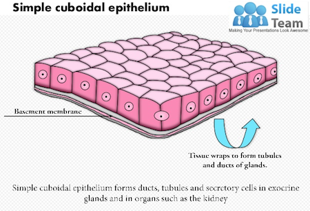
Single layer of cube- like cells common in glands and their ducts
Forms walls of kidney tubules
Covers the ovaries
Cuboidal
This epithelium consists of a single layer of cuboidal cells resting on an undetectable basement membrane
The height of each epithelial cell is nearly equal to the width of the cell
The cells are tightly packed, forming a continuous wall of cells
The Nucleus is spherical and sits at the center of the cell
The nuclei of adjacent cells are relatively close together, certaininly closwer than in a simple squamous epithelium, aligned in a single row

Simple Columnar
Single layer of tall cells
Often includes goblet cells, which produce mucus
Lines digestive tract

Columnar
The Epithelial containing a single layer of columnar cells ( the cell’s height are significantly greater than the width) and rests on a basement membrane, cells are tightly packed, forming a continuous sheet of cells that effectively form the protective lining of the stomach
The nucleus is generally ovoid and sits at the lower to mid portion of the cell

Pseudo stratified
Single layer, but some cells are shorter than others often looks like a double cell layer, sometimes ciliated, the respiratory tract may function in absorption or excretion
Cilia of the respiratory epithelium beat constantly towards the pharynx and nasal passages, creating a continuous flow of mucus in that direction
Presence of goblet cells secreting mucus over the surface of the epithelium

Stratified Squamous
Cells at the free edge are flattened found as a protective covering where friction is common (Skin, Mouth and Esophagus)


Transitional Epithelium
Shape of cells depends on the amount of stretching
Lines organs of the urinary system (ureter, bladder, part of the urethra)
Glandular Epithelium
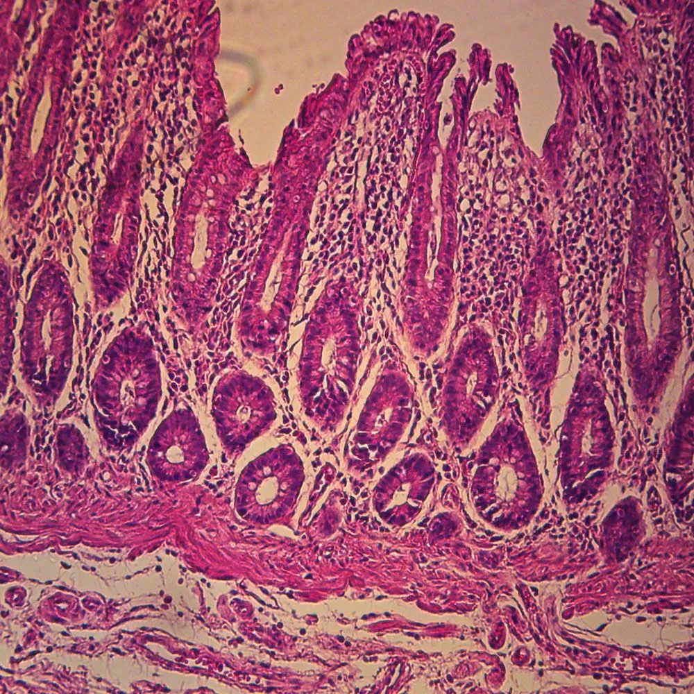
Gland- one ore more cells that secretes a particular product
Two major gland types
Endocrine- ductless and secretions are hormones
Exocrine- Empties through ducts at the epithelial surface Includs oil and sweat glands.
Connective Tissue
Found everywhere in the body
Includes the most abundant and widely distributed tissues
Functions of Connective
Binds body Tissue
Supports the body
Provides protection
Where is connective located?
Commonly found under epithelium
Special characteristics
Variations in blood supply
Vascularized (rich with blood vessels)
Avascularized( no blood vessels)
affects the ability to repair itself
Extracellular matrix (non living material that surounds cells)
Matrix main elements(individual numbers, variables, expressions, or characters organized inside its grid of rows and columns)
Ground substance
inorganic mostly water along with adhesion protiens and mostly polysacharide molecules
Collagen Fibers
Thick, appear parallel and are strong, not stretchy (in skin) usually pink under microscope
Elastic Fibers
Cells spread apart, stretchy reticular fine fibers in soft organs such as spleen (support network thin fibers)
Collagen
Most abundant protein in our body
Type 1 Collagen
mostly abundant in bones, tendons, skin and ligaments
Type II
mostly abundant in cartilage (Especially hyaline and articular)
Type III
Abundant with reticular fibers in soft tissues
Reticular fiber - fine collagenous fibers that form a delicate internal support system
Fine delicate thin fibers that forms supporting network Found in soft organs like the spleen
Lining cells
Within a protective layer called the Lacuna
Cell that makes the fibers
Osseous Tissue
Composed of Bone cells in Lacunae (Cavities)
Hard Matrix of Calcium Salts( Make rigid)
Large numbers of collagen fibers
Used to protect and support the body
Matrix of Bone
Innorganic material salts provide bone’s hardness
Organic collagen fibers provide bone flexability
in demineralization
Denature colagen by heating and bones will be brittle
Bone is not completely solid since it had small spaces for vessels and red bone marrow
Spongy bone had many such spaces -compact bone has very few such spaces


Hyaline Cartilage
The most abundant type of cartilage, characterized by a glossy, semi-translucent matrix containing fine Type II collagen fibers and chondrocytes situated inside lacunae. It provides smooth, flexible support and reduces friction; found on the articular surfaces of long bones, the costal cartilages of the ribs, the nose, trachea, larynx, and embryonic skeleton.

Elastic Cartilage
A flexible variety of cartilage containing a dense network of elastic fibers within its extracellular matrix, allowing it to withstand repeated bending while maintaining its shape; found in the external ear (auricle) and the epiglottis.

Fibro Cartilage
he strongest and most durable type of cartilage, composed of dense parallel bundles of thick Type I collagen fibers and chondrocytes housed in lacunae. It provides exceptional tensile strength, cushioning, and shock absorption to withstand heavy compression; found in intervertebral discs, the pubic symphysis, and the menisci of the knee.

Dense Connective Tissue
class of connective tissue characterized by tightly packed collagen fibers with relatively few cells (mainly fibroblasts) and minimal ground substance, providing high tensile strength and resistance to pulling forces; categorized into regular (found in tendons and ligaments), irregular (found in the dermis of the skin and joint capsules), and elastic connective tissue (found in the walls of large arteries).

Adipose Tissue
Matrix is areolar
Tissue in which fat globules predominate
Many cells contain large lipid deposits
Insulates the body
Protects some organs
Serves as a site of fuel storage

Reticular Connective
Delicate Network of Interwoven fibers
Forms stroma (Internal supporting network) of lymphoid organs
Lymph roles Spleen and bone marrow
Blood
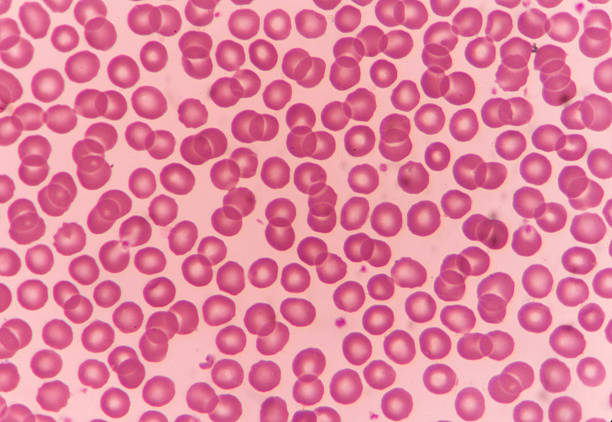
Bone cells surrounded by a fluid matrix
Fibers are visible during clotting
functions as the transport vehicle for materials
Epithelial membrane
An epithelial membrane is a thin layer of tissue composed of an epithelial tissue sheet bound to an underlying layer of connective tissue
Serous membrane
a thin, double-layered tissue that lines closed body cavities not open to the outside world and covers the organs inside them
Found as the paricardial sack which prevents friction when the heart beats, pleural membranes around the lungs, mesentaries attaching the intestines
Mucous membrane
a moist, soft-tissue layer that lines body cavities, tracts, and organs open to the external environment
Forms structure of GI tract and Respiratory passageway
Muscle Tissue
40% of your body weight
Fibers contract to produce tension
associated tissues or tendons
Cells have the ability to contract ( contractability)
Have the ability to respond to a stimulus (irratibility)
Muscle cells (myocytes) have the ability to contract
Function
Locomotion
Other body movements
Posture and Body support
Heat Production

Skeletal Muscle
Location= attached to bones via tendons
Voluntary movement (unique)
cells in bundles
long and cylindrical
Fibers are striated-Alternating light and dark bands
Each myofibril is multinucleated
Short and intense reaction

Smooth muscle
Walls of internal organs (except heart)
Involuntary movement
Non striated
Long, spindle shape
Single nucleus(uninucleated)
Two layers at 90 degrees
Constrict of dilate lumen
Propels substances along
Stracts like draw string bag
Smooth muscle can tense and relax but had greater elastic properties than striated muscle
Thi is important in organ systems like the urinary bladder where contractile tone must be preserved
Actin and myosin form continous chains with the smooth muscle cell which are achored at the dense bodies

Cardiac Muscle
Striations
Involuntary
One nucleus
Intercalated disks (unique) link cells together
Communication so all cells contract together in a rhythym
Deep center
Bundles branched
Heart muscle

Nervous Tissue
Neuron= Specialized cell of Nervous System
Cells with the ability to:
Respond to Stimuli (irritability)
Transmit impulses (conductivity)
Ability to transmit impulses through an electrochemical impulse

Parts of a Neuron:
Dendrite, Cell Body, Axon
Cell Body
Also known as the soma, this part of the neuron contains the nucleus, which controls the cell’s activities and contains the genetic material. The soma also contains organelles that support and maintain the neuron’s functioning
Axon
This part of the neuron looks like a long tail and sends messages from the cell. The axon terminal is the end part of the axon that makes a synaptic connection with another cell
Dendrites
These parts of the neuron look like branches of a tree and receive messages for the cell. Dendrites branch as they move towards theirtips and have leaf like structures on them called spires
Tissue Repair
Depends on Vascularization of Tissue Repair
Vascularization of tissues
The body's process of restoring architecture and function after an injury, occurring through two main mechanisms: regeneration (replacing lost tissue with the same cell type) or repair/scarring (replacing lost tissue with fibrous connective tissue)
Regeneration
Repaired by same type of cells, cells continue to function normally
On Epithelial tissue
Exhibits rapid mitosis with original function retained
Although avascular, vessels are nearby
Occurs in: Skin, Respiratory tracts, liver, glands, mucous membranes and blood vessels
Fibrosis
Fibrosis tissue knits damaged parts together (scar tissue)
Poor vascularization with slow repair and non functional tissue occurs in connective tissue layers not bone, adipose and areolar

Areolar Tissue
Areolar connective tissue is the most common type of loose connective tissue in vertebrates, acting as the body's universal packing material to bind, support, and cushion organs.
Wound Healing
Hemostasis, Inflammation, Granulation, Regeneration or Fibrosis
Hemostasis
Timeline: Begins immediately after injury (lasts from minutes to a few hours).
What happens: Blood vessels constrict (vasoconstriction) to reduce blood loss. Platelets stick together and form a plug, while clotting proteins like fibrin create a stable seal (blood clot) over the wound.
Goal: Stop bleeding and create a temporary barrier against bacteria.
Inflammation
Timeline: Lasts about 1 to 5 days (or up to a week).
What happens: Blood vessels dilate (vasodilation) to let immune cells enter the site. White blood cells (neutrophils and macrophages) clear out bacteria, dead tissue, and foreign debris.
Signs: Common symptoms include redness, warmth, swelling, and pain.
Goal: Prevent infection and prepare the tissue for new growth.
Proliferation
Timeline: Lasts from about 3 to 21 days (several weeks).
What happens: The body rebuilds the damaged area. Fibroblasts build new collagen and an extracellular matrix, while new blood vessels grow (angiogenesis) to supply oxygen and nutrients. Granulation cells make new capillaries, fibroblasts Macrophages digest original clog (phagocytosis) and scar tissue comes where the clot was
Goal: Close the wound and restore tissue structure.
Vasodilation increased( more blood vessels)
Regeneration or Fibrosis
Tissue repair after an injury results in either regeneration (restoring normal structure and function) or fibrosis (forming excessive scar tissue), depending on the severity of the damage, the type of tissue, and cellular signals.
Acute
a sudden break in the skin or soft tissue that progresses through normal healing stages and typically heals within 8 to 12 weeks (often much faster, within 1 to 4 weeks) with minimal scarring such as an abrasion or cut
Chronic
an injury that fails to progress through normal healing or does not show significant improvement within 4 weeks to 3 months, often becoming stuck in a prolonged inflammatory state (veneous and and aretorial ulcers)
Hypertrophic
Extra connective tissue forms within the original wound area
Keloid Scar
Extra connective tissue extends beyond the original wound
Body Membranes
Epithelial membrane
Cutaneous membrane
Serous membrane
Connective tissue membrane
Mucous membranes
moist, soft-tissue linings that cover internal cavities, organs, and tracts which are open or exposed to the external environment
Respiratory tract: Nose, sinuses, throat, trachea, and lungs
Digestive tract: Mouth, lips, esophagus, stomach, and intestines
Reproductive and urinary tracts: Uterus, urethra, and rectum
Sensory cavities: Conjunctiva of the eyes and the middle ear / Eustachian tube
Cutaneous membrane
Skin ,an epithelial tissue membrane that forms the body's outer protective boundary formed by keratinized stratified squamous epithelium
Connective Tissue Membrane
ules
surounding joints
in synovial joints
Integumentery System
The integumentary system is the body's largest organ system, forming a physical barrier between the internal body and the external environment.
Skin Functions
Protects deeper tissues from
Mechanical damage
Chemical damage
Bacterial damage
Thermal Damage
Ultraviolet radiation
Desiccation- prevents drying out or lysis because of keratin
Aids in heat regulation
Aids in excretion of urea and uric acid
Synthesizes vitamin d
Epidermis
The outermost, avascular (no blood vessels) cellular layer made of stratified squamous epithelium. It includes sub-layers like the stratum corneum and stratum basale.
Dermis
Deep connective tissue The thick inner connective tissue layer containing blood vessels, nerves, hair follicles, and sweat glands.
Hypodermis
A deeper layer of adipose and areolar tissue that anchors the skin to underlying muscle and bone while providing insulation. [1, 2, 3, 4]
Stratum Basale
Location: Deepest layer, attached to the basement membrane.
Cells: Contains stem cells (basal cells) that constantly divide to produce new keratinocytes, along with melanocytes (pigment producers) and Merkel (tactile) cells. [1, 2]
Stratum spinosum
Location: Just above the stratum basale.
Cells: Keratinocytes linked together by desmosomes (giving a spiny shape under a microscope); contains immune-defending Langerhans (dendritic) cells.
Function: Provides strength and flexibility.
Stratum Granulosum
Location: Above the stratum spinosum.
Cells: Cells flatten out and fill with keratohyalin granules and lamellar bodies.
Function: Marks the transition where cells die and release waterproofing lipids.
Stratum lucidum
Location: Present only in thick skin (palms of hands and soles of feet) between the granulosum and corneum.
Cells: Thin, translucent layer of dead, flattened keratinocytes
Stratum corneum
Outermost surface of the skin.
Cells: Composed of 20 to 30 layers of dead, fully keratinized, scale-like cells (corneocytes) that constantly shed.
Function: Acts as the primary barrier against pathogens, UV rays, and water loss.
Dermis Papilarry layer
Pain receptors capilary loops
Reticular layer
Blood vessels Glands Nerve receptors

Blister
A bubble of fluid underneath the outer layer of skin, Friction causes the layers of skin to seperate and fill with fluidn
Frostbite vs Hypothermia
Hypothermia is a fancy way of saying you have an abnormallt low body temperature
core temperature of 95 or less
lead to cardiac and respiratory failure
Frostbite is frozen body rissue, causing injury by freezing, often affects your hands and feet
Burn
tissue damage caused by heat, chemicals, electricity, sunlight, or radiation
Associated dangers (Dehydration immeadiate)
Circulatory ahock (Life threatening after 24 hours

First Degree Burn
affects only the outer layer of your skin, called the epidermis. like sunburn

Second degree
damages both the outer layer of skin (epidermis) and part of the inner layer (dermis), causing deep redness, swelling, fluid-filled blisters, and a shiny, moist surface that is very painful to the touch. No pain sometimes due to damaged receptors
Third degree
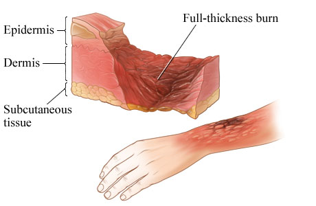
a severe, full-thickness injury that destroys the top layer of skin (epidermis), the middle layer (dermis), and reaches down into the deep fatty layer (hypodermis).

Fourth degree
A fourth-degree burn is a critical, life-threatening injury that penetrates past all skin layers into fat, muscle, tendons, and bone.
Critical Burn
Burns are only considered critical if over 25% of the body has 2nd degree burns
If there are over 10% third degree
There are 3rd degree on hands and faces
Rule of 9s
Way to determine the extent of burns Body is divided into 11 areas, 10 = 9% pubic area is 1%
Skin Cancer
Cancer = abnormal cell mass Skin cancer most common
Benign
does not spread, distinct smooth regular borders
malignant
Moves to other parts of the body grows faster than a benign tumor