Reproductive system
 Call Kai
 Learn
 Practice Test
 Spaced Repetition
 Match
 Flashcards
 Knowt Play
1/65

There's no tags or description
Looks like no tags are added yet.
66 Terms
gonads
Function: to make gametes ( sex cells )
in males = spermatozoa
in females =ova ( egg cells )
Also the site of sex hormone production: testosterone, estrogen, and progesterone
have one copy of chromosomes, hence called haploid cells ( 23 chromosome: 22 autosomes, code for all our traits, 23 sex chromosomes)
When the cell is fertilized, you get a
diploid cells ( 46 chromosome ), zygote ( fertilized egg)

gametogenesis
The making of gametes occurs through meiosis

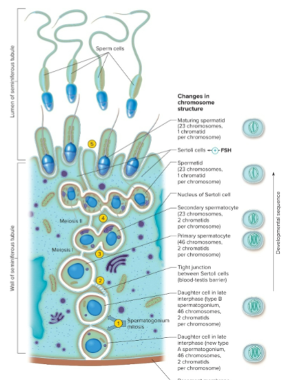
primary spermatocytes & primary oocytes are called
germ cells

explain this photo


spermatogonia
diploid stem cells, go through differentiation and mitotic division


primary spermatocytes
has double the chromosomes & undergoes meiosis 1 ( has double bc it goes through M1 and M2 ) 92 chromosomes in total
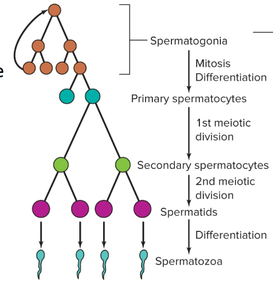

secondary spermatogenesis
goes through meiosis 2 → creating spermatids

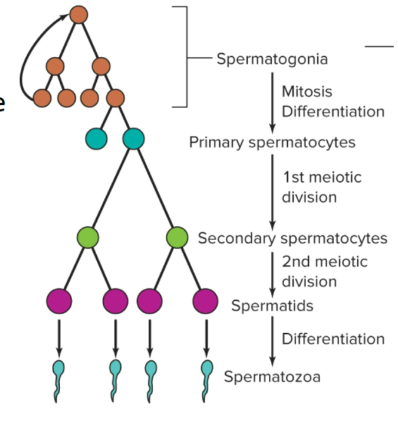
spermiogenesis
The Turing of spermatid into actually spermatids what is released into the seminiferous tubules (still not mature yet)

spermiogenesis
The Turing of spermatid into actually spermatids what is released into the seminiferous tubules (still not mature yet)
sex differentiation
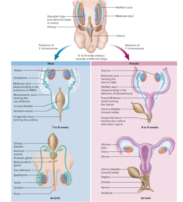
Primitive gonads are undifferentiated, neither male or female
In utero, cells will have a Wolffian duct, Müllerian duct, and gonadal ridges
Y chromosomes will have the SRY gene, which makes a protein called the testis-determining factor
is not present; testosterone & AMH are not produced, and Müllerian ducts develop into the female reproductive tract; gonadal ridges develop into ovaries
testiest determining factor
causing the differentiation of our gonadal ridge, causing it to form into the testes, and once they are formed, they will start secreting testosterone & anti-Mullerian hormone ( AMH), which causes the Müllerian ducts to degenerate
cryptorchism
if the testes fail to descend end are loc in the upper pelvic cavity

androgens
Hormones that create a masculinizing effect include testosterone
Mainly secreted by the testes, but they are also secreted by the adrenal gland just not as potent (which is secreted in females )
Dihydrotestosterone (5-alpha-reductase) is the more potent form of an androgen; it is what testosterone is converted into
estrogen
made by females in their ovaries & secreted by the placenta during pregnancy, but the ovaries make a small amount of testosterone in females
Some of the testosterone will be converted into estrogen by aromatase (enzyme)
Male testes secrete a small amount of estrogen (aromatase does the same here)
progesterone
a steroid hormone just like testosterone, secreted by the ovaries and the placenta during pregnancy

How are gonads stimulated to perform their function(Explain this photo )
Neurons in the hypothalamus release kisspeptin, which stimulates the neurons that make gonadotropin-releasing hormone

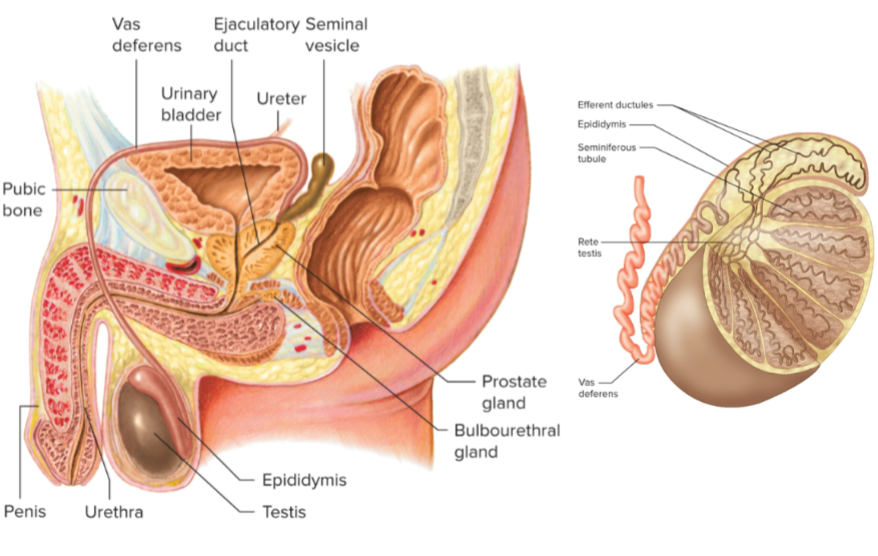
explain this


seminal vesicles
provides fructose for providing energy to the sperm cells,
alkaline fluid ( used to neutralize the acidic environment of the vaginal canal), prostaglandins help protect the sperm cell from the female’s immune system, also enhances the motility of the sperm cell as they’re swimming through the female reproductive tract,
fibrinogen, a gel-like constituent of seminal fluid

prostate gland
Release zinc to make sure the process of spermatogenesis occurs accurately ( support spermatogenesis)
low zinc level = low sperm count
Prostate-specific antigen ( PSA) is an enzyme that helps break down prostatic secretions in the male reproductive tract
As males grow older, their prostate gland enlarges, which can cause it to secrete more PSA. An abnormally high level of PSA can indicate cancer

bulbourethral glands
secret fluid before ejaculation even begins pre-ejaculation fluid, which cleans the urethra bc it is shared by both semen and urine, also helps lubricate it

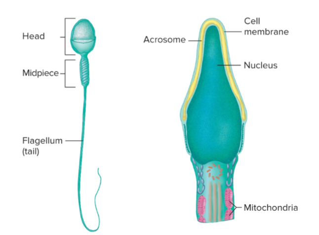
acrosome
is a cap on the sperm cell that contains hydrolytic enzymes that have to be released over the oocyte to break down the outer shell of the oocyte for fertilization to take place


midpiece
has an abundance of mitochondria where fructose is used to make ATP for their journey
Sertoli cells
Respond to FSH, bc they have FSH receptors; when FSH from the pituitary reaches these cells, it causes cell production
Also makes androgen-binding proteins, which bind to testosterone, allowing testosterone to protect sperm cells and provide longevity to sperm cells
Also makes inhibin, a hormone that inhibits FSH production
Leydig cells
are found between seminiferous tubules and have receptors for LH
When LH is secreted from the anterior pituitary, it binds to Leydig cells and stimulates them to secrete testosterone
Some testosterone diffuses across the Sertoli cells and activates sperm production
Testosterone & FSH are important for sperm production


structure
Sertoli cells create a structure bound by tight junctions, creating a barrier between developing sperm cells and blood outside the seminiferous tubules ( aka blood testes barrier )
Protecting the developing sperm cell from being attacked by the male immune system, marijuana, nicotine, and cocaine can pass through this barrier and affect sperm production

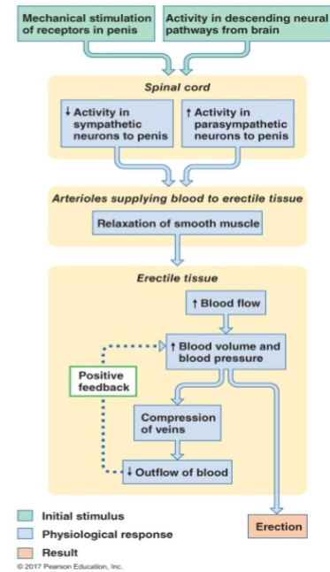
erection ( explain pic)
Can come from mechanical stimulation or stimulation from the brain \controlled by the autonomic NS; both are spinal reflexes
Parasympathetic neurons secrete nitric oxide, which binds to the arterioles of the penis, causing vasodilation, allowing a large amount of blood to enter the penis erection)
vessels that drain the penis are compressed, preventing blood from leaving the penis ( an erection is maintained through a positive feedback loop )
An erection is a parasympathetic response

transport of sperm
As sperm cells reach the lumen of the seminiferous tubule, they are not fully mature or motile yet
Sertoli cells secrete a fluid that creates pressure in the lumen of the seminiferous tubules, which helps move the sperm cell closer to the epididymis
Maturation takes 2 weeks
Ejaculation is under the control of the sympathetic nervous system, becomes more active at the time of ejaculation, and causes contraction of the smooth muscle of the epididymis, the smooth muscles of the vas deferens and ejaculatory ducts, which helps the sperm move through the male reproductive tract (go-go squeeze)
Increased sympathetic activity causes the seminal vesicles and the prostate to secrete their product ( add products to sperm cell)
Ejaculation: the discharge of seminal fluid from the urethra

andropause
will continue making sperm cells all their life
testosterone grows dramatically during puberty
around 30, testosterone reaches its peak
After 40, testosterone levels will decline slowly
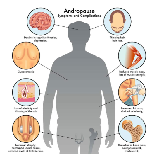
Low testosterone levels cause
Symptoms of middle age ( aka andropause )
not going to have all the symptoms
lower energy levels
depression
libido is going to be low
cognitive concentration issues
might start accumulating more body fat
hair loss
losing muscle
low testosterone =
lower sperm count and quality

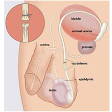
vasotomy
a procedure where the vas deferens is cut and tied ( cauterized hem), there is still pleasure, just no sperm
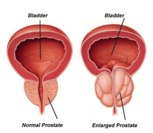
benign prostatic hypertrophy ( BPH)
A condition where the prostate gland enlarges ( seen in elderly men), age-related in this instance, not cancerous
can press and squeeze on the urethra, causing retention of urine; feel like they need to pee, causing them to need to pee more often, dribbling of urine
have to void completely; complete loss of control of the sphincter


Explain Pic ( how the oocyte moves)
have a limited numbler of gamtes only fertial a couple of days out of the mount
dmatesa are developed by the follicle found in the ovaries, and when matured released into the uterus tube ( aka fallopian tube)
in the fallopian tube where the oocute enconters sperm cell ad fertilication takes place
the fertilized eff traves dowbthe tube and reaches the uterus ant attaches tothe inner ling pf te uterus


endometrium
the inner most layer of the uterus, if their is no pregancy thus loses it superfical layer ( what women shead evey mounth


Myometrium
The middle layer of the smooth muscle contracts during childbirth, helping push the baby out

cervix
most inferior portion of the uterus helps hold the developing fetus inside the uterus
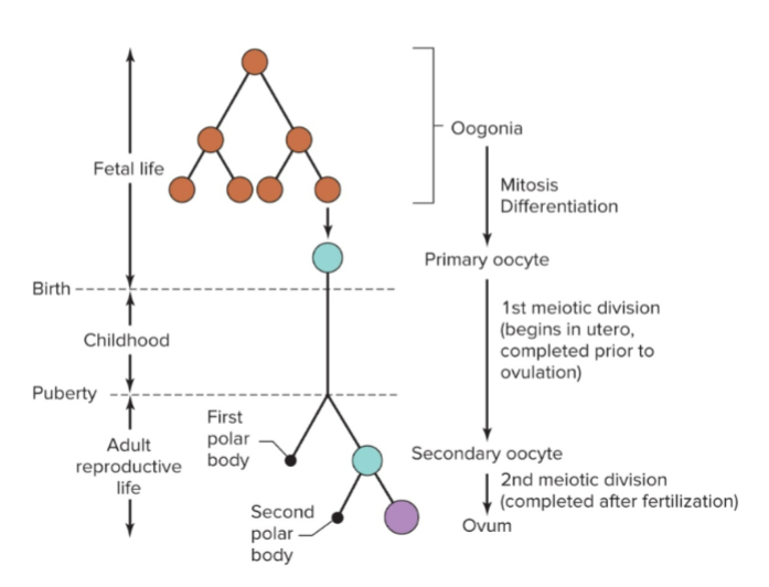
During fetal life, we have
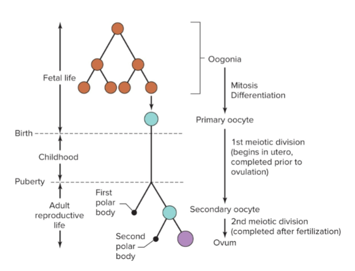
Oogonia ( stem cells) divide by meiosis and start differentiating to create 2- 4 million primary oocytes


Primary oocytes begin meiosis during fetal life, but
stop in meiosis ( prophase 1 ), aka meiotic arrest)
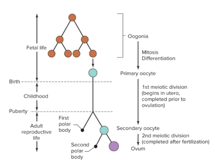
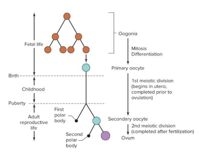
Half of the oocytes go through
Apoptosis (programmed cell death)


When a female is born, she has
1-2 million primary oocytes ( will have 46 chromosomes) housed in the primordial follicles


Between birth & puberty, these primary oocytes will go through
atresia ( degeneration ), where around 400,00 primary oocytes remain in the ovaries, but only 400 will get released
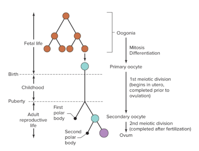

When females begin puberty, it’s due to an increase in
hormones, and each month before a female ovulates, there is a surge of LH that triggers meiosis to continue creating secondary oocytes


One secondary oocyte gets all
The cytoplasm components survive, mature, are released from the ovaries, and go through atresia
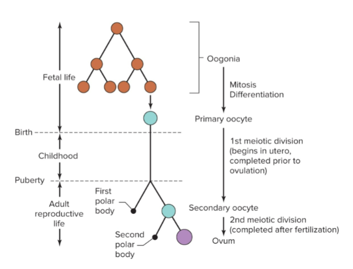
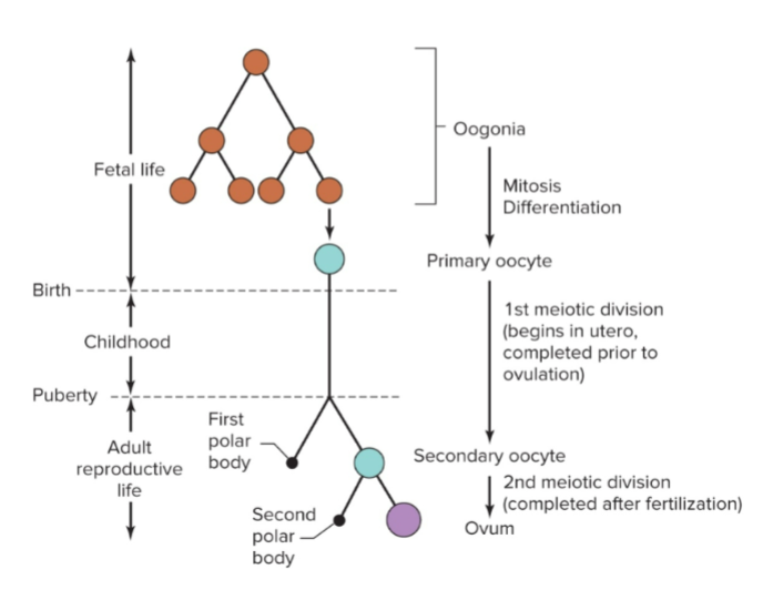
After the secondary oocyte gets fertilized,
it completes meiosis 2 bc it was a secondary oocyte; it will have another polar body, which undergoes atresia


Explain this pic
We have a set number and use all oocytes, compared to males
pre-made primary oocytes
only get one gamete, and it’s a cyclical event
only one oocyte matures and, if fertilized, gets to complete mitosis
only need one oocyte
happens every month
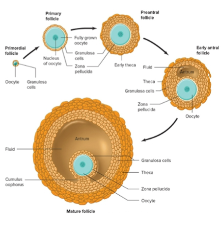
follicle growth
At the beginning of every menstrual cycle, under the influence of FSH and LH, 10-12 primordial follicles begian the process of development but only one will fyll develop and become mature oocyte and get fertilized
primary follicle is a single layer of squmamous cell ( graulosa cells ) it is surround and support the developing oocytes ( the same proimary oocytes aresete in meiosis 1)


explain this picture
As the oocytes grow, they will start to proliferate and start to secrete something called zona pellucida (a glycoprotein that makes the shell) that wraps around and covers the actual oocytes
Granulosa cells have receptors for FSH, which stimulates them to secrete estrogen from the developing oocyte
The outermost granulosa cells differentiate and become theca cells, which have receptors for LH, which stimulates them to release testosterone (androgens); testosterone is converted to estrogen → causing estrogen to increase slowly
Estrogen nourishes the follicle
FSH and estrogen are needed for oogenesis
As the oocyte continues to grow, a small fluid-filled space will start to grow, called the antrum; the fluid is called follicular fluid ( bc its in the follicles ) and accumulates more fluid until it eventually pushes against the oocyte, pushing it closer against the wall of the follicle


development of the oocyte
A dozen prenatal follicles begin development in a month, but only one will become dominant and reach full maturation; other follicles will go through atresia
99% of primordial follicles a person is born with will go through atresia
Mature follicles = Graafian follicle ( which gets ovulated) , before ovulation, the oocyte will finish its first round of meiosis

fraternal twins
aka dizygotic twins
Females mature 2 oocytes in one month, and can be fertilized by two different sperm, basically separate pregnancies at the same time
Not identical; each has their own amniotic sac
identical twins
aka monozygotic twins
One egg fertilized by one sperm divides into two cells and develops in the same amniotic sac; identical twins are always the same sex
ovarian cycle
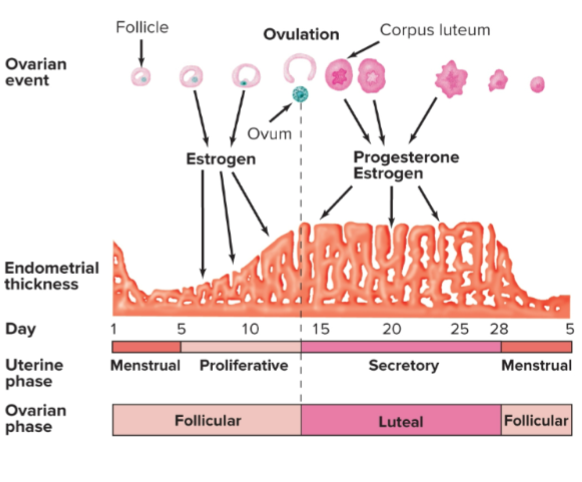
events that take place during the menstrual cycle

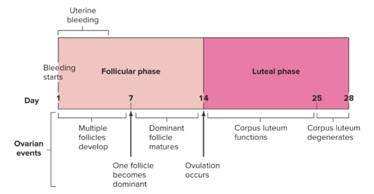
follicular phase
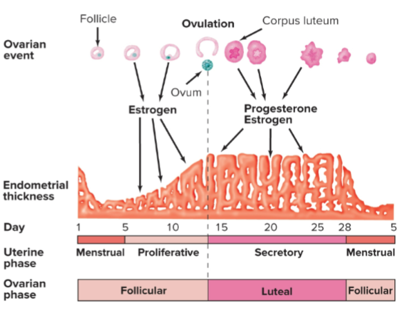
Beginning on the first day of menstruation all the way to the day of ovulation
first 4-5 days, when the female is menstruating and follicles are developing in the ovaries under the influence of FSH
No pregnancy, so follicles have to mature (multiple follicles have to mature )
Mid-follicular phase: one of the follicles becomes dominant and mature and ovulates on day 14
Finalizing maturation and becomes a Graafian follicle, and is ovulated


luteal phase
Ovulation to the next menstruation, typically around 28 days, but can vary
Named after the corpus luteum
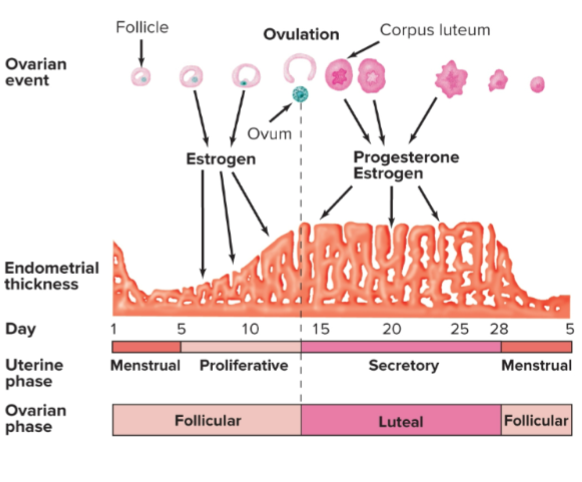
Day 14: an oocyte is released, but we still have remnants of the follicle, aka corpus luteum, which contains granulosa cells that will start secreting estrogen and progesterone
Granulosa cells also secrete inhibin
Progesterone production is important in the luteal phase bc it affects the endometrium (innermost lining of the uterus ), helping thicken the uterus; the new layer is called the functional layer
also causes angiogenesis ( the formation of new blood vessels)
helps create a nice, inviting layer in case implantation occurs, inviting and ready for the early-stage embryo (prepping for implantation )
If no implantation, the corpus luteum degenerates and stops releasing hormones, becoming the corpus albicans ( no progesterone or estrogen)
Levels of progesterone and estrogen drop, causing another menstruation cycle

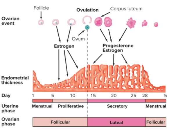
concentration of hormones during the menstrual cycle
At the beginning of a menstrual cycle, LH & FSH start to rise → stimulating the follicles in the ovary to grow, helping the development of the granulose and the theca cells to develop for us
theca cells respond to LH and start to secrete testosterone, which is converted to estrogen by granulosa cells ( which themself also secrete estrogen )
estrogen levels increase during the follicular phase
estrogen helps thicken the endometrium in the first half of the cycle, where the functional layer is starting to replenish itself imp in helping to rebuild
an increase of estrogen will negatively feedback into the anterior pituitary to slow down the secretion of FSH and LH
Inhibin release by granulosa cells negatively feeds back into the anterior pituitary to slow down FSH
As estrogen increases, it has a positive effect of LH to increase in the blood; the sharp increase, aka LH surge, happens on day 13, which is going to induce ovulation; when ovulation occurs, LH drops bc its not needed anymore
After ovulation, there is an initial decrease in estrogen but a rise in progesterone; then estrogen eventually picks back up
Estrogen & progesterone work to thicken the endometrium even more, and with an increase in progesterone → angiogenesis in the new functional layer occurs
In the luteal phase, a rise in inhibin bc the corpus lutum us secreting that inhibin to prevent menstruation even if you are pregnant
Progesterone inhibitory effect on FSH & LH during the luteal phase
Estrogen, progesterone, and inhibin all negatively feedback to the anterior pituitary to inhibit the production of FSH and LH during the luteal phase

mensural phase
Begins when menstruation occurs, around 5 days. It is also related to the follicular phase of the ovarian cycle. The functional layer is shed, severing blood vessels, which causes bleeding in the endometrium

proliferative phase
Begins when menstruation ends until ovulation; estrogen increases, thickening the uterine lining; cervical mucus becomes thinner, helping facilitate the entry of the sperm cells
Ovulation occurs quickly ( half an hr)
When the Graafian follicle leaves the ovary


secretory phase
Considered in the luteal phase
Estrogen and progesterone are being secreted by the corpus luteum, making it thick, plush, and very hospitable
Endometrial glands get bigger and secrete a fluid that contains a lot of glycogen that acts like an energy source for the embryo
angiogenesis
cervical mucus gets thicker and creates a plug, preventing microorganisms from reaching the embryo; if there is fertilization, it protects the developing embryo
If fertilization does not occur, the corpus luteum degenerates to the corpus albicans, which doesn’t secrete estrogen & progesterone, causing their levels to drop
- removing the inhibitory effects of FSH & LH, allowing them to rise again
allowing a new batch of follicles to begin the follicular phase again

progesterone
a steroid hormone that causes the thickening of the endometrial lining, creating a new functional layer
increasing secretion within the endometrium
angiogenesis
cervical mucus thickening
calming effect on the myometrium smooth muscle; it is unable to contract unnecessarily (relaxing it), important for implantation

fertilization
Once an oocyte is ovulated, it is only viable for 24 to 48 hrs.
Takes place in the uterine tube, specifically in the ampulla ( middle region )
sperm that survive and get to the egg start moving through the granulosa cells
sperm cells have protein heads that bind to the zona pellucida; once bound, the acrosome on the sperm cell will release enzymes over the zona pellucida that will create an opening for the head of the sperm to enter the oocyte
After the sperm enters the zona pellucida, it causes changes in it, making it harder to prevent other sperm cells from entering it
midpiece and tail stay outside of the zona pellucidum and meiosis II happens, and it’s all done


early embryonic development
Fimbriae create currents that pull ovulated eggs into the tube; peristaltic movement helps move the egg down the reproductive tract
When the fertilized egg goes through mitosis
after meiosis, it replicates through mitosis, identical cells
Trophoblast cells get sticky and stick to the uterine wall; cells that make a cavity and inner mass of cells
blastocyte inner layer + trope blasts outer layer


fetal and maternal tissues in the formation of the placenta
Trophoblast cells grow into the endometrium, forming chorionic villi & projection that have lots of blood vessels; in the myometrium, they come very close to the maternal blood vessels
Blood vessels close enough for waste/ nutrients to exchange between mom and baby; no physical contact


placenta has two parts
The maternal part and the fetal placenta with chorionic villi in endometrial tissue (embryonic tissue and chorionic villi )
umbilical cord contains two umbilical arteries and one umbilical vein, helping carry blood to and from the fetus
Amniotic sac: the fetus is growing in a sac filled with amniotic fluid, which helps cushion the embryo against physical trauma

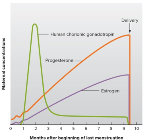
concentration of hormones during pregnancy
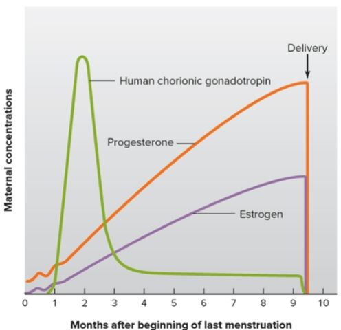
HCG is secreted by the trophoblast at the time of implantation; communication with the corpus luteum they don’t degenerate continue secreting estrogen and progesterone
HCG is picked up by the maternal blood and urine; confirms pregnancy 7 to 14 days post-implantation, as detected in home pregnancy kits
corpus luteum continues its release of HCG into the placenta; it is fully formed. Somewhere after the first 10 weeks, the placenta is fully formed and will take over the secretion of estrogen and progesterone
Corpus luteum degenerates, causing a drop in plasma levels of HCG
When progesterone & estrogen drop in level, it will induce the contraction of the uterus
Will also affect mood in women, causing postpartum depression of baby blues

parturition
giving birth
Amniotic sac ruptures
onset of labor: uterus starts contracting, starting from the top in a wave-like fashion, moves down to push the baby down against the cervical wall, eventually causing the cervix to dilate
abdominal muscles also contracting, helping dilate the muscle further, pushing on the cervical canal
pressure in the uterus positively feeds back to the posterior pituitary to release oxytocin → more oxytocin causes more contractions, pushing the baby in the cervical canal → causing more oxytocin to be released, making contractions greater and stronger
Contraction causes detachment of the endometrium, causing contraction of the uterus, delivering the placenta
Decreased levels of oxytocin


expulsion
feats passing through the birth canal
