topic 2A: membranes and transport
 Call Kai
 Learn
 Practice Test
 Spaced Repetition
 Match
 Flashcards
 Knowt Play
1/50

There's no tags or description
Looks like no tags are added yet.
51 Terms
single vs double membrane organelles
SINGLE
ER
vacuole
DOUBLE
chloroplast
mitochondria
nucleus
functions of cell membrane
to control movement of substances into / out of the cell / organelle
for many chemical reactions to take place
flexible to allow cells to change shape (e.g. in phago/endo/exocytosis)
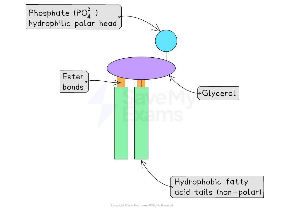
structure of phospholipid
hydrophilic (polar) head:
phosphate (PO43−) + glycerol
hydrophobic (non-polar) tails:
2 fatty acids (saturated — straight ; unsaturated — bent)
functions of membrane proteins
GATED CHANNELS
channel proteins
movement of ionic, polar, charged substances across membrane
by making pores / channels in proteins
some can be open or shut based on cell conditions
ACTIVE CARRIER SYSTEMS
active transport through carrier proteins
requires energy
RECEPTOR PROTEINS
makes cells sensitive to certain hormones
ENZYMES
catalyse certain reactions
GLYCOPROTEINS
recognition markers
for cells to recognise each other

how proteins are embedded into bilayer
middle section of protein:
hydrophobic non-polar R-groups ⇒ interact with hydrophobic non-polar tails
top & bottom:
hydrophilic polar R-groups ⇒ interact with hydrophilic polar tails
(if channel protein) inner tunnel:
hydrophilic polar R-groups ⇒ allow polar + water-soluble substances to pass through
substance type transport mechanism
small non-polar / lipid-soluble
simple diffusion
e.g. O2/ CO2/ steroid hormones / vitamin A, D, E, K
small uncharged polar
e.g. water
simple diffusion (slower bc polar) / facilitated diffusion
charged ions
e.g. K+/ Na+/ Cl−/ Ca2+
facilitated diffusion
large polar
e.g. glucose / amino acids
facilitated diffusion
macromolecules
vesicles (-cytosis)
*active transport: for going against conc. gradient (x non-polar molecules bc not needed)

gorter and grendel model
PROPERTIES
phospholipids arranged in a bilayer
APPROVED
no. of phospholipids from RBC membranes (bilayer) have twice the area of that of plasma membrane (monolayer)
DISPROVED
missing explanation for locations of proteins
missing explanation for molecules insoluble in lipids moved in / out of cell

davison and danielli’s model
PROPERTIES
globular proteins arranged in layers above and below bilayer
APPROVED
membranes were effective at controlling movement of substances in / out of cells
hydrophobic tails repel water-soluble substances but not small non-polar ones
outer protein layers hold fragile lipids tgt, preventing membrane from breaking apart and leaking cell contents
protein-lined, water-filled narrow pores allowed selective entry of small water-soluble molecules
electron micrographs showed membrane had two dark lines with a lighter band between
proteins appear darker than phospholipids
DISPROVED
electron micrographs of centre of membrane showed globular structures scattered throughout
research showed proteins were globular, varied in size and had hydrophobic parts
cannot interact with cell env. / cytoplasm / hydrophilic head

fluid mosaic model
PROPERTIES
fluid nature of phospholipids
phospholipids and proteins free to move laterally
some proteins fixed in place (to cytoskeleton)
maintain cell shape
keep specific functions localised
proportion of fatty acids are unsaturated fatty acids
↑ fluidity ⇒ movement of proteins
presence of cholesterol
↑ stability + strength of membrane ⇒ harder for passage of molecules
APPROVED
shows proteins extending into centre of membranes
membrane proteins are free to move within the bilayer
factors affecting fluidity of membrane
EXTERNAL
chemicals
↑ fluidity
affect pH ⇒ denature proteins
e.g. alcohol ⇒ dissolve phospholipids
temperature
↑ temp ⇒ ↑ KE ⇒ move faster / vibrate more ⇒ break interactions between phospholipid molecules ⇒ ↑ fluidity
INTERNAL
cholesterol
↓ fluidity
↑ interaction with fatty acids of phospholipids
unsaturated vs saturated fatty acid tails
unsaturated ↑ fluidity
C=C double bond ⇒ bent ⇒ less packed

why models are built and improved
evolved as technological development over time
IV & DV of investigation of membrane permeability by beetroot cells
IV:
temp. of water bath
range between 0-70 °C
DV:
amount of dark purple-red pigment (betalain) leaking out of cell
larger amt: higher permeability
measured by
qualitative: naked eye
quantitative: colorimeter
investigating membrane permeability
prepare 8 water baths pre-set to a range of temperatures between 0-70 °C
take 8 test tubes and add 10 cm3 of distilled water to each tube
place each one into different temperatures of the water baths
check & record actual temperatures of each bath
use cork borer to cut 8 beetroot cylinders and trim them all to 1 cm
rinse cylinders thoroughly with water until water runs clear
pat dry with paper towel
add one beetroot cylinder to each test tube and leave in the water bath for 15 mins
shake tubes once
use forceps to remove the cylinder from each test tube
set colorimeter to a blue/green filter and percentage transmission
zero the colorimeter using blank cuvette filled with distilled water
transfer liquid from each test tube in turn into a colorimeter cuvette
place each into colorimeter and take percentage transmission reading
record in table/plot graph
higher absorbance ⇒ more pigment released ⇒ greater membrane permeability
increasing accuracy for investigating membrane permeability for beetroot cells
leave test tubes without beetroot cylinders in water bath for 5 mins
equilibrate ⇒ ensure at correct temp. before experiment begins
use of cork borer
ensure uniform size & shape
rinse cylinders until water runs clear
remove excess surface pigment from the cut cells at the edge
excess pigment will distort transmission readings (higher absorbance/lower transmission) ⇒ inaccurate
gently pat dry with paper towel
no leftover surface pigment
no extra mass
zero colorimeter with blank cuvette filled with distilled water
repeat experiment 5 times
diffusion definition
net movement of substances from high concentration to low concentration
along concentration gradient
continues until it reaches equilibrium (uniform distribution/same conc.)
facilitated diffusion
charged / ionic / polar molecules
channel / carrier proteins
facilitated diffusion
charged / ionic / polar molecules
channel / carrier proteins
channel protein properties
allow one particular molecule to pass through (shape & charge)
may open if there is electrical change across membrane

carrier protein mechanisms
specific for particular / groups of molecules (shape of carrier & substance)
change shape to pass substances & return to original shape after

osmosis definition
net movement of free water molecules through a partially permeable membrane down a water potential gradient
osmotic concentrations
isotonic
Ψin environment = Ψin cytoplasm/vacuole
hypotonic
solute conc. lower
Ψin environment > Ψ in cytoplasm/vacuole
hypertonic
solute conc. higher
Ψin environment < Ψ in cytoplasm/vacuole
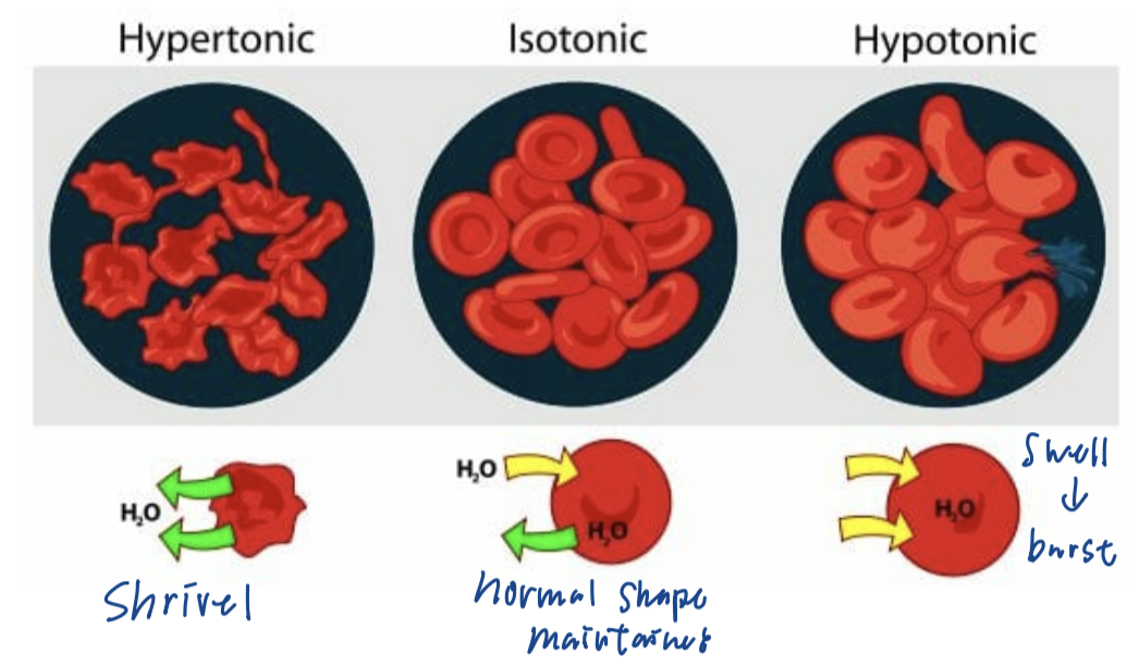
animal cells in different solutions of osmotic conc.
HYPERTONIC
shrivelled
ISOTONIC
normal
HYPOTONIC
swell and burst
plant cells in different solutions of osmotic conc.
HYPOTONIC
texture: rigid
appearance: turgid (cell wall press against membrane)
turgor pressure support stem & leaves
ISOTONIC
normal
HYPERTONIC
texture: flaccid
appearance: plasmolysed (cell wall separated from membrane)

hyper vs hypo
hyper: more
hypertonic: higher conc.
hypo: less
hypotonic: lower conc.
active transport properties
involves specific carrier proteins
only picks up similar substances
compete for space on carrier
requires energy (ATP)
from rsp
ATP hydrolysed to release energy catalysed by ATPase
for changing shape of carrier protein
cyanide: prevents production of ATP
against concentration gradient

endocytosis
intake of materials (too large for simple diffusion / membrane proteins) into cells through formation of vesicles to surround it
phagocytosis
engulf large solid particles
e.g. WBC engulf bacteria
pinocytosis
ingest extracellular fluids & dissolved solutes

exocytosis
emptying of membrane-bound vesicle
vesicle containing large particles fuse with cell surface membrane to release contents

endo & exocytosis
possible bc of fluid nature of membrane
require ATP
fick’s law of diffusion
rate of diffusion = thickness⋅of⋅exchange⋅membrane⋅or⋅barriersS.A.×conc.diff
calculate rate at which substance of a given size will diffuse at a given temperature
↑ rate: ↑ surface area / ↑ concentration difference / ↓ diffusion distance
gas exchange importance
to obtain oxygen for aerobic respiration
to remove carbon dioxide produced from respiration
reason for gas exchange system in large organisms
SMALL ORGANISMS:
large S.A. to vol. ratio ⇒ rapid exchange of gases by diffusion through outer body surface
LARGE ORGANISMS:
smaller S.A. to vol. ratio + long distance from outside to reach cytoplasm of cells + higher metabolic rate (higher demand of O2 ; more CO2 & waste produced)
insufficient gas exchange by diffusion
e.g. lungs in human / gills in fish / tracheal system in insects / leaves in plants
properties of mammalian gas exchange system
large surface area
give sufficient gas exchange to supply all needs of organsisms
compensate relatively small S.A. to vol. ratio
rich supply of blood capillaries
rapid transport of blood
maintain steep conc. gradient
thin layers
short diffusion distance
water film / moist surface on air sacs
dissolves gases as diffusion takes place with gases in solution
permeable surfaces
allow free passage of respiratory gases
pressure difference of gas between two sides of membrane
human gas exchange system
nasal cavity / mouth
larynx
trachea
bronchi
bronchioles
alveoli

nasal cavity
main route which air enters the gas exchange system
hair: traps large particles
rich blood supply: warm incoming air
moist surfaces: moisten incoming air
mucus-secreting cells: trap particles and pathogens
cilia: beating constantly and sweeps dust to pharynx
mouth
alternative route where air enters respiratory system
without cleaning, warming and moistening effects of nasal route
epiglottis
flap of tissue that closes over glottis when food is swallowed
prevents food from entering gas exchange system
larynx
voice box
uses flow of air across it to produce sounds
trachea
major airway to the bronchi
filter incoming air
mucus-secreting cells: trap particles & pathogens
cilia: beat constantly & sweep mucus and dust away from lungs to larynx and pharynx
cartilage
incomplete rings (C-shaped)
prevent trachea from collapsing
allows food to be swallowed and moved down the oesophagus
bronchi
lead to lungs
similar in structure to trachea but narrower
with non-C shaped cartilage
mucus-secreting cells and celia present but less
bronchiole
ends in alveoli
some gas exchange may occur
cilia present
alveoli
main site of gas exchange
intercostal muscles
between ribs
moves air into and out of lungs to maintain steep conc. gradient for rapid gas exchange

pleural membrane
surround the lungs and line the chest cavity to form a sterile sealed unit
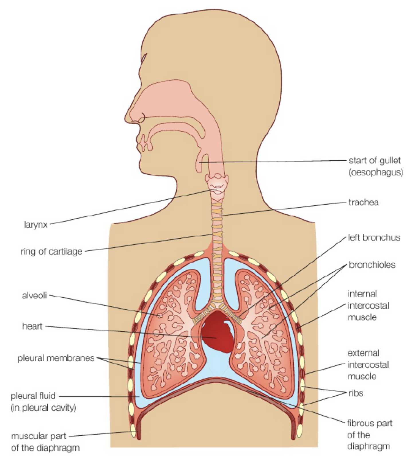
pleural cavity
space between pleural membranes
filled with thin layer of lubricating fluid
allows membranes to slide easily with breathing movements

diaphragm
broad sheet of tissue made of tendon and muscle
gas exchange in air sacs
blood enter air sacs (↑CO2 ↓O2)
O2 dissolves in water film and diffuses from air sac to blood while CO2 from blood to air sacs

air sacs structure
liquid lung surfactant
on inner walls of alveolar sacs
mixture of lipids and proteins
prevent collapse of air sacs
water film surface tension pull toward eo ⇒ surfactant ↓ surface tension ⇒ stop air sacs from sticking tgt & collapse ⇒ makes breathing easier
water film
monolayer of phospholipids on top
alveolus surface
folded to increase surface area
elastic tissue
between walls (epithelial cells) of capillary & air sacs
stretched when inhaling
recoils during exhalation to force air out of lungs

percentage of gas in inspired, alveolar, and expired air

inhalation (ventilation)
active, energy-using process
muscles of diaphragm contract to lower and flatten diaphragm
intercostal muscles contract to raise rib cage upwards and outwards
volume of chest cavity increases and pressure in chest cavity decreases
pressure in chest cavity becomes lower than that in atmosphere
air is forced into lungs

exhalation (ventilation)
can be passive (normally) / active (coughing)
diaphragm muscles relax and diaphragm returns to dome shape
intercostal muscles relax and rib cage moves downwards and inwards
elastic fibres of air sacs return to normal length
forced exhalation: abdominal muscles contract to force diaphragm upwards
volume of chest cavity decrease and pressure in chest cavity increase
pressure in chest cavity becomes higher than that of the atmosphere
air is forced out of lungs
