Ch 16 Innate Immunity: Non-Specific Defenses of The Host - flashcards
 Call Kai
 Learn
 Practice Test
 Spaced Repetition
 Match
 Flashcards
 Knowt Play
1/73

Earn XP
Description and Tags
25m run
Name | Mastery | Learn | Test | Matching | Spaced | Call with Kai | Chat |
|---|
No analytics yet
Send a link to your students to track their progress
74 Terms
What is the immune system, and what is it protecting the host from?
The system that protects the human host from pathogens.
Define immunity.
The ability to fight off pathogens and prevent disease.
Compare host resistance and host susceptibility (in terms of immunity and disease outcome).
Host resistance = host has immunity → no disease.
Host susceptibility = host lacks immunity (lacking resistance) → yes disease.
What happens if host defenses are successful vs. NOT successful?
Successful → host has immunity (resistance) → no disease.
Not successful → host becomes diseased (susceptibility) → yes disease.
If a pathogen is present, does disease always result? Explain both outcomes.
Pathogen present → disease may or may not occur depending on host defenses.
No pathogen present → no disease.
List the 5 key characteristics of innate immunity.
1) Host defenses present at birth — this is why it's "innate," important for survival of newborns.
2) AKA "non-specific" defenses of the host.
3) No specific recognition of microbes — gets rid of all pathogens (all bacteria, viruses, fungi, etc.) = "not picky"
4) No immunological memory response.
5) Faster immune response.
What lines of host defense make up innate immunity, and how many total lines of host defense are there?
Composed of the 1st & 2nd Lines of Host Defenses (3 lines total).
List the 5 key characteristics of adaptive immunity.
1) Host defenses that develop later to handle a specific microbe — NOT born with this.
2) AKA "specific" defenses of the host.
3) Specific recognition & response to a specific microbe — will discriminate ("picky").
4) Has immunological memory = immune system has encountered the same pathogen before.
5) Slower immune response than innate immunity (but still fast).
What line of host defense makes up adaptive immunity?
Composed of the 3rd Line of Host Defenses.
What are the 3 categories of the 1st Line of Defense, and # of EX. in each?
Physical Barriers (5 examples)
Chemical Barriers (4 examples)
Biological Barriers (1 example)
What are the 5 components of the 2nd Line of Defense?
Formed Elements.
Phagocytosis.
Inflammation.
Fever.
Antimicrobial Substances (2 examples).
What are the 2 components of the 3rd Line of Defense, and what must happen for disease to occur?
Humoral Immunity.
Cell-Mediated Immunity.
A pathogen must breach all 3 lines of host defense to cause disease.
List & define 5 examples of physical barriers (1st Line of Host Defense).
Intact skin = non-broken skin.
Mucous membranes = line all body cavities; produce mucus.
Ciliary escalator = present in respiratory tract.
Lacrimal apparatus = involves eyes & tears.
Saliva, urine & vaginal secretions = (no added detail on this slide).
Define intact skin, and what are its 5 protective properties?
Intact skin = non-broken skin, no injuries
Closely packed epithelial cells (flat/tight).
Continuous layering — multiple layers at the epidermis.
Keratin protein in top layer — reinforces skin, protects underlying epidermis layers.
Dryness — no moisture → no pathogens.
Shedding of top layer — pathogens fall off.
— 1st Line: Physical barrier
Which two layers of skin are shown blocking a pathogen from reaching the blood vessel (Figure 16.1)?

What are mucous membranes, and how do they protect the host?
Epithelial layer that lines gastrointestinal, respiratory & genitourinary tracts (lines all tubes & body cavities).
Secrete viscous fluid called mucus (produced by mucous cells) — traps pathogens so they can be gotten rid of.
— 1st Line: Physical barrier
What is the ciliary escalator, and how does it work?
Epithelial cells of the lower respiratory tract (airways) have cilia.
Cilia move together in one direction, sweeping mucus up & out of the body — via cough → spit out or swallow.
— 1st Line: Physical barrier
What is the lacrimal apparatus, and how does it protect the host?
Protects the eyes, tears provide continuous washing action (flushing) that removes pathogens from the eye.
— 1st Line: Physical barrier
How do saliva, urine, and vaginal secretions protect the host?
Flushing mechanism that washes away pathogens, preventing pathogen accumulation from the mouth, urethra, and reproductive tract.
— 1st Line: Physical barrier
What are chemical barriers, and what are the 4 examples?
Chemical barriers, chemicals produced by the body.
Chemical factors of the skin.
Lysozymes/Enzymes.
Gastric juices.
Blood transferrin’s, circulating proteins in the blood.
— 1st Line: Chemical barrier
How does the pH of skin protect against pathogens?
Skin pH is slightly acidic (skin goal ~5-6), most bacteria prefer neutral pH 7 and don't like acidic conditions.
— 1st Line: Chemical barrier
How does skin salinity protect against pathogens? (Bacteria only)
Most bacteria are NOT halophiles, salinity creates a hypertonic/salty environment.
Water leaves the bacterial cell, causing the bacteria to shrink/die.
— 1st Line: Chemical barrier
What are lysozymes, where are they found, and how do they kill bacteria? (Bacteria only)
Enzymes found in body secretions (sweat, tears, saliva), break chemical bonds in peptidoglycan, destroying the bacterial cell wall.
No peptidoglycan → no cell wall → bacteria die.
— 1st Line: Chemical barrier
What are gastric juices, and how do they destroy pathogens? (Bacteria only)
Produced by the stomach, contain enzymes & acid, destroys most bacteria by breaking down the cell wall.
— 1st Line: Chemical barrier
What are blood transFerrins, and how do they inhibit bacterial growth? (Bacteria only)
Proteins that bind to iron (Fe), "remove iron.”
Iron (a micronutrient) is necessary for bacterial growth, so removing it leaves no iron available for bacterial growth.
— 1st Line: Chemical barrier
What is Normal Microbiota (N.M.), and what are its two types?
Normal Microbiota, no harm to host (non-pathogenic).
Two types: commensal microbes vs. beneficial microbes.
— 1st Line: Biological barrier
How do opportunistic microbes differ from Normal Microbiota?
Opportunistic microbes are NOT Normal Microbiota, they are pathogenic microbes that can cause harm.
— 1st Line: Biological barrier
What are commensal microbes, and what's an example?
Microbe benefits while the host does NOT benefit (but host is not harmed).
Microbes provide nothing to the host.
Ex: bacteria on skin.
— 1st Line: Biological barrier
What are beneficial microbes, and what's an example?
Both microbe & host benefit
Microbes provide something to the host
Ex: vitamin K-producing bacteria in the Colon (E. coli)
— 1st Line: Biological barrier
What are opportunistic microbes? (Bacteria only)
Microbes that act as pathogens under certain circumstances.
Used to be Normal Microbiota that got removed from their usual environment.
Ex: E. coli present in the urethra.
— 1st Line: Biological barrier
What is competitive exclusion, and what does it accomplish? (Bacteria only)
AKA "microbial competition.”
Normal microbiota compete with invading pathogens, leading to decreased populations of pathogenic bacteria.
— 1st Line: Biological barrier
How do normal microbiota defeat invading pathogenic bacteria? (Bacteria only)
Take up all the nutrients, no nutrients left for invaders.
Take up all the space for growth (normal microbiota multiply), no space left for invaders.
Produce substances harmful to invading pathogens.
— 1st Line: Biological barrier
1st Line done,
What are the 5 barriers of the 2nd line & how many examples?
Formed Elements in the blood.
Phagocytosis.
Inflammation.
Fever.
Antimicrobial Substances. (2)

What are the formed elements in blood, where are they created, and via what process?
Cells and cell fragments suspended in plasma.
Created in red bone marrow stem cells via hematopoiesis.
What are the 3 formed elements, and what are their functions?
Erythrocytes = RBC, contain Hb (hemoglobin), binds to O2
Leukocytes**** = WBC, involved in immune function
(both cells)Thrombocytes = PLT, involved in blood clotting upon injury (cell fragment)
— 2nd Line: Formed Elements
Which formed elements are cells vs. cell fragments?
Cells: erythrocytes, leukocytes
Cell fragments: platelets (thrombocytes) — from the megakaryocyte parent cell
— 2nd Line: Formed Elements
Hematopoiesis
Hematopoiesis
what he wants us to know:
The parent cell.
The platelets are cell fragments.
Leukocytes are our focus.
— 2nd Line: Formed Elements

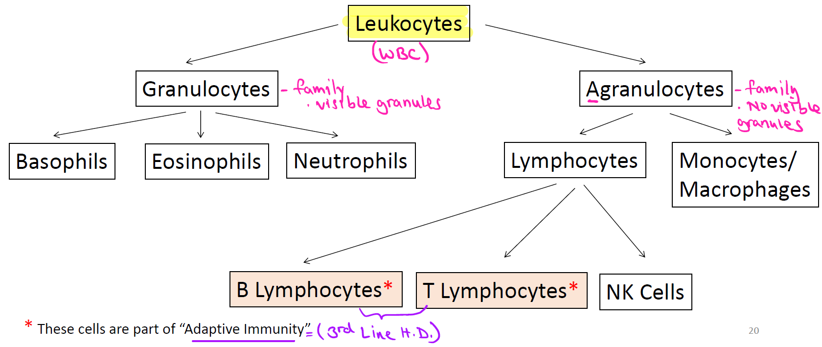
What are the two families of leukocytes (WBC), and what distinguishes them?
Granulocytes, family with visible granules:
Includes basophils, eosinophils, neutrophils.
Agranulocytes, family with no visible granules:
Includes lymphocytes, monocytes/macrophages.
— 2nd Line: Formed Elements

What are the three types of lymphocytes, and which are part of adaptive immunity?
B Lymphocytes* → adaptive
T Lymphocytes* → adaptive
NK Cells → part of innate immunity (2nd Line H.D.)
— 2nd Line: Formed Elements
Define granulocytes (the granulocyte family).
Leukocytes with granules in their cytoplasm that are visible with a light microscope.
— 2nd Line: Formed Elements
What are basophils, and what do they release?
Release histamine granules, involved in allergic responses (allergy symptoms/sxs).
— 2nd Line: Formed Elements

What are eosinophils, and what are they effective against?
Toxic against parasites and worms.
— 2nd Line: Formed Elements
What are neutrophils, what is their function, and when do they work? (Bacteria only)
Phagocytic, a type of phagocyte.
Engulfs pathogens.
Work in early stages of infection.
First type of phagocyte to arrive at site of injury, called the "first responder.”
— 2nd Line: Formed Elements

Define agranulocytes (the agranulocyte family).
Leukocytes with granules in their cytoplasm that are NOT visible with a light microscope.
— 2nd Line: Formed Elements
What are monocytes, and what do they become?
Monocytes travel in blood, mature into macrophages in tissues, where they become phagocytic cells (an example of a phagocyte).
— 2nd Line: Formed Elements
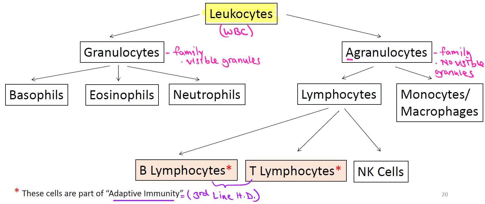
What are the 3 types of lymphocytes, and which two are involved in adaptive immunity (3rd Line H.D.)?
T cells* → adaptive immunity.
B cells* → adaptive immunity.
NK cells → innate immunity.
— 2nd Line: Formed Elements

List the 5 leukocyte types in order from highest to lowest:
The abundance of each type of white blood cell in a sample of 100 white blood cells (in a normal state).
*Don’t memorize number, just know order*
NLMEB (Never Let Monkeys Eat Bananas)

Define phagocytosis and phagocyte.
Phagocytosis: ingestion (engulfing) of microbes or other substances (solids) by a phagocyte.
Phagocyte: non-specific host cell capable of phagocytosis, part of "us" (belongs to host).
— 2nd Line: Phagocytosis
What are the 3 examples of phagocytes, and where is each found?
Neutrophil: 1st responder, in blood.
Macrophage: in tissue, comes from monocyte.
Dendritic: cell, in skin cells.
— 2nd Line: Phagocytosis

What are the 4 phases of phagocytosis, and what happens in each?
Chemotaxis:
Release of chemical signals (cytokines).
→ Attracts phagocytes (phagocyte travels toward pathogen).
Adherence (binding):
- Attachment of phagocyte to surface of the pathogen.
Ingestion (engulf):
- Endocytosis of pathogen → forms a phagosome.
- Phagosome merges with lysosome (has digestive enzymes) → forms a phagolysosome inside phagocyte.
Digestion:
- Pathogen is digested inside the phagolysosome.
— 2nd Line: Phagocytosis
What are the 3 ways pathogens evade phagocytosis, and what is the example genus/species for each? (Bacteria only)
Capsule: pathogen too big to be engulfed.
- Ex: Streptococcus pneumoniae.
Leukocidins: a pore-forming toxin (poison) capable of killing phagocytes.
- Ex: Genus Staphylococcus.
Mycolic acid: a waxy lipid found in cell wall of some bacteria, inhibits lysosome (digestive) enzymes of phagocyte → bacteria multiply inside phagocyte & hide from immune system (bacteria protected).
- Ex: Genus Mycobacterium
What is inflammation due to, and what is its overall goal?
Due to infection, goal is to rid of pathogen (by neutrophils & macrophages) so tissue can heal.
— 2nd Line: Inflammation
What are the 5 signs & symptoms of inflammation, and what causes each?
Pain, due to release of certain chemicals (cytokines), damages nerve endings.
Redness (erythema), increased blood flow to affected area (Neutrophils & monocytes).
Immobility, from local loss of function in severe inflammation (tissue is damaged).
Swelling (edema), due to accumulation of fluids outside of blood vessel.
Heat, increased blood flow to affected area.
— 2nd Line: Inflammation

What are the 6 steps of the process of inflammation?/
Can involve phagocytosis
Tissue or cells get damaged/injured due to infection (caused by pathogen).
Cytokine chemical released.
Cytokines promote chemotaxis of phagocytes (Ex: neutrophils & monocytes/macrophages).
Phagocytes squeeze through blood vessel & move to site of tissue injury.
Phagocytosis of invading pathogens begins.
Tissue gets repaired.
Neutrophil (first responder).
Monocyte in blood → Macrophage in tissue.
— 2nd Line: Inflammation / Phagocytosis
What is fever due to, and what is its goal? (Bacteria only)
Due to bacterial infection, goal is to rid of pathogen (by phagocytes).
— 2nd Line: Fever
What triggers a fever, and how does it develop? (steps 1-4 BACTERIA ONLY)
Toxins released by some bacteria (Ex: gram(-) →induce cytokine release.
Cytokines bind to hypothalamus receptors (body's thermostat), increasing the set point for body temperature.
The "new" set point makes the person feel cold (chills), so they increase body temperature.
Once the "new" set point is reached, the person experiences abnormally high body temperature (fever).
— 2nd Line: Fever
How does a fever resolve? (steps 5-6 BACTERIA ONLY)
As pathogens are eliminated, toxins & cytokines get eliminated too.
Body thermostat resets, body temperature falls, decline of fever is called "crisis."
— 2nd Line: Fever
What is the general sequence/progression of a fever?
Chills → fever → crisis
What are the 3 consequences (outcomes) of fever due to infection?
Increases metabolic rate (catabolism), more ATP made → breaking ATP → release energy.
Enhances immune response, promotes phagocytic activity.
Induces antimicrobial substances, makes certain cells release substances that destroy pathogens.
What are the 2 examples of antimicrobial substances, and what do they both involve?
Complement System.
Interferons.
Both involve proteins produced by the human host.
— 2nd Line: Antimicrobial Substances
What is the complement system?
Proteins (complement proteins) found in blood that enhance/"compliment" the immune system in destroying pathogens.
— 2nd Line: Antimicrobial Substances
What is complement activation, and what 2 forms do complement proteins exist in?
Complement proteins act in a cascade (complement activation): one active complement protein activates the next inactive one in the chain.
see img.
— 2nd Line: Antimicrobial Substances

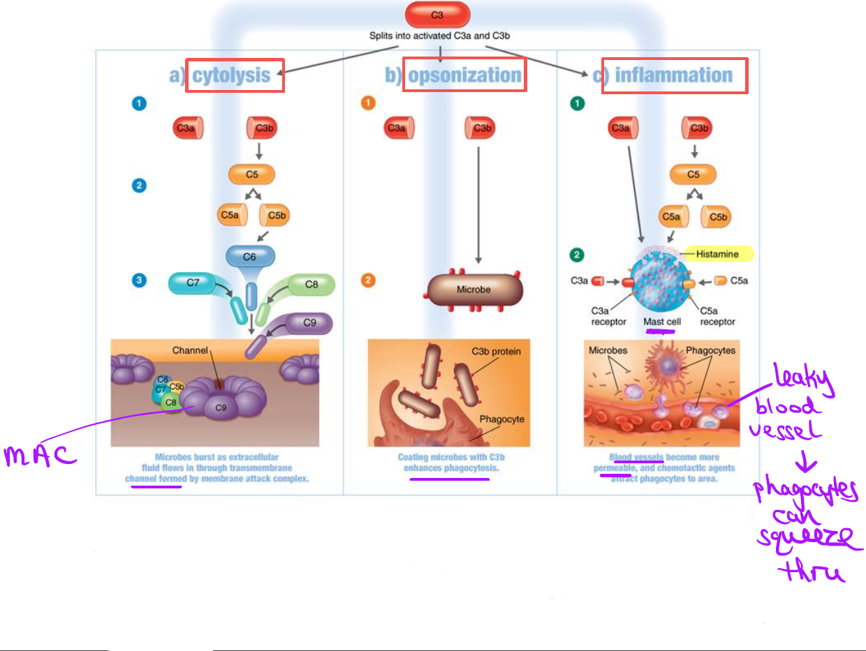
What are the 3 outcomes (consequences) of complement activation?
Opsonization (To "coat the surface" of something)
Inflammation
Cytolysis

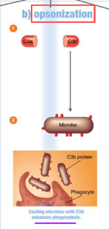
What is opsonization?
Complement proteins coat surface of pathogen.
Promotes attraction/attachment of phagocyte to pathogen.
— 2nd Line: Antimicrobial Substances

What happens during the inflammation outcome of complement activation?
Complement proteins bind mast cells (also released by basophil).
Mast cells release histamine → increases blood vessel permeability (leaky BV).
Phagocytes squeeze through blood vessel and arrive to site of injury.
— 2nd Line: Antimicrobial Substances

What is cytolysis, and how does it destroy the pathogen?
Complement proteins create a membrane attack complex (MAC), inserted into the pathogen's cell wall
MAC creates a hole/channel in the pathogen's cell wall/cell membrane → fluid enters the pathogen → pathogen bursts!
— 2nd Line: Antimicrobial Substances

What protein starts the complement cascade, and which specific proteins form the MAC in cytolysis?
— 2nd Line: Antimicrobial Substances
Complement protein
"What are the 3 ways bacteria evade the complement system?" (Bacteria only)
Capsule production, complement proteins cannot bind easily (they prefer to bind the cell wall or cell membrane).
Inhibition of MAC formation, enzymes from bacteria prevent MAC assembly = no channel formed.
Inactivation/destruction of complement proteins, bacteria produce protease (an enzyme).
What two things can trigger complement activation?
A pathogen directly, or an antibody-antigen reaction.
What is an Interferon (IFN) & **Antiviral protein (AVP)?
When a cell gets infected by a virus, it sends out a warning flare (interferon) to its neighbors before it dies. The neighboring cells catch that flare and basically "lock their doors" (make antiviral proteins) so if the virus tries to spread to them next, it can't get in as easily.
IFN = the warning signal.
AVP = the gun.
— 2nd Line: Antimicrobial Substances
What are interferons (IFNs), where do they come from, and what does their name mean?
Small proteins produced by some animal host cells upon animal viral infection, "interfere" with viral infection of animal host cell.
— 2nd Line: Antimicrobial Substances
What is the antiviral action of interferons?
Cause uninfected neighboring animal cells to heighten their anti-viral defenses, this protects them against potential/future viral infection.
— 2nd Line: Antimicrobial Substances

What are the 4 steps of the interferon mechanism of action?
Animal virus enters animal host cell and multiplies.
Infected animal host cell produces IFNs (interferons), which serve as a signal to uninfected neighboring animal cells for potential virus infection.
IFNs released by the virus-infected animal host cell bind to the cell membrane of the uninfected neighboring animal cell, causing it to make anti-viral proteins (AVPs).
Newly released viruses that infect the neighboring animal cell have AVPs waiting to destroy the virus (pathogen).
— 2nd Line: Antimicrobial Substances
Mechanism of Action of IFNs.
Host Cell 1 makes interferon but is still killed by the virus, the interferon it released protects the uninfected neighboring cell (Host Cell 2), not itself.
— 2nd Line: Antimicrobial Substances
