ch14 a&p exam 2
 Call Kai
 Learn
 Practice Test
 Spaced Repetition
 Match
 Flashcards
 Knowt Play
1/227

There's no tags or description
Looks like no tags are added yet.
228 Terms
What are the four major regions of the brain?
The cerebrum, diencephalon, brainstem, and cerebellum. Together they process sensory information, coordinate movement, regulate vital functions, and support memory and consciousness.
What are the three parts of the brainstem?
The midbrain, pons, and medulla oblongata, arranged from superior to inferior.
What is the cerebrum responsible for?
The cerebrum is the largest brain region and is responsible for conscious thought, intelligence, memory, sensory processing, and voluntary skeletal muscle control.
has a left and right side (cerebral hemispheres)
cerebral hemispheres are highly folded and covered by
cerebral cortex which is a thin layer of gray matter and contain gyri (rounded elevation) for increase surface area
What is the purpose of sulci and fissures of the cerebral cortex
Sulci (shallow grooves) and fissures (deep grooves) separate gyri; fissures divide major brain regions.

What is the diencephalon?
A region connecting the cerebral hemispheres with the brainstem. It includes the thalamus, hypothalamus, and epithalamus and helps regulate sensory, autonomic, and endocrine functions.

What is the primary function of the cerebellum?
To coordinate skeletal muscle activity, balance, posture, and the timing of movements so that actions are smooth and precise.
second largest brain region, has 2 hemispheres covered by cerebellar cortex (gray matter)

What is the neural tube?
The embryonic structure that develops into the central nervous system, including the brain and spinal cord.

What are the three primary brain vesicles?
The prosencephalon (forebrain), mesencephalon (midbrain), and rhombencephalon (hindbrain).

What are the five secondary brain vesicles?
The forebrain divides into the
▪ Telencephalon
▪ Diencephalon
The hindbrain constricts, forming the
▪ Metencephalon
▪ Myelencephalon.
▪ The midbrain remains undivided - mesencephalon

What does the telencephalon develop into?
The two cerebral hemispheres and associated lateral ventricles.

What does the diencephalon develop into?
The thalamus, hypothalamus, epithalamus, and associated third ventricle.
What does the rhombencephalon develop into?
metencephalon (turn into pons and cerebellum)
myelencephalon (medulla oblongata)

What does the mesencephalon develop into?
The midbrain and cerebral aqueduct.

What does the metencephalon develop into?
The pons and cerebellum, associated with the fourth ventricle.

14. What does the myelencephalon develop into?
The medulla oblongata, associated with the inferior portion of the fourth ventricle.

What are the ventricles of the brain?
Four interconnected, fluid-filled chambers lined by ependymal cells that contain cerebrospinal fluid (CSF).

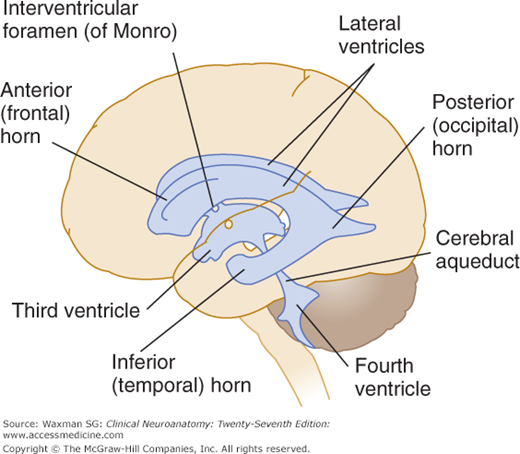
what are the ventricles in the ventricular system
The two lateral ventricles.
The third ventricle.
The fourth ventricles
Where are the lateral ventricles located?
One lateral ventricle is located within each cerebral hemisphere. They are separated by the septum pellucidum (thin plate of brain tissue)

What are the three horns of each lateral ventricle? and the lobes it is located in
The anterior horn (extends to the foramen of Monro), posterior horn (extends into the occipital lobe), and inferior horn (extends into the temporal lobe)
Where is the third ventricle located?
Within the diencephalon, between the thalamic regions.
lateral ventricles communicate/drain into the third ventricle and they are connected through the Foramen of Monro

What are the interventricular foramina?
Passageways called the foramina of Monro that connect the lateral ventricles to the third ventricle.

What is the cerebral aqueduct?
A narrow canal within the midbrain connecting the third ventricle to the fourth ventricle.
aka aqueduct of Sylvius

Where is the fourth ventricle located? and funx
Between the brainstem and cerebellum. It receives CSF from the cerebral aqueduct and communicates with the central canal and subarachnoid space.

What is the central canal?
A narrow, CSF-containing passageway through the spinal cord that is continuous with the ventricular system.
What are the four major protective mechanisms of the brain?
The cranial bones, cranial meninges, cerebrospinal fluid, and blood-brain barrier.
What are the three cranial meninges from superficial to deep?
Dura mater, arachnoid mater, and pia mater, they are continuous with spinal meninges

What is the dura mater?
The tough outer meningeal membrane. In the cranium, it contains periosteal and meningeal layers that support and protect the brain.
What are the periosteal and meningeal layers of cranial dura?
The periosteal layer fused to the periosteum of the cranial bones; the meningeal layer lies deeper and forms dural folds
there is no epidural space in the cranium unlike the spinal cord

What are the three major dural folds of the meningeal layer of the dura?
The falx cerebri, tentorium cerebelli, and falx cerebelli.
purpose is to provide extra stabilization and support for the brain

where is the dural venous sinnus found and what is the purpose
Dural venous sinuses lie within the dural folds
Brain veins drain into these sinuses, which then carry venous blood to the neck veins

What is the falx cerebri?
A sickle-shaped dural fold extending into the longitudinal fissure that separates and stabilizes the two cerebral hemispheres.
where does the falx cerebri attach and what sinuses run within this dural fold?
Its lower portions attach anteriorly to the crista galli and posteriorly to the internal occipital crest.
Dural venous sinuses - Superior and inferior sagittal sinuses run within this fold.
• The posterior margin of the falx cerebri meets the tentorium cerebelli.

What is the tentorium cerebelli and what sinus runs through
A dural fold separating and protecting the cerebellum from the overlying cerebral hemispheres.
transverse sinus runs along the occipital bone.
▪ This sinus drains venous blood from the back of the head.

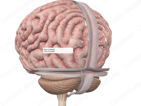
What is the falx cerebelli and what sinus runs through?
A dural fold separating the two cerebellar hemispheres along the midline, inferior to the tentorium cerebelli
Occipital sinus runs through the posterior margins of falx
cerebelli
what is the subdural space
a space between the dura mater and the arachnoid mater

What is the arachnoid mater?
The thin, spider-web-like middle meningeal membrane that covers the brain without closely following every gyrus and sulcus.

What is the subarachnoid space?
The space between arachnoid mater and pia mater containing cerebrospinal fluid and connective tissue trabeculae.

What is the pia mater?
The innermost meningeal membrane that closely adheres to the brain surface (anchored by astrocytes) and accompanies blood vessels entering nervous tissue.

What is cerebrospinal fluid (CSF)? and what does it contain
A clear fluid filling the brain's ventricles and surrounding the CNS. It provides buoyancy, cushioning, and transport of dissolved substances.
mostly water, also contains proteins, electrolytes, and glucose
What are the three major functions of CSF?
Supporting the brain through buoyancy, cushioning it against trauma, and transporting nutrients, chemical messengers, and metabolic wastes.
How does CSF reduce the effective weight of the brain?
By suspending the brain in fluid, reducing its effective weight from approximately 1,400 grams to about 25-50 grams.
Where is CSF produced?
Primarily by the choroid plexuses associated with the ventricles.

What is the choroid plexus?
A specialized structure containing capillaries and modified ependymal cells that produces CSF and regulates its chemical composition.
what is the purpose of ependymal cells that surround the choroid plexus capillaries
Ependymal cells with microvilli and tight junctions surround
choroid plexus capillaries.
▪ CSF begins as filtrate leaking from capillaries.
▪ Ependymal cells modify, secrete, and cleanse CSF, removing wastes and adjusting composition.
What is the complete pathway of CSF circulation?
Lateral ventricles → interventricular foramina → third ventricle → cerebral aqueduct → fourth ventricle → subarachnoid space → arachnoid villi and granulations → venous sinuses.

What are arachnoid villi and granulations?
Projections of arachnoid tissue into dural venous sinuses that allow CSF to return to the bloodstream.
arachnoid granulations are large clusters of arachnoid villi in adults and absorb CSF into venous circulation
What is hydrocephalus?
Abnormal accumulation of CSF due to impaired circulation or drainage, potentially increasing pressure and damaging brain tissue.
Why can hydrocephalus enlarge an infant's head?
The cranial bones have not fully fused, allowing increased fluid pressure to expand the skull.
What is the blood-brain barrier (BBB)?
A protective barrier formed mainly by tight junctions between CNS capillary endothelial cells that controls the passage of substances from blood into brain tissue.
What role do astrocytes play in the BBB?
Astrocytes help maintain endothelial tight junctions and regulate capillary permeability through their interactions with CNS blood vessels.
Which substances can readily diffuse across the BBB?
Lipid-soluble substances such as oxygen, carbon dioxide, ammonia, steroids, and small alcohols can cross cell membranes relatively readily.
Why do ions and large water-soluble (like urea) molecules require transporters at the BBB? What are some ions that cannot diffuse
They cannot freely pass through endothelial lipid membranes and tight junctions, so their entry depends on regulated channels or transport mechanisms.
Hydrogen (H⁺), sodium (Na⁺), potassium (K⁺), and ammonium (NH₄⁺) ions
Why is the BBB important when administering medication?
Some drugs cannot cross it effectively, limiting their ability to reach brain tissue. Certain medications may require alternative delivery methods, including intrathecal administration (via lumbar puncture to reach the CSF)
What are the three major structural components of each cerebral hemisphere?
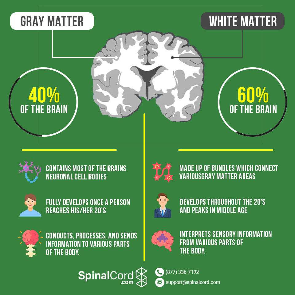
The superficial cerebral cortex, internal white matter, and deep gray matter collections called basal nuclei.
What is gray matter composed of and found?
Neuron cell bodies, dendrites, neuroglia, and primarily unmyelinated neural processes involved in processing information.
located in the cerebral cortex and in deeper basal nuclei
]What is white matter composed of and found?
Primarily myelinated axons that transmit information between different regions of the central nervous system.
lies deep to the cerebral cortex and around the basal nuclei.
The basic pattern of the CNS is a central cavity surrounded by x matter, external to which is y matter.
x = gray
y = white
What are the five cerebral lobes?
The frontal, parietal, temporal, occipital, and insular lobes.

longitudinal fissure
separates the cerebral hemispheres
transverse cerebral fissure
separates the cerebral hemispheres from the cerebellum

What is the central sulcus?
A major groove separating the frontal and parietal lobes and dividing the primary motor cortex from the primary somatosensory cortex.
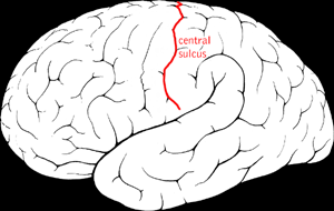
parieto-occipital sulcus
major groove separating the occipital lob from the parietal lobe

What is the lateral sulcus?
A deep groove separating the temporal lobe from portions of the frontal and parietal lobes.
forms the floor of the insular lobe

What is the precentral gyrus?
A gyrus immediately anterior to the central sulcus in the frontal lobe that contains the primary motor cortex.

What is the postcentral gyrus?
A gyrus immediately posterior to the central sulcus in the parietal lobe that contains the primary somatosensory cortex.

Where is the insula located?
Deep within the lateral sulcus, covered by portions of the frontal, parietal, and temporal lobes.

Which cranial fossae contain the frontal and temporal lobe?
The frontal lobes lie in the anterior cranial fossa, while the anterior portions of the temporal lobes occupy the middle cranial fossa.
What structures are located in the posterior cranial fossa, and where are the occipital lobes?
The posterior cranial fossa houses the brainstem and cerebellum, while the occipital lobes are located well superior to the posterior cranial fossa.
what are the three basic regions of the cerebral hemispheres
A superficial cerebral cortex of gray matter, which looks gray in fresh brain tissue.
Internal white matter.
Basal nuclei, islands of gray matter situated deep within the white matter – subcortical gray matter.

What is the cerebral cortex?
The superficial layer of gray matter covering the cerebral hemispheres that supports conscious sensation, voluntary movement, reasoning, memory, and complex behavior.
aka executive center of the NS and conscious mind
What are the three major functional categories of cerebral cortex?
Motor areas, sensory areas, and association areas (coordinates the incoming and outgoing data from the sensory and motor areas)
What is contralateral control?
Each cerebral hemisphere primarily controls motor activity and processes sensory information from the opposite side of the body.
What is cerebral lateralization?
Functional specialization of the left and right cerebral hemispheres for different tasks.
Motor Areas of the Cerebral Cortex include
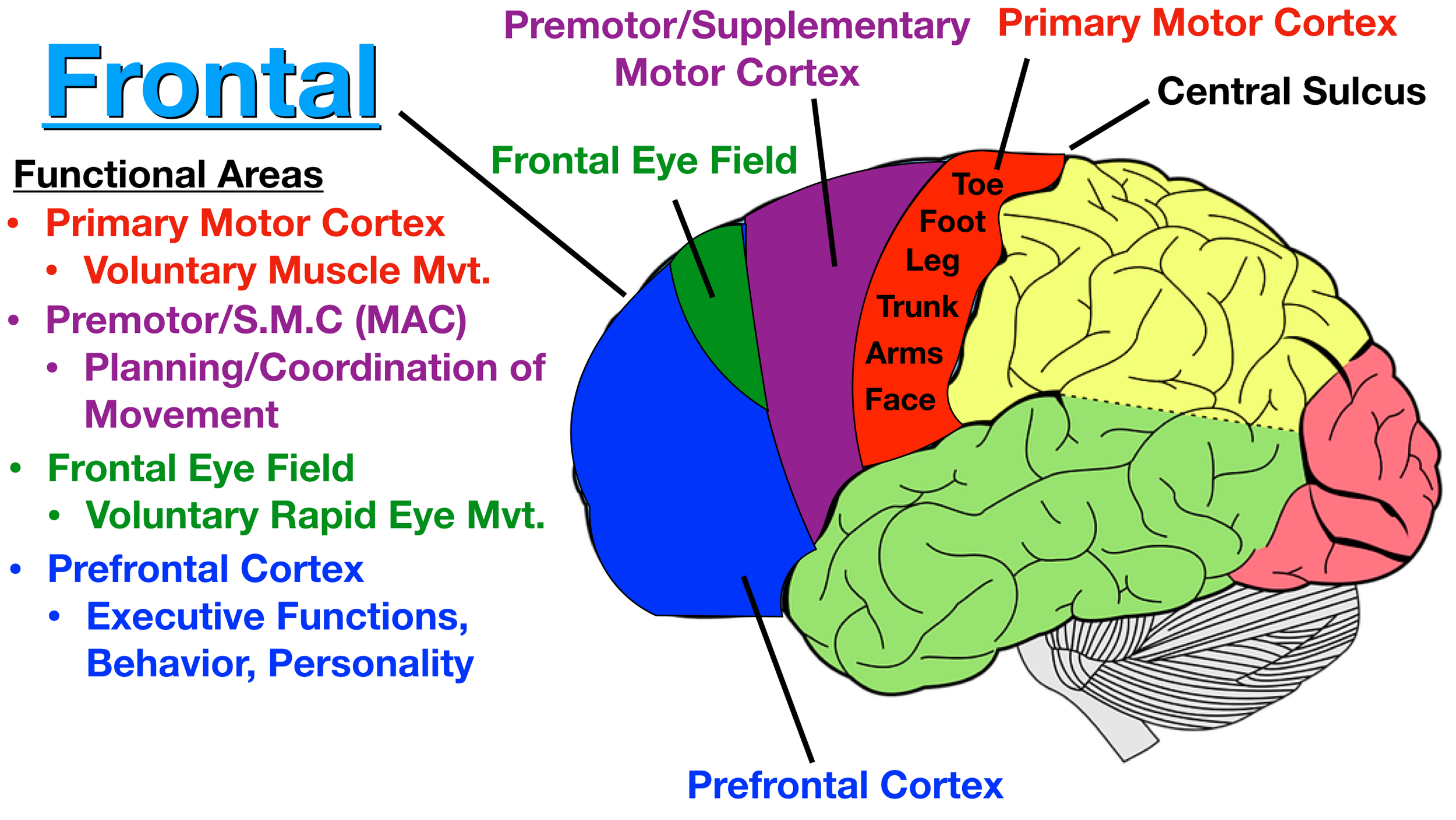
Primary motor cortex.
Premotor cortex.
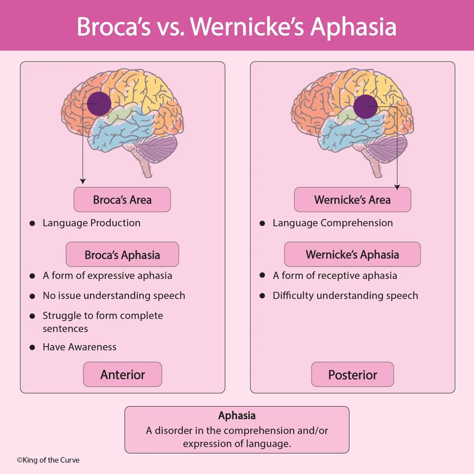
Broca’s area (Speech area).
The frontal eye field
lies in the posterior part of the frontal lobe
Where is the primary motor cortex located?
In the precentral gyrus of the frontal lobe, immediately anterior to the central sulcus.
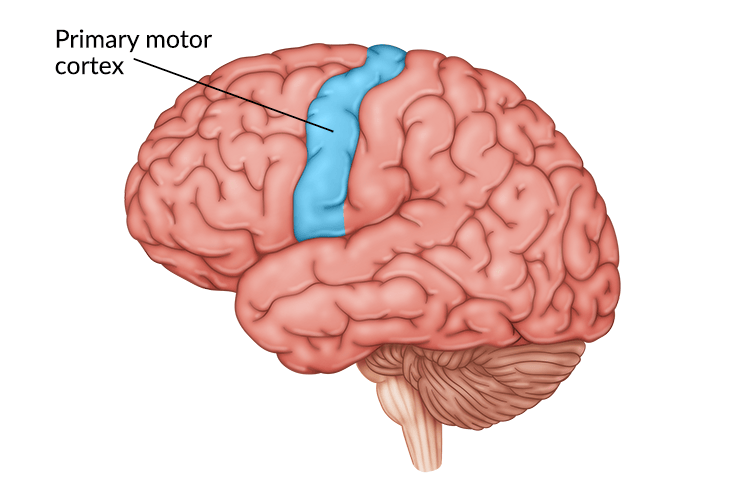
What is the function of the primary motor cortex?
It generates voluntary motor commands controlling precise skeletal muscle movements.
What are pyramidal cells?
Large motor cortical neurons whose axons contribute to descending motor pathways involved in voluntary movement.

What are corticospinal tracts?
Descending pathways that carry voluntary motor commands from the cerebral cortex toward the spinal cord, pyramidal cells
68. What is the motor homunculus?
A body map in the primary motor cortex where the size of each represented region reflects the precision and complexity of its motor control.

Why are the hands and face large on the motor homunculus?
They require highly precise, coordinated movements and therefore occupy disproportionately large cortical regions.
What can damage to the primary motor cortex cause?
Loss or impairment of voluntary movement, often affecting the opposite side of the body.
Where is the premotor cortex located?
Anterior to the primary motor cortex in the frontal lobe.

What is the function of the premotor cortex?
It plans and organizes complex, learned movement sequences such as typing or playing a musical instrument.
What is the difference between the primary motor cortex and premotor cortex?
The premotor cortex plans and coordinates movement sequences; the primary motor cortex generates commands for voluntary muscle contraction.
What is the anterior association area/prefrontal cortex responsible for?
Most complex region
Reasoning, judgment, planning, personality, working memory, problem-solving, and complex decision-making.
these functions develop slowly
75. Where is Broca's area located and what does it do?
This is the motor speech area
Usually in the left frontal lobe; it coordinates the muscle activity needed for speech production.
regulates breathing and vocalization patterns
coordinates respiratory, laryngeal, and pharyngeal muscles
cheeks, lips, and jaw

What is Broca's aphasia?
A disorder involving slow, effortful, fragmented speech with simplified grammar, associated with damage to Broca's area or nearby regions.
What is the frontal eye field?
A frontal cortical area that controls voluntary eye movements and helps direct visual attention.
smooth following of moving objects
79. What sensations are processed by the primary somatosensory cortex and where is it located?
Receive info from sensory receptors about touch, pressure, pain, temperature, vibration, and proprioceptive information about body position.
located in the postcentral gyrus of the parietal lob, just posterior to the primary motor cortex

What is the somatosensory homunculus?
A body map in the primary somatosensory cortex where larger representations correspond to regions with greater sensory sensitivity.
What is proprioception?
The sense of body position and movement derived from receptors in muscles, tendons, and joints.
what are sensory association areas
Sensory association areas are cortical regions that monitor and interpret the information that arrives at the sensory areas of the cortex.
▪ Somatosensory association cortex.
▪ Visual association area.
▪ Auditory association area.
What is the function of the somatosensory association cortex and location
It integrates sensory information to recognize an object's shape, size, texture, and other physical characteristics.
Integrate sensory inputs relayed to it via the primary somatosensory cortex to produce an
understanding of an object being felt
posterior to the primary somatosensory cortex and has
many connections with it

What is the function of the primary visual cortex and location?
It receives and processes visual information originating from the retina.
In the occipital lobe, with much of it located along the calcarine sulcus.

What is the function of the visual association area?
It interprets visual information using previous experiences to recognize objects, patterns, and spatial relationships.

What is the difference between damage to the primary visual cortex and visual association area?
Primary visual cortex damage may cause blindness; visual association damage may preserve sight but impair recognition of what is seen.
What is the function of the primary auditory cortex and location
It processes auditory signals, including characteristics such as pitch, loudness, and sound location.
Located along the superior portion of the temporal lobe near the lateral sulcus.
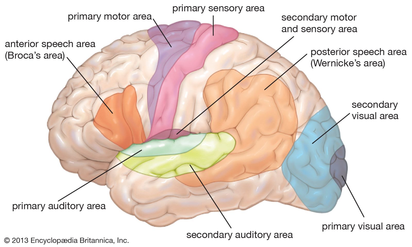
What is the function of the auditory association area?
It interprets sounds by comparing incoming auditory information with previously experienced sounds like a scream, music, thunder, memories of sounds, etc

Where is Wernicke's area located and what does it do?
In the posterior superior temporal region; it is involved in understanding spoken and written language.