Practicum 3
 Call Kai
 Learn
 Practice Test
 Spaced Repetition
 Match
 Flashcards
 Knowt Play
1/111

There's no tags or description
Looks like no tags are added yet.
112 Terms
Rectus
straight eye muscle

Oblique
slanted eye muscle

cornea

aqueous humor
in the lens
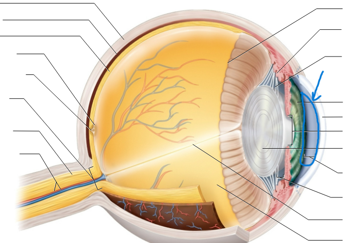
iris

pupil
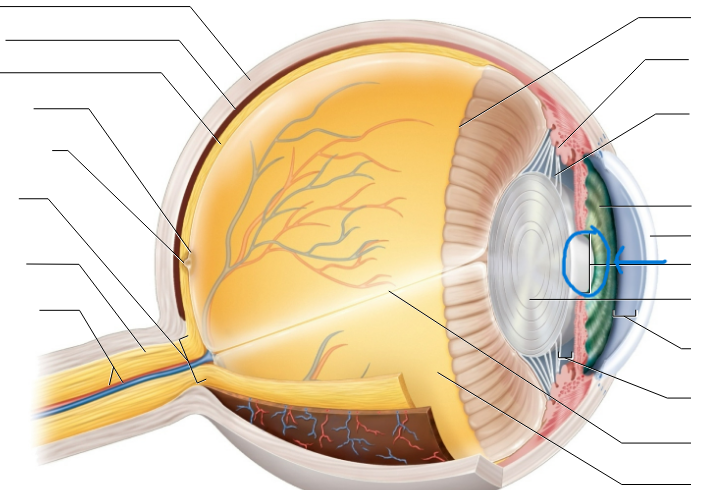
lens

suspensory ligament
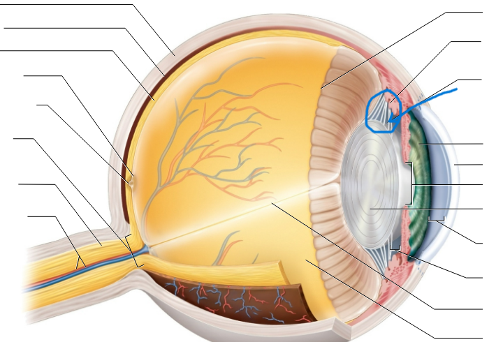
ciliary body

vitreous humor
in the eyeball
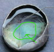
retina

optic disc

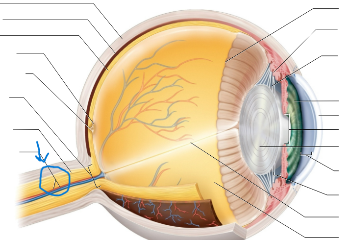
optic nerve on eye
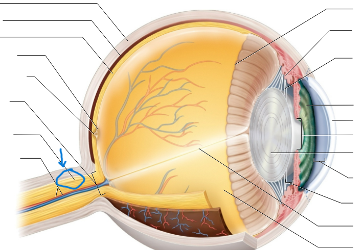
central artery/vein
macula lutea

fovea centralis

choroid

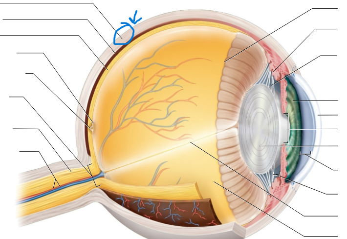
sclera
Central Sulcus
separates the frontal and parietal lobe

Lateral Sulcus

Longitudinal fissure

Transverse fissure
separates the cerebellum and the temporal-occipital lobe

Parieto-occipital sulcus
separates the parietal lobe and the occipital lobe

Precentral gyrus/ Primary Motor Cortex

Postcentral gyrus = Primary somatosensory cortex

Frontal lobe
cognition

Parietal lobe
sensory

Temporal lobe
auditory and speech

occipital lobe
vision

insula
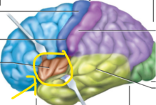
Broca’s area
speech production
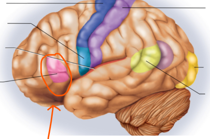
Wernicke’s area
speech comprehension

Cerebellum
balance and equilibrium

Cerebellar lobes

Flocculonodular lobe (flocculus and nodulus)
Folia (gray matter)

arbor vitae (white matter)

Cingulate gyrus

Corpus callosum

Thalamus
relay station

Hypothalamus
visceral control center
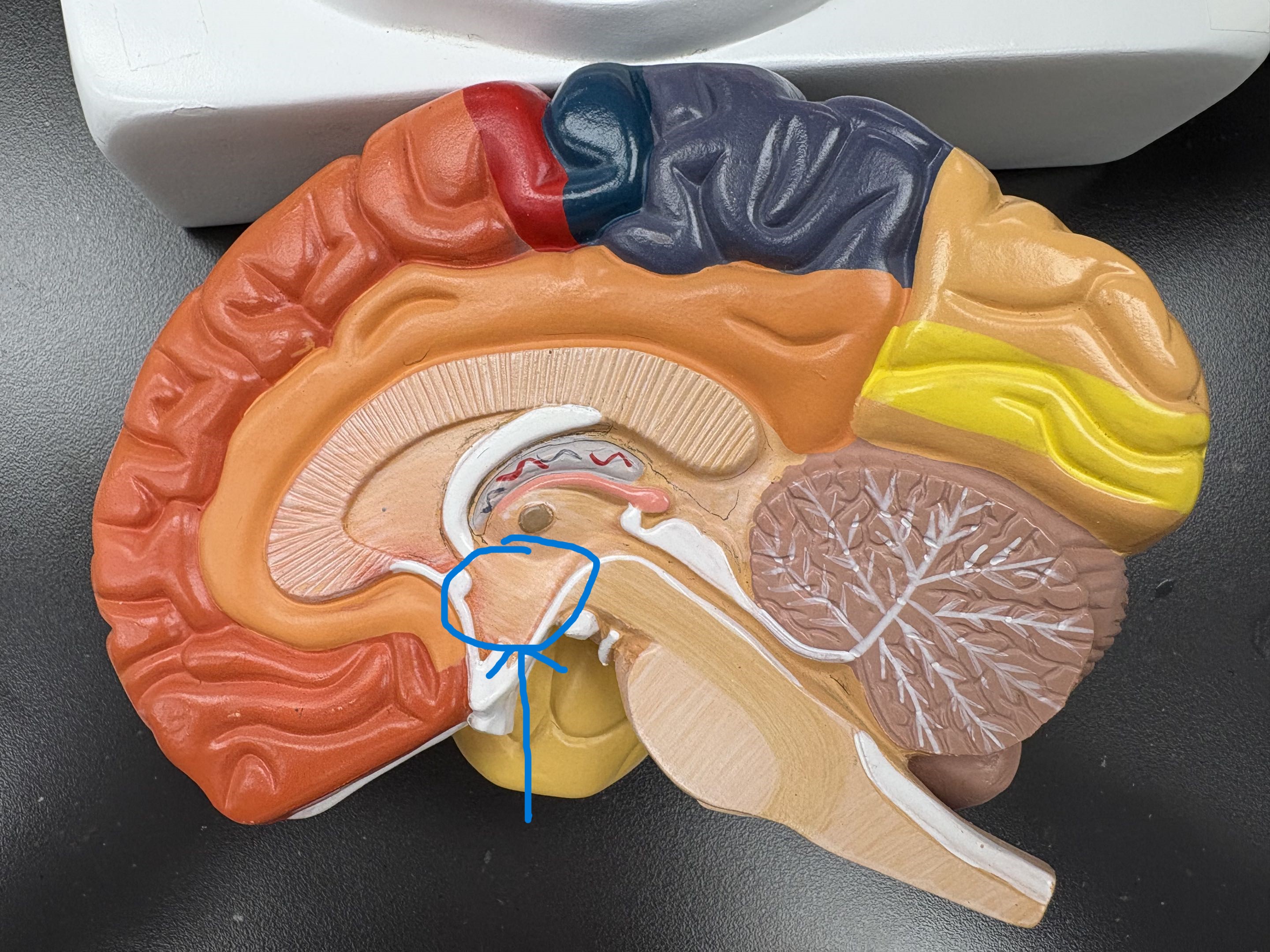
Epithalamus
pineal gland, habenula

Pituitary gland

Internal capsule
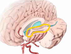
Corpus striatum
caudate, putamen, globus pallidus

Midbrain

Superior colliculi
visual reflex

Inferior colliculi
auditory reflex

Pons

Medulla oblongata
pyramids, olives (vital centers)

Lateral ventricle

Interventricular foramen

Third ventricle
a narrow, slit-like, fluid-filled cavity located in the center of the brain within the diencephalon

Cerebral aqueduct

Fourth ventricle
a diamond-shaped, fluid-filled cavity located within the hindbrain of the central nervous system

Central canal

Choroid plexus
tuft of capillaries that produces cerebrospinal fluid

Olfactory
I, sensory, smell

Optic
II, sensory, vision
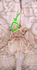
Oculomotor
III, motor, moves eyes

Trochlear
IV, motor, moves eyes
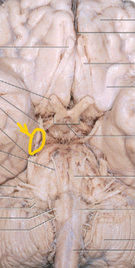
Trigeminal
V, b, sensory to face; chewing

Abducens
VI, motor, moves eyes

Facial
VII, both, facial muscles; taste

Vestibulocochlear
VIII, sensory, equilibrium, and hearing

Glossopharyngeal
IX, both, swallowing; taste

Vagus
X, both, parasympathetic motor; taste
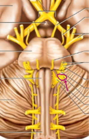
Accessory
XI, motor, head and neck muscles
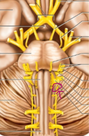
Hypoglossal
XII, motor, moves tongue

Dorsal root
afferent signals

Dorsal root ganglion
sensory neuron cell bodies

Ventral root
efferent signals

Spinal nerve
mixed (afferent and efferent)

Dorsal ramus

Gray matter
dorsal and ventral horns; gray commissure

White matter
dorsal, ventral and lateral funiculus

Cervical Enlargements

Conus medularis
under the lumbar enlargement and above the cauda equina
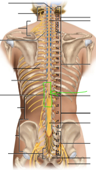
Cauda equina
under the conus medularis

Filum terminale

Musculocutaneous nerve
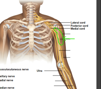
axillary nerve

radial nerve

median nerve

ulnar nerve

Dura mater
“tough film like sheet over brain”

arachnoid mater
web-like holding the meninge together

pia meter
outer layer of the spinal cord

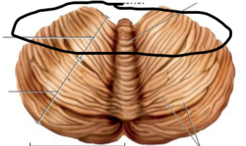
Cerebellar posterior lobe

Cerebellar anterior lobe
pineal gland

habenula

ventral ramus

Lumbar Enlargements

hyaloid canal

ora serrata

Rod
photoreceptor is more numerous and has a rhodopsin visual pigment, scotopic (good for low light/night vision)

Cone
photoreceptor that has a photopsin visual pigment, photopic (bright light/day vision)
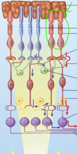
pigment epithelium
a single, tightly packed layer of pigmented cells located in the outermost part of the retina

auricle
