Human Anatomy Exam 3
 Call Kai
 Learn
 Practice Test
 Spaced Repetition
 Match
 Flashcards
 Knowt Play
1/226

There's no tags or description
Looks like no tags are added yet.
227 Terms
Joints/Articulations
Where two or more bones meet
Synarthrosis
No movement
- Sutures
- Gomphosis
- Synchondrosis
- Synostosis
Amphiarthrosis
Little movement
- Syndesmosis
- Symphysis
Diarthrosis
Free movement
- Fluid filled cavity
- Synovial fluid
- Articular capsule
Synchondrosis
Immovable joint
Cartilaginous bridge between 2 articulating bones
Gomphosis
Immovable joint
Fibrous joint between teeth & jawbones
Synostosis
Immovable joint
Bony fusion joint between 2 bones
Syndesmosis
Slightly movable joint
Ligaments that connect two bones
Symphysis
Slightly movable joint
Bones separated by a wedge/pad of cartilage
Components of synovial joints
- Fibrous joint capsule
- Synovial membrane
- Articular cartilages
- Joint cavity containing synovial fluid
Fibrous joint capsule location
Outermost lining

Synovial membrane location
Innermost lining

Articular cartilages location
Cover ends of bones

Joint cavity location
Hollow area

Accessory structures of synovial joints
- Bursae
- Fat pads
- Menisci
- Ligaments
Bursae function
- Fluid filled pockets
- Cushioning, reduce friction
Fat pads function
- Packing material
- Absorb shock
Menisci function
- Fibrous cartilage
- Reduce friction
Ligaments function
- Support
- Strength
- Reinforce
Circumduction
Circular movement

Rotation
Pivot around central axis

Abduction
Away from midline

Adduction
Towards midline

Flexion
Decrease angle between bones

Extension
Increase angle between bones

Elevation
Move body part superiorly

Depression
Move body part inferiorly

Protraction
Move body part anteriorly

Retraction
Move body part posteriorly

Pronation
Rotate forearm so palm faces posterior/down
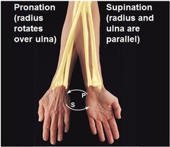
Supination
Rotate forearm so palm faces anterior/up

Opposition
Move thumb to touch the tips of other fingers on the same hand

Inversion
Turn sole of foot medial/in

Eversion
Turn sole of foot lateral/out

Dorsiflexion
Lift foot so toes approach shin

Plantar flexion
Depress foot to elevate heel

Plane joint
- Nonaxial movement
- Gliding

Hinge joint
- Uniaxial movement
- Flexion & extension

Pivot joint
- Uniaxial movement
- Rotation

Condylar joint
- Biaxial movement
- Flexion & extension
- Adduction & abduction
- Oval articular surfaces

Saddle joint
- Biaxial movement
- Flexion & extension
- Adduction & abduction
- Concave & convex articular surfaces

Ball-and-socket joint
- Multiaxial movement
- Flexion & extension
- Adduction & abduction
- Rotation

Dislocation/Luxation
Complete separation of bones that form a joint

Temporomandibular joint made of
- Mandible condylar process
- Temporal bone mandiublar fossa

Temporomandibular joint type
- Hinge joint
- Side to side movements
Temporomandibular joint ligaments
- Stylomandibular ligament
- Lateral ligament

Knee joint made of
- Medial menisci
- Lateral menisci
- Fat pads
Knee joint ligaments
- Fibular collateral ligament
- Tibial collateral ligament
- Anterior cruciate ligament (ACL)
- Posterior cruciate ligament (PCL)
ACL
- Connect tibia to lateral edge of intercondylar fossa of femur
- Prevent anterior sliding of tibia

PCL
- Connect tibia to medial edge of intercondylar fossa of femur
- Prevent forward sliding of femur or backward displacement of tibia

Knee joint articular capsule
Extends from distal femur to proximal tibia & includes patella (NO fibula)
Shoulder joint type
- Ball and socket
- Free movable
Shoulder joint articulation
Head of humerus with glenoid cavity
Shoulder joint ligaments
- Coracoclavicular
- Acromioclavicular
- Coracohumeral
Shoulder joint bursae
- Pockets with articulation, filled with synovial fluid
- Protect & prevent friction between joints
Rotator cuff
Shoulder joint muscles
Intervertebral discs
Pads of fibrous cartilage between vertebrae
Intervertebral discs made of
- Anulus fibrosus (outer)
- Nucleus pulposus (inner)
- Vertebral endplate (inferior & superior)

Herniated disc
When soft internal center of intervertebral disc pushes through outer layer and presses on nerves

Arthritis
- Inflammation of joints
- Damage to articular cartilages

Bursitis
Inflammation of bursa sacs
Structural organization of skeletal muscle
- Endomysium = wrap individual fibers
- Perimysium = wrap individual fascicles
- Epimysium = wrap all fascicles

Sarcolemma
Muscle cell membrane
Sarcoplasm
Muscle cytoplasm
T-tubules
Invaginations of sarcolemma

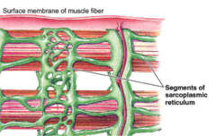
Sarcoplasmic reticulum
Muscle modified endoplasmic reticulum
Myofibrils
Long cylindrical units responsible for contraction

Parallel muscle
- Parallel muscle
- Parallel muscle with bands
- Wrapping muscle
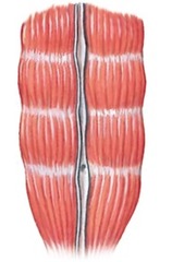
Convergent muscle
- Raphe (tendinous sheet)
- Shell like

Pennate muscle
- Unipennate (1 feather)
- Bipennate (2 feathers)
- Multipennate (many feathers)

Circular muscle
Donut shape

Muscle origin
- Where muscle begins
- Remains stationary
Muscle insertion
- Where muscle ends
- Moves
Agonist muscle
Muscle that accomplish certain action

Antagonist muscle
Muscle acting against agonist

Synergistic muscle
Group of muscles working together
Fixator muscle
Muscle that holds 1 bone in place relative to the body while a distal bone is moved by other muscle
Sarcomere
Unit of contraction

Neuromuscular junction structures
- Axon terminal
- Synaptic cleft
- Motor end plate
Motor neuron
Nerve cells that control muscle movement
Muscular atrophy
When muscle is not used for a long period of time, reduction in muscle size & power occur
Muscular hypertrophy
- Increase of contractile activity
- Increase in size of muscle
- Change in ATP production
- Bodybuilding
4 groups of axial muscles
1. Head & Neck
2. Vertebral column
3. Thorax & Abdominal wall
4. Pelvic floor
Origin, Insertion, Action, Location: Frontalis
O: Epicranial aponeurosis
I: Skin of eyebrows, bridge of nose
A: Raise eyebrows & wrinkle forehead

Origin, Insertion, Action, Location: Occipitalis
O: Superior nuchal line
I: Epicranial aponeurosis
A: Tense & retract scalp

Origin, Insertion, Action, Location: Temporoparietalis
O: Fascia around external ear
I: Epicrnail aponeurosis
A: Move auricle of ear

Origin, Insertion, Action, Location: Orbicularis oculi
O: Medial margin of orbit frontal & maxilla
I: Skin around eye lids
A: Closes eyes
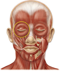
Origin, Insertion, Action, Location: Corrugator supercilii
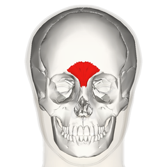
O: Arch of frontal bone
I: Eyebrow
A: Wrinkles eyebrows

Origin, Insertion, Action, Location: Procerus
O: Lateral nasal cartilages
I: Aponeurosis at bridge of nose & forehead
A: Wrinkle & move nose, horizontal wrinkles frowning
Origin, Insertion, Action, Location: Nasalis
O: Maxilla & alar cartilage of nose
I: Bridge of nose
A: Dilate nostrils

Origin, Insertion, Action, Location: Orbicularis oris
O: Maxilla & mandible
I: Lips
A: Compress & pulse lips

Origin, Insertion, Action, Location: Buccinator
O: Alveolar processes of maxilla & mandible
I: Fibers of orbicularis oris
A: Compress cheeks

Origin, Insertion, Action, Location: Risorious
O: Fascia associated to masseter muscle
I: Angle of mouth
A: Contracting with smiling

Origin, Insertion, Action, Location: Levator labii superioris
O: Zygomatic bone & infraorbital margin of maxilla
I: Skin upper lip
A: Opens lips

Origin, Insertion, Action, Location: Depresor labii inferioris
O: Body of mandible lateral to middle line
I: Skin of lower lip
A: Depress lower lip

Origin, Insertion, Action, Location: Zygomaticus
O: Zygomatic bone
I: Skin at corners of mouth
A: Smiling

Origin, Insertion, Action, Location: Platysma
O: Fascia covering superior parts of pectoralis major & deltoid
I: Mandible & skin of cheek
A: Open mouth, tense skin (neck)

Origin, Insertion, Action, Location: Mentalis
O: Incisive fossa of mandible
I: Skin of chin
A: Protrude lower lip

Dimple
Zygomaticus major
6 extra-ocular muscles & movements
1. Superior oblique = down & lateral
2. Inferior oblique = up & lateral
3. Superior rectus = up
4. Inferior rectus = down
5. Medial rectus = medial, toward nose
6. Lateral rectus = lateral, toward ear
