Eye Anatomy
 Call Kai
 Learn
 Practice Test
 Spaced Repetition
 Match
 Flashcards
 Knowt Play
1/26

There's no tags or description
Looks like no tags are added yet.
27 Terms

Iris
Colored part of eye
Controls size of pupil (pupil constricts and dilates in response to light)
Pupillary constrictor receives PARASYM innervation
pupillary dilator receives SYM innervation
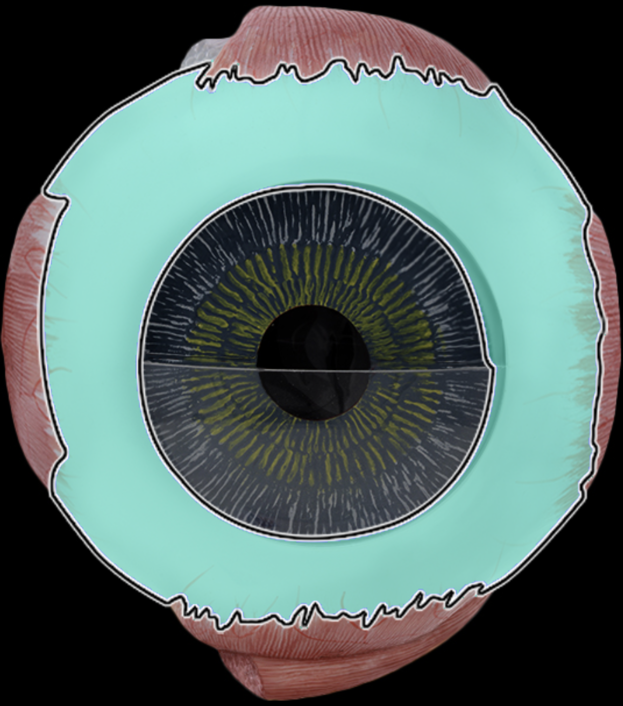
Sclera
(fibrous layer)
Dense collagenous c.t.
Protects eye
Maintains shape of eye
"White" of the eye

Pupil
light enter eye
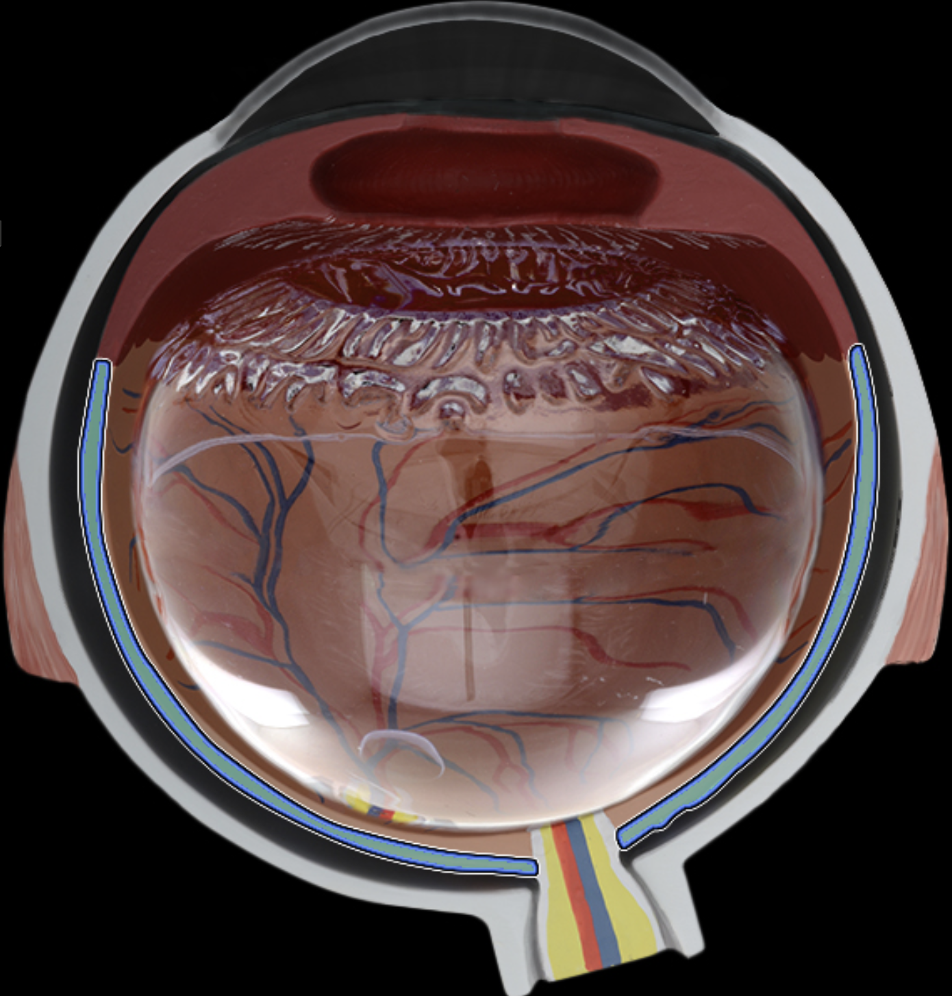
Choroid
Highly vascular layer
Contains dark brown pigment - melanin
Blood supply to outer part of neural retina
Prevent light reflection within eye

ciliary body
b/w choroid + iris
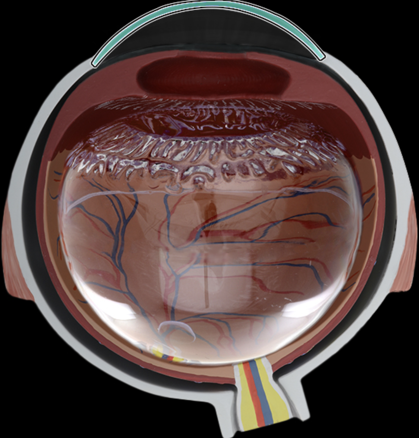
Cornea
Transparent c.t. layer of anterior eye
Site for light refraction
Protection of anterior eye

Lens
posterior to iris
Biconvex lens
lens fibers (cells)
Light refraction
Focuses light onto neural retina

optic nerve (CN 2)
Vision
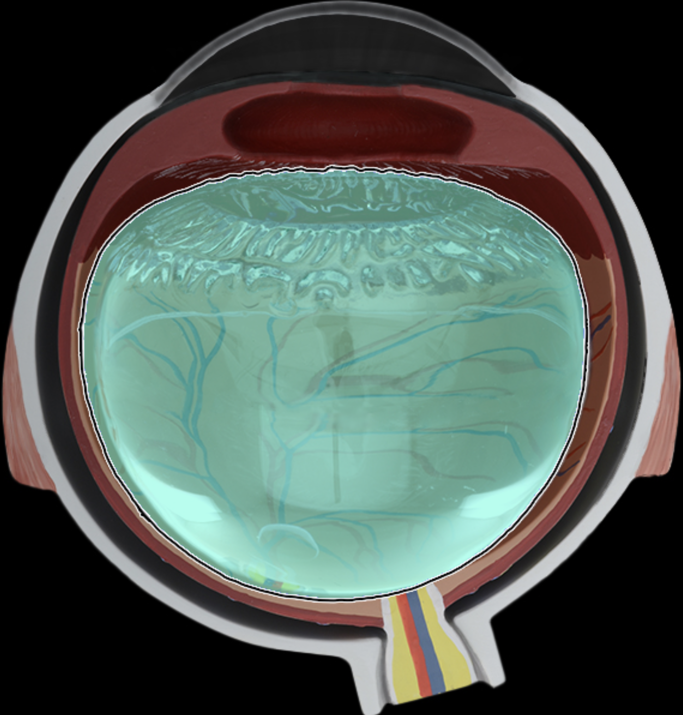
posterior cavity
Posterior to lens
Maintain normal intraocular pressure and shape of the eye
Maintain lens and retina in place
Refraction of light
(Vitreous chamber)

retina
2 layers
Outer pigmented layer immediately inside choroid
Inner neural layer contains photoreceptors + associated neurons
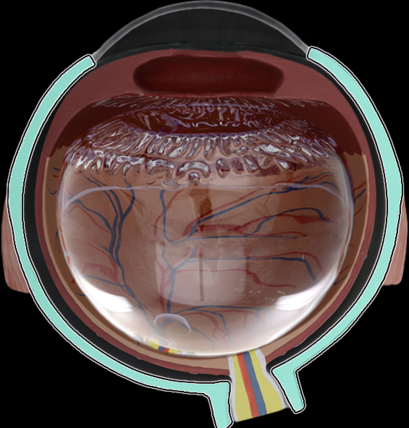
Sclera
(fibrous layer)
Dense collagenous c.t.
Protects eye
Maintains shape of eye
"White" of the eye

anterior cavity
b/w cornea + lens
Aqueous humor produced in posterior chamber
enters anterior chamber
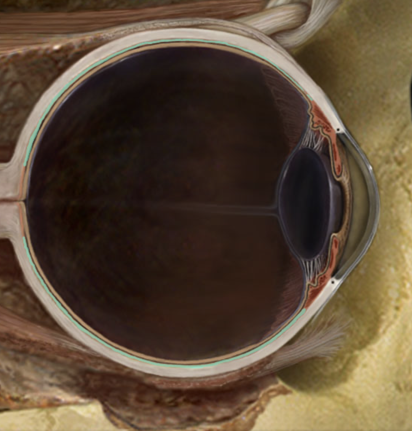
Choroid
Highly vascular layer
Contains dark brown pigment - melanin
Blood supply to outer part of neural retina
Prevent light reflection within eye

ciliary body
b/w choroid + iris
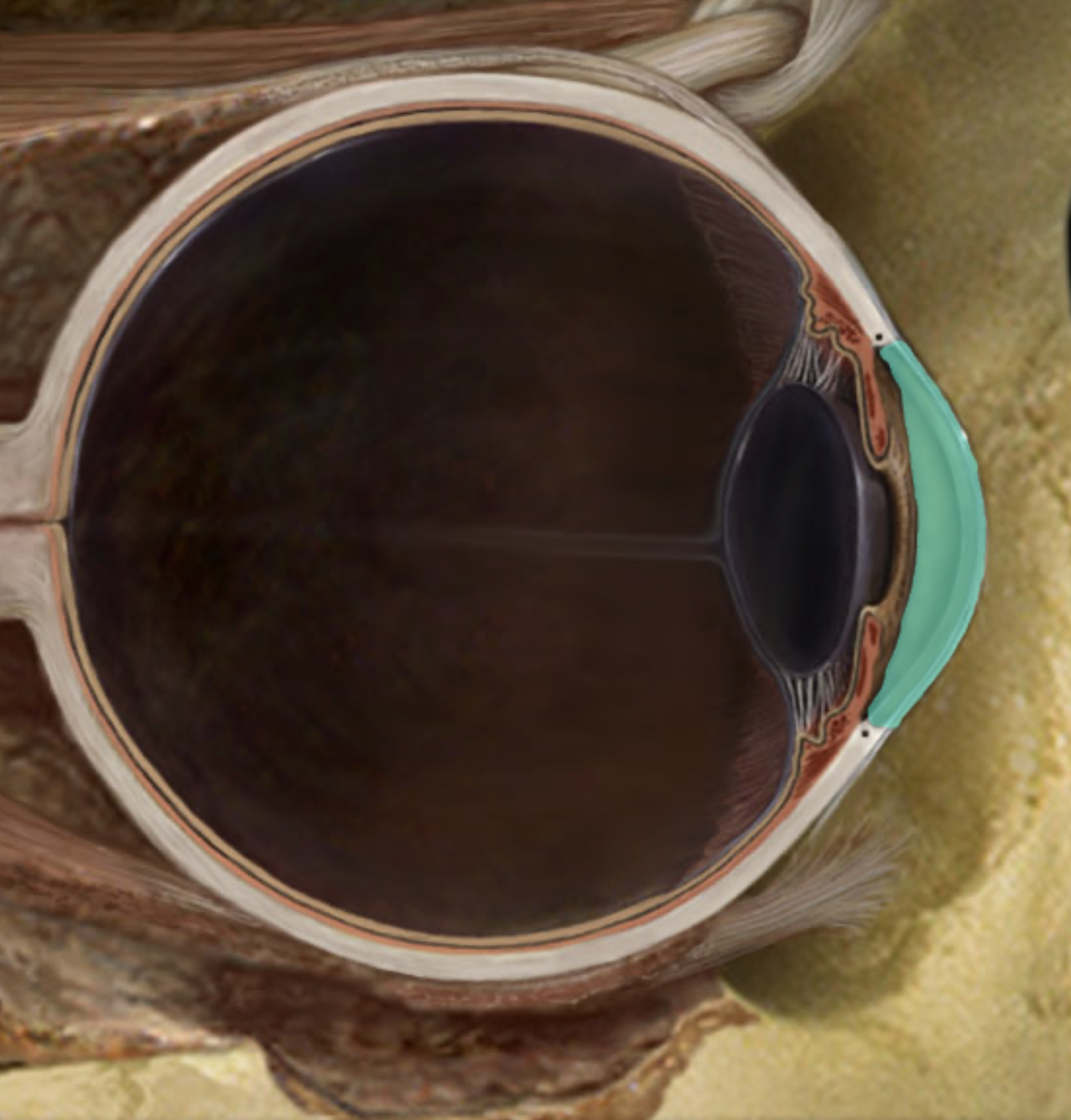
Cornea
Transparent c.t. layer of anterior eye
Site for light refraction
Protection of anterior eye

Iris
Colored part of eye
Controls size of pupil (pupil constricts and dilates in response to light)
Pupillary constrictor receives PARASYM innervation
pupillary dilator receives SYM innervation

Lens
posterior to iris
Biconvex lens
lens fibers (cells)
Light refraction
Focuses light onto neural retina

Optic disc
At junction w/ optic nerve
Description:
Composed of optic fibers (axons of retinal ganglion cells) that form optic nerve
NO photoreceptors in disc = “blind spot”

optic nerve (CN 2)
Vision

posterior cavity
Posterior to lens
Maintain normal intraocular pressure and shape of the eye
Maintain lens and retina in place
Refraction of light
(Vitreous chamber)
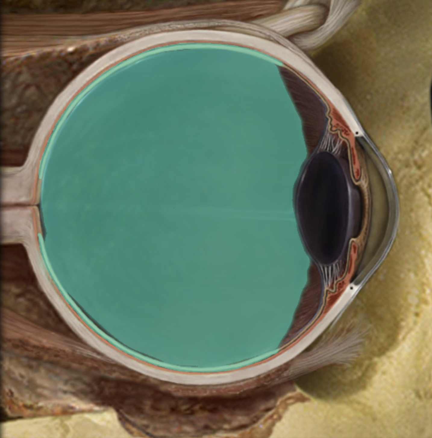
retina
2 layers
Outer pigmented layer immediately inside choroid
Inner neural layer contains photoreceptors + associated neurons

Suspensory ligaments of lens
b/w ciliary body and lens
Transparent, elastic fibers
Suspends lens from ciliary body
Connects ciliary muscle to lens
Permits thickness of lens to change with contraction/relaxation of ciliary muscle
Contraction of ciliary muscle > relaxes suspensory ligaments > lens thickens
Relaxation of ciliary muscle > tenses supsensory ligaments > lens gets thinner
(Zonular fibers)

eye
Fibrous tunic - cornea + sclera
Vascular tunic - choroid, ciliary body, iris
Neural tunic - retina + pigment epithelium
Optical components collect and focus light
Neural components process and transmit light-driven signals to brain

optic nerve (CN 2)
Vision
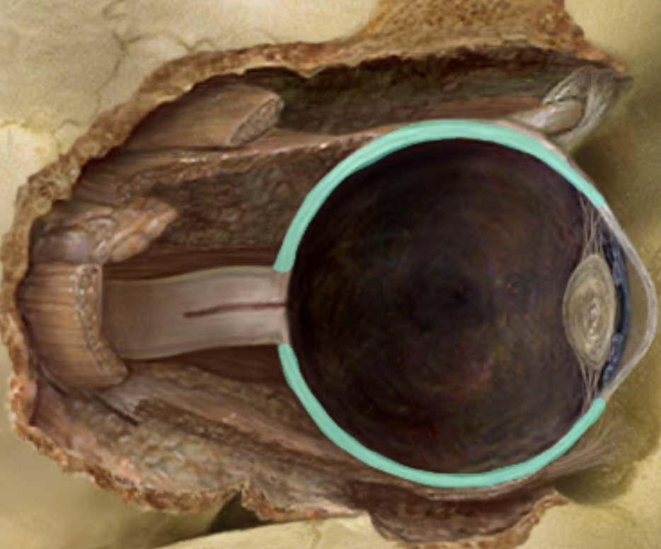
Sclera
(fibrous layer)
Dense collagenous c.t.
Protects eye
Maintains shape of eye
"White" of the eye

Lens
posterior to iris
Biconvex lens
lens fibers (cells)
Light refraction
Focuses light onto neural retina