Skin Disorders of Pigmentation and Melanocytes
 Call Kai
 Learn
 Practice Test
 Spaced Repetition
 Match
 Flashcards
 Knowt Play
1/15

There's no tags or description
Looks like no tags are added yet.
16 Terms

Define:
Macule :(<5 mm); Patch: (>5 mm)
Papule :(<5 mm); Nodule: (>5 mm)
Plaque:
Vesicle: (<5 mm); Bulla: (>5 mm)
Blister:
Pustule:
Wheal:
Scale:
Lichenification:
Excoriation:
Onycholysis:
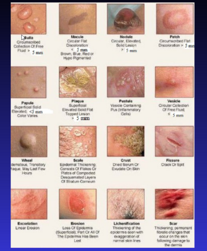
The Skin: clinical terms
Macule :(<5 mm); Patch: (>5 mm)
Circumscribed flat and discoloured lesion
Papule :(<5 mm); Nodule: (>5 mm)
Elevated dome-shaped or flat-topped lesion
Plaque:
Elevated flat-topped lesion,
usually >5 mm across (may be caused by coalescent papules).
Vesicle: (<5 mm); Bulla: (>5 mm)
Fluid-filled raised lesion
Blister:
Common term used for vesicle or bulla
Pustule:
Discrete, pus-filled, raised lesion
Wheal:
Itchy, transient, elevated lesion w/ variable blanching and erythema formed as result of dermal edema
Scale:
Dry, horny, plate-like excrescence; often due to imperfect cornification (i.e., keratinization).
Lichenification:
Thickened and rough skin characterized by prominent skin markings;
usually result of repeated rubbing in susceptible persons
Excoriation:
Traumatic lesion characterized by breakage of epidermis, causing raw linear area (i.e., a deep scratch)
Onycholysis:
Separation of nail plate from nail bed


Define:
Hyperkeratosis:
Parakeratosis:
Hypergranulosis:
Acanthosis:
Papillomatosis:
Dyskeratosis:
Acantholysis:
Spongiosis:
Hydropic swelling (ballooning):
Exocytosis:
Erosion:
Ulceration:
Vacuolization:
Lentiginous:
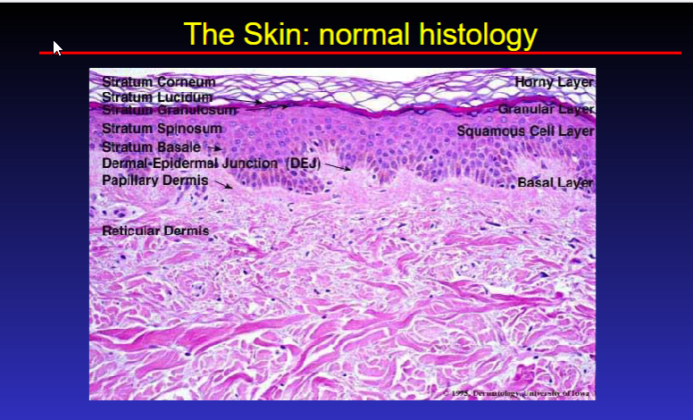
Histopathologic terms
Hyperkeratosis:
Thickening of stratum corneum,
often w/ qualitative abnormality of keratin
Parakeratosis:
Keratinization w/ retention of nuclei in stratum corneum.
On mucous membranes, normal
Hypergranulosis:
Hyperplasia of stratum granulosum,
Often due to intense rubbing.
Acanthosis:
Diffuse epidermal hyperplasia
Papillomatosis:
Surface elevation caused by hyperplasia and enlargement of contiguous dermal papillae
Dyskeratosis:
Abnormal keratinization occurring prematurely w/in individual cells or groups of cells below the stratum granulosum.
Generally the same as DYSPLASIA
Acantholysis:
Loss of intercellular connections -> loss of cohesion btw keratinocytes
Spongiosis:
Intercellular edema of epidermis
Hydropic swelling (ballooning):
Intracellular edema of keratinocytes
Exocytosis:
Infiltration of the epidermis by inflammatory or circulating blood cells
Erosion:
Discontinuity of the skin exhibiting incomplete loss of the epidermis
Ulceration:
Discontinuity of skin exhibiting complete loss of epidermis and lower portions
Vacuolization:
Formation of vacuoles within or adjacent to cells;
often refers to basal cell- basement membrane zone area.
Lentiginous:
A linear pattern of melanocyte proliferation w/in epidermal basal cell layer.

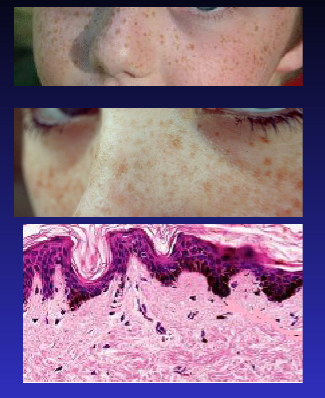
Freckle (Ephelis)
Describe Freckle (Ephelis)
What is it?
Description
Course
Cause?
Freckle (Ephelis)
What is it?
Commonest pigmented lesion of childhood in light- skinned
Description:
Small macules (one-few mm) on sun-exposed area
(skin, neck)
Course:
Starts age 5 years
Fade and darken winter & summer
Cause:
Increased melanin transfer to basal keratinocytes
no increase in melanocyte number
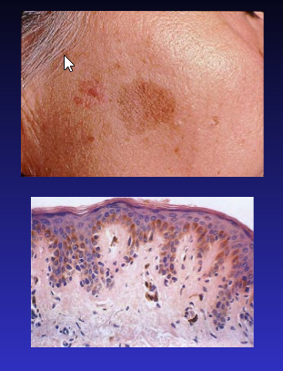
Lentigo
Describe Lentigo
Description:
Histology:
Cause
Lentigo
Description:
Small, sharply circumscribed, pigmented macule surrounded by normal skin
Histology:
linear non-nested melanocyte hyperplasia ;
elongated thin rete ridges
Cause:
unknown
NOTE: ‘Lentigo’ means “pigmented with clear edge”
Describe Melanocytes
What is it?
Embryo:
Melanocyte: keratinocytes
Histo Description:
Describe Nevus cells
What are they?
Histo:
Melanocytes
What is it?
Melanin-producing cells
@ bottom epidermis layer
Embryo:
Neural crest cells
Melanocyte: keratinocytes
1: 10
Histo Description:
Oval dendritic cells,
Smaller than keratinocytes
S 100 positive
Nevus cells
What are they?
Modified melanocytes
No dendrites
Larger
Histo: S 100 positive
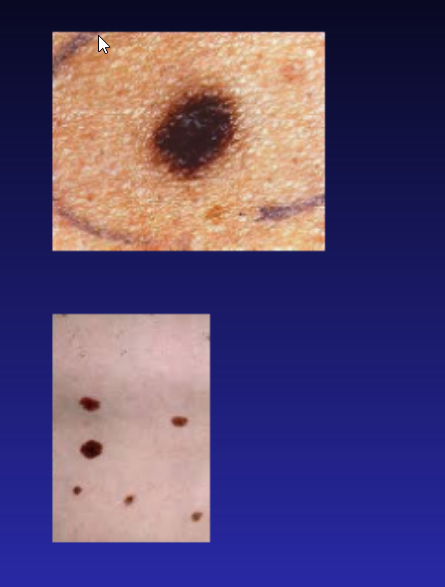
Melanocytic Nevus: Mole
Describe Melanocytic Nevus:
AKA
What are they?
Epi:
MOA:
Different Types:
Clinical Description:
Melanocytic Nevus: Mole
What are they?
Common benign neoplasm of melanocytes
Epi:
congenital : About 1% born with moles
fair skin> dark skin
MOA:
Mutation in RAS pathway
Different Types:
Based on Location:
Junctional:
@ junction of epi/dermis
Compound:
@ junction + w/in dermis
Intradermal:
confined to dermis:
Clinical Description:
Tan to brown
Uniform in color
Small ( <6 mm )
Macules or papule
Well-defined rounded borders


Dysplastic nevus
Describe Dysplastic nevus
Clinical Description (2)
Histo: (2)
Epi: (3)
Causes: (2)
Dysplastic nevus
Clinical Description
Larger than 5 mm
Flat or raised plaques
Histo:
Single layer of nevus cells at basal epidermal layer
Dermis & epidermis may be involved
Epi:
Often familial
Marker of melanoma risk
Cytologic atypia
Causes:
RAS and BRAF mutation may be present
List the Variants of melanocytic nevi (good to know)
