Lecture 6 - Vasculature and Vascular Mechanics
 Call Kai
 Learn
 Practice Test
 Spaced Repetition
 Match
 Flashcards
 Knowt Play
1/28

There's no tags or description
Looks like no tags are added yet.
29 Terms
What are the three main structures in terms of vascular structure?
veins
arteries
microcirculation structures: arterioles, capillaries, venules
Describe the characteristics of veins
Lowest pressure
Thin muscled walls
Highly distensible
Capable of stretching, expanding etc.
High resistance
As blood flows through the system, blood ends up through veins, pressure drops, increasing resistance (resistance is highest in arterioles though)

Describe the characteristics of arteries
Highest pressure
Strong, muscular walls
Provides low resistance to flow

Describe the characteristics of microcirculation structures
arterioles → main site of vascular resistance
capillaries and venules → sites for gas and nutrient exchange
what is compliance?
how readily a structure deforms/expands when force is applied
Reciprocal of compliance is stiffness
The more its able to stretch, the less stiff it is
Reflected by increase in volume in response to increasing transmural pressure
Compliance = delta V / delta P
Ex. high compliance in vein when a high volume of blood enters vein, but small change in pressure caused
What is elasticity?
how readily a structure returns to its original volume/size after removal of external force
Deformed elastic structures store elastic potential energy
how compliant are veins?
very compliant, large changes in volume cause small changes in P
Able to accommodate for huge changes in volume, but cause small changes in p
As volume increases in graph, pressure slowly increases (graph)

how compliant are arteries?
less compliant then veins, will still expand in response to increase in pressure (in contrast to a rigid pipe)

describe the P-V graph in terms of vascular compliance and the compliance formula
???

How elastic are arteries?
Arteries are highly elastic but not compliant
How is arterial BP maintained?
Due to high elasticity of arteries,
During systole, blood enters aorta, stretching of aortic walls stores elastic potential energy
Heart pumps blood into aorta faster than it drains it out
If aorta was not compliant, SBP would shoot up
SBP is kept from increasing further as aorta expands to hold excess blood
During diastole, aortic walls recoil, squeezing blood, sustaining aortic pressure
Required to maintain constant blood flow

How does left ventricle pressure decrease to near 0 in diastole, while aortic pressures do not? Why?
LV pressure drops after heart finishes contraction (so all blood is ejected into aorta) and the ventricle relaxes
aorta is now full with blood, so Paorta > Pventricle and aortic elasticity (recoil) maintains constant blood flow out of aorta

Why does the pressure oscillation decrease along the arterial system?
Elasticity
Aortic recoil preserves pressure, decreasing magnitude of oscillation
Vascular resistance
Blood encounters resistance as it flows resulting in p
As blood moves away from the heart and flows through circulatory system, resistance begins to increase, dropping pressure
what is the pulse pressure equation, and when is it at its peak? when does it drop?
Pulse pressure = SBP - DBP
SBP in aorta is similar to SBP in LV
Peak aortic pressure during mid systole (rapid ejection), drops as ejection decreases and blood drains out of aorta
Left ventricle pressure drops lower than aorta, what happens?
Aortic valve closes → dicrotic notch
Aortic elasticity (recoil) causes momentary rise in pressure
Aorta continues to recoil during diastole, p drops to 80 mmHg
pulse p magnitude is determined by?
stroke volume (positive effect)
arterial compliance (negative effect)
Compliance dec → aortic distension dec (inc. SBP) → dec elastic recoil (dec DBP)
What conditions alter pulse pressure waves?
arteriosclerosis (specifically, artherosclerosis)
aortic valve stenosis
how does atherosclerosis affect pulse pressure?
Lesions develop in aortic walls (plaque)
Arteries become less compliant (increased stiffness) (hardened arteries)
SBP increases due to less compliance **
DBP decreases (sometimes or unchanged ) due to less elasticity (less recoil)
Increased pulse pressure (amplitude)
Based on graph, pressure increase from peak to dicrotic notch, and curve would decrease more than graph around diastolic decline

how does aortic valve stenosis affect pulse pressure?
Incomplete opening of aortic valve
Diameter of aortic valve opening is reduced
Increases resistance to flow into aorta
Inc. resistance results in pressure building up in LV and pressure drop in aorta dec SV (dec. SBP)
SBP decreases
Can be a small dec. in recoil (dec or unchanged DBP)
Smoothened or absent dicrotic notch
Dec. pulse pressure (amplitude)

Define internal intravascular pressure (Pi)
force of blood against artery walls

define external pressure (Pt)
pressure exerted on walls from external tissue and interstitial fluid

Define transmural pressure
(delta P) = Pi-Pt
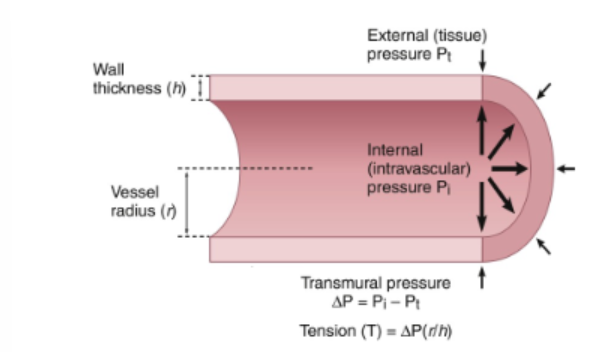
Define tension
internal pulling force within wall that resists the net distending pressure across the wall to prevent tear
Tension = delta P x radius/wall thickness

Tension and larger arteries
Larger arteries with greater radius experience greater tension in vessel wall
Must have stronger, thicker walls reinforced with collagen, elastic tissue and smooth muscle

Hypertension
Chronic increase in Pi causes inc in tnesion
Hypertrophic remodelling increases vessel wall thickness and decrease radius to decrease tension, prevents tear under high pressure
describe venous compliance
Veins distend easily which helps keep the pressure in the venous system low
The venous system can accommodate large changes in blood volume with only small changes in pressure (blood loss, transfusion, etc.)
The venous system is a blood reservoir and by contracting or relaxing, can adjust how much blood is available in active circulation
Veins distend easily which helps keep the pressure in the venous system low
The venous system can accommodate large changes in blood volume with only small changes in pressure (blood loss, transfusion, etc.)
During sympathetic stimulation, veins squeeze blood out of veins and into heart to ↑ venous return, ↑ SV and ↑ CO
During sympathetic inhibition, veins relax, allowing blood to pool, reducing venous return to heart
describe the venous blood reservoir
Veins hold about 65% of total volume of blood
By regulating venous tone (constriction or dilation), blood in the reservoir can be redistributed to perfuse more/fewer capillary beds as necessary
With sympathetic stimulation, ↑venous constriction, ↑venous return, ↑SV, ↑ CO and ↑ blood to microcirculation
Even with ↑ venous return, total blood volume in arteries changes very little. Why?
↓ compliance, cannot accommodate much larger volumes
Arteries are high, flow conduits rather than reservoirs (flow conduit = fast)
How are venous pressures affected by gravity?
Right atrial pressure (central venous pressure) is normally very low (0 mmHg) → there is no “sucking power”
Blood in veins can only return to heart if pressure in veins is greater than 0
When lying, veins are located at same level to heart, therefore pressure from heartbeat pushes blood
When standing, hydrostatic pressure (pressure exerted by fluid due to gravity) is exerted
Increases with fluid height
With height, there is more downwards pressure (hydrostatic pressure) causing blood to pool in legs
To overcome this, skeletal muscles contract by squeezing veins to push blood upward to the heart
When muscles relax, blood may pool back down → to prevent this, one-way valves lock blood in place, to prevent backflow and venous pooling
Gravity alters venous pressure based on changes in venous return/position of the vessel