Palmer - Spinal Anatomy Final Exam Objective (Spring 2026) (Beveridge)
 Call Kai
 Learn
 Practice Test
 Spaced Repetition
 Match
 Flashcards
 Knowt Play
1/30

Earn XP
Description and Tags
Credit to Sara Gehling (saragehling on Quizlet)!
31 Terms
How many synovial joint surfaces are on a typical cervical vertebrae?
8
Which ligaments attach to the C2 vertebral body?
ALL
PLL
tectorial membrane (PLL continued)
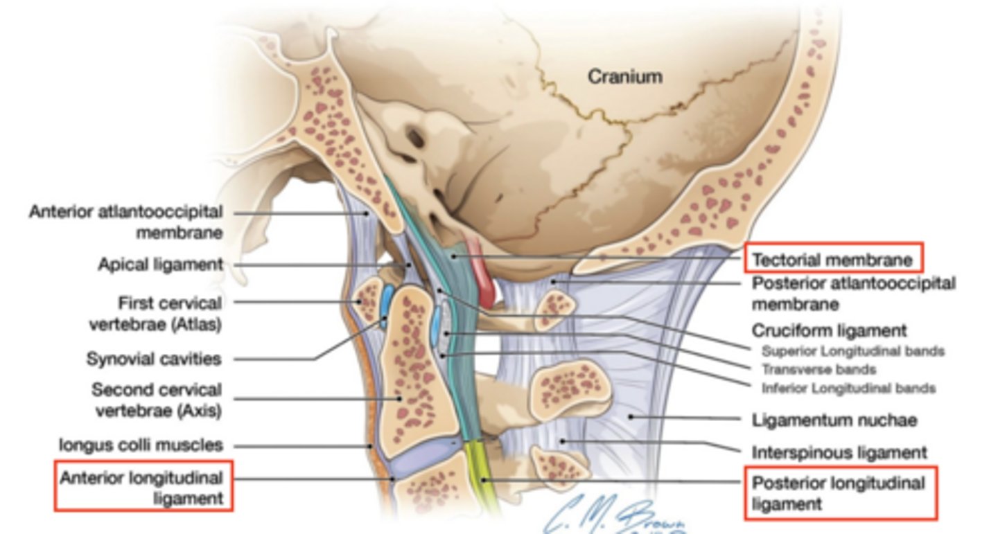
Describe the median atlantoaxial joint including its boundaries
synovial pivot (diathrosis trochoid) joint
Boundaries:
ant: anterior arch of C1
post: transverse ligament of C1
lat: lateral masses of C1
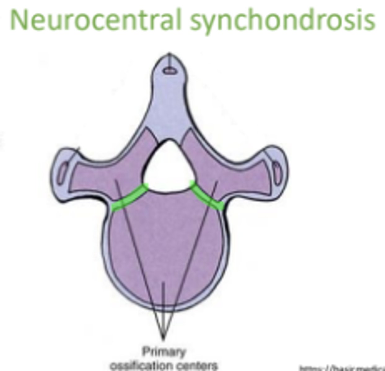
Describe the neurocentral synchondrosis
Synchondrosis between centrum & neural arch centers of ossification on each side;
Fully ossifies around 8 y.o.
Unilateral ossification can cause scoliosis
Describe the ponticulus posticus including its attachment sites
an accessory bone;
attachment: lateral mass & arcuate rim
superior to the groove/sulcus for the vertebral artery (potential source of vertebral a. compression)
complete ossification: arcuate foramen
more likely in females
forms b/w 2-7 y.o.
Understand the structural and functional classification of the ligaments forming the
anterior and posterior boundaries of the spinal canal
PLL (ant boundary) & ligamentum flavum (post boundary)
structural classification: fibrous syndesmosis
functional classification: amphiarthrosis
Describe the classification of fibrous joints
(Fibrous joints)
suture: dense fibrous CT; b/w skull bones
syndesmosis: fibrous CT; parallel bones; ligaments (ALL, PLL, LF)
gomphosis: Conical process into a socket; teeth roots & mandible alveolus
schindylesis: Ridged bone articulating in groove of neighboring bone: vomer & sphenoid
Which muscles attach to the T1 TVP?
longissimus (thoracis, cervicis, capitis)
semispinalis (cervicis, capitis)
multifidus
rotator longus/rotator brevis
intertransversarii
levator costorum brevis
How many synovial joint surfaces are located on T10, T11, and T12 vertebral body?
2 (costal facets)
Describe costal notches found on the sternum
1st costal notch: cartilaginous synchondrosis, separated from articular surface of 1st rib by hyaline cartilage
2nd-7th costal notches: synovial plane, stabilized by radiate sternocostal ligaments
Ribs 1-7 costal cartilages attach to costal notches
Describe costovertebral joints (location and total joint surfaces)
joint formed when the ribs articulate with the thoracic vertebrae
2 types of costovertebral joints:
1) costocentral (4; on body) - synovial plane
2) costotransverse (2 on arch) - synovial plane
Describe ossification centers in the sternum
1° ossification:
manubrium: 1-3
sternebrae 1 & 2: 1
sternebrae 3 & 4: 2
2° ossification:
xiphoid process
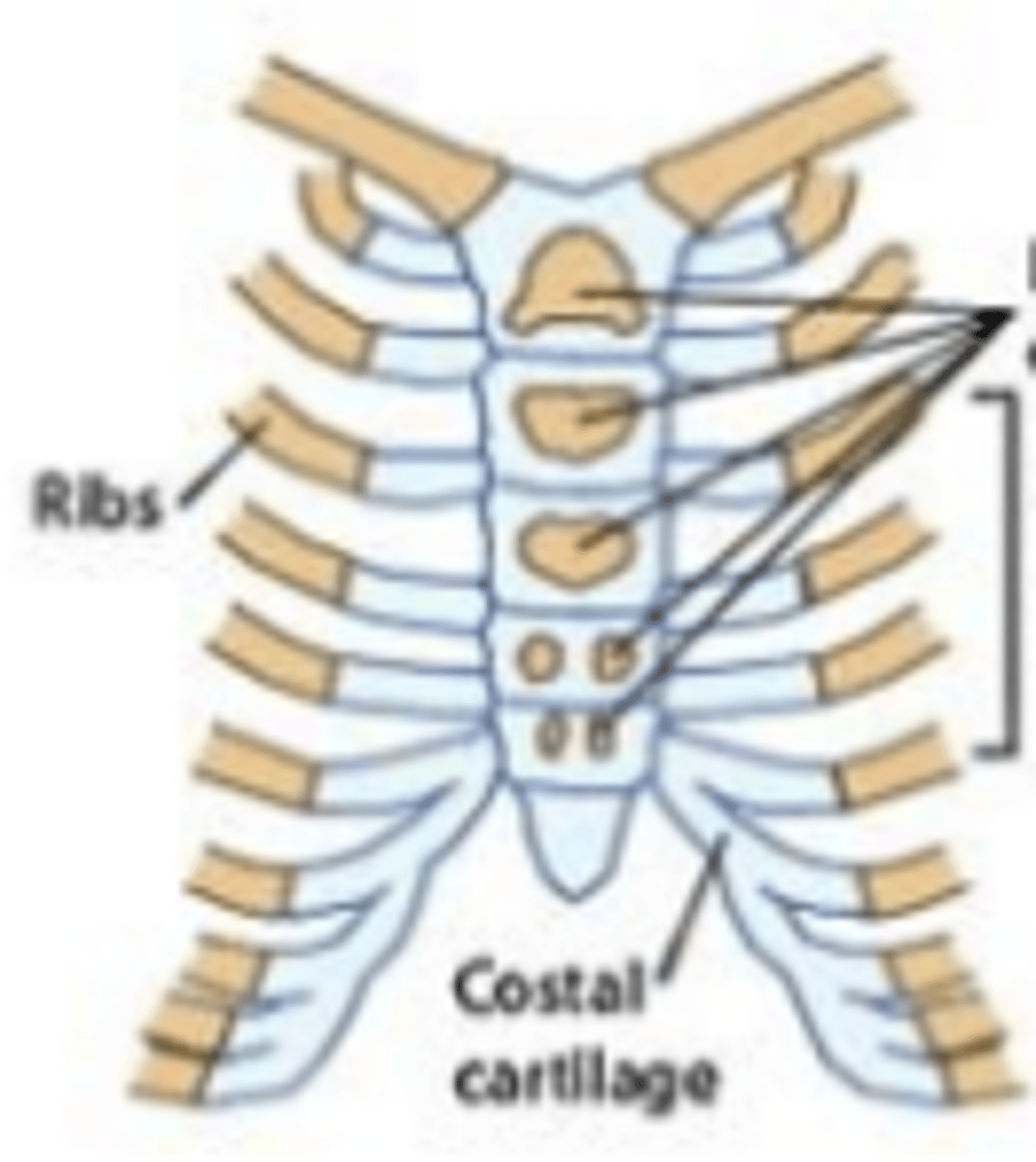
Describe joints associated with the manubrium
manubriosternal symphysis: manubrium and sternum, cartilaginous (amphiarthrosis) symphysis
sternoclavicular joint: synovial saddle
sternochondral joint: cartilaginous synchondrosis
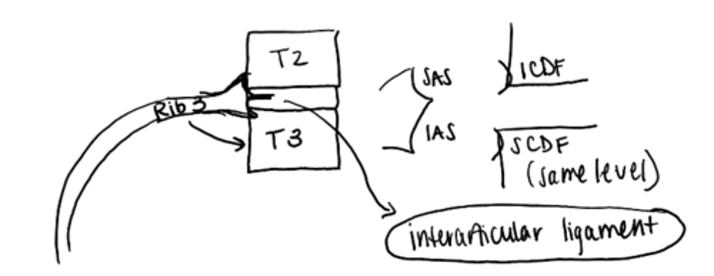
Describe superior and inferior costal demi facets including where they articulate
synovial plane joint
sup. costal demi facet: articulates with inf. articular surface of rib at same level (i.e. T3 sup. demi facet with Rib 3 inferior articular surface)
inf. costal demi facet: articulates with sup. articular surface of rib below (i.e. T2 inf. demi facet with Rib 3 inferior articular surface)
Describe para-articular processes
T10 lamina (most common)
ossification of ligamentum flavum
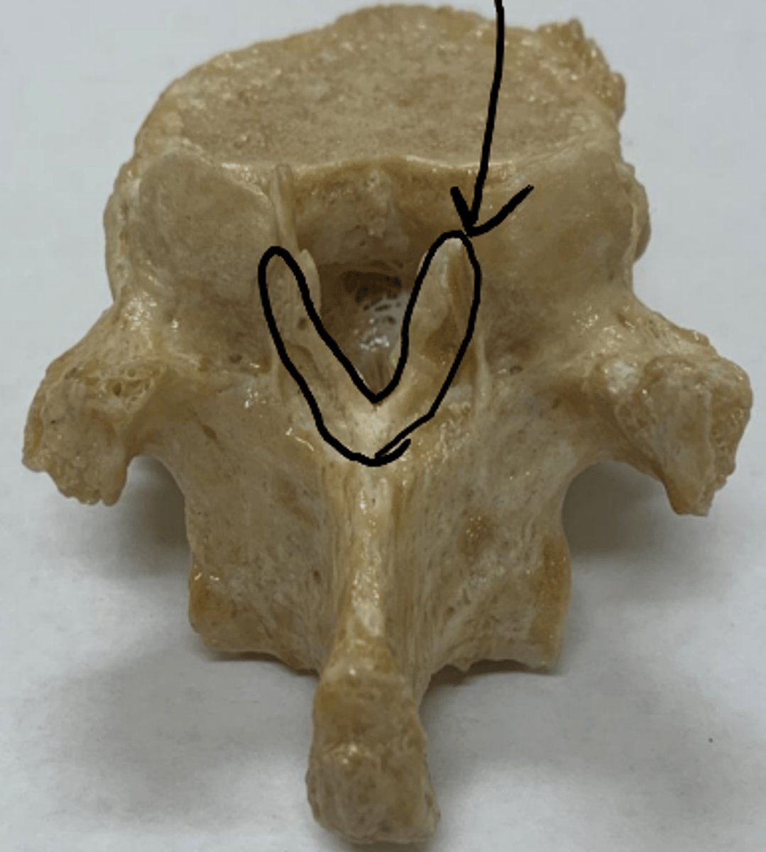
Understand and describe where spinal nerves exit between the intervertebral foramina
C1-C8 named for vertebrae below
(i.e. C2 nerve b/w C1 & C2 vertebrae)
T1-L5 named for vertebrae above
(i.e. T2 nerve b/w T2 & T3 vertebrae)
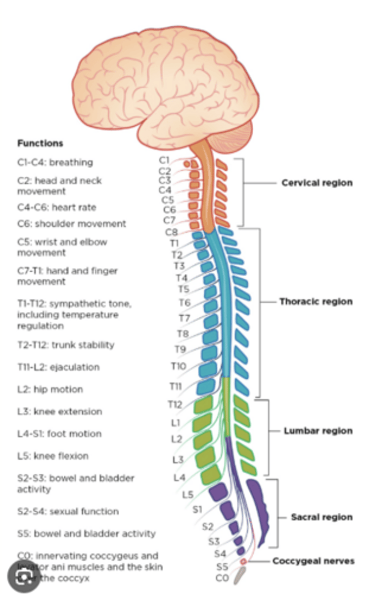
Describe the superior and inferior articular processes/facets in the lumbar spine
Sup - BUM
Inf - FoLD
Describe cervicalization at T1
T1 becomes cervical-like:
- T1 rib small or absent
- T1 has a transverse foramen
- T1 sup. art. facet BUM
- T1 uncinate process
Describe the subdental synchondrosis
subdental synchondrosis:
- bilateral primary centers of ossification for the dens
- cartilaginous synchondrosis
- fuses between 4-7
- persists beyond age 7 -> os odontoideum
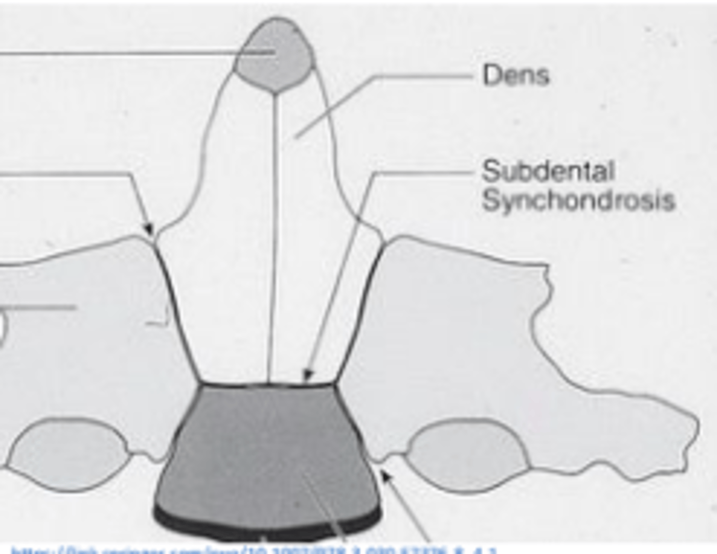
Describe the tip of the dens synchondrosis at C2
tip of the dens synchondrosis:
- secondary center of ossification for tip of the dens
- cartilaginous synchondrosis
- fusion before age 12
- persists after age 12 -> terminal ossicle
Describe the sacral hiatus
inferior opening of sacral spinal canal
Between median and intermediate sacral crests
Contents:
• Cauda equina
• Spinal meninges
• Co1 spinal nerve
• S5 (sometimes S4) sacral spinal nerve exits
Describe Assimilation of the Atlas
assimilation of atlas = occipitalization of atlas
- congenital
- partial or complete fusion with occiput
- anterior arch and foramen magnum fused
- occipital condyles fused to lateral masses of C1
- posterior arch fused to foramen magnum
Describe cervical and lumbar ribs
lumbar ribs:
- more common
- elongated transverse processes
- 7% of population
cervical ribs:
- dorsalization of C7
- can be found as high as C4
- usually at C7
Describe lumbar laminae
broad
thick
distinct spaces b/w segments -> easy to access subarachnoid space (L3-L4 lumbar spinal tap)
Where are ciliary bodies, ciliary glands, and meibomian glands located?
ciliary body: behind iris/ in posterior chamber - secretes aqueous humor
ciliary gland: at the base of eyelashes - secretes aqueous humor
meibomian gland: within tarsus - secretes meibum
Describe the supraorbital and infraorbital artery and where they come from
supraorbital a.:
internal carotid a. -> ophthalmic a. -> supraorbital a.; exits supraorbital foramen
infraorbital a.:
internal maxillary a. -> infraorbital a.; exits infraorbital foramen
Describe innervation to the parotid gland
- secretomotor
- parasympathetic
- CN 9: glossopharyngeal n.
- pre-ganglionic-> lesser petrosal n. (foramen ovale)
- otic ganglion (synapse)
- post-ganglionic -> auriculotemporal n. V3
Describe the tectorial membrane (membrana tectoria)
superior continuation of PLL (above C2)
Describe the ligamentum nuchae
runs from occiput to C7 spinous process
limits flexion of cervical spine
anterior & posterior layer (aka lamellar/funicular)
Describe the ligamentum flavum and its superior continuations
- connect adjacent laminae (C2-S1)
- homologue of posterior atlanto-occipital/ atlantal-axial ligament
- thickest in lumbar region
(thinnest in cervical)
- ossification: most common in thoracic/thoracolumbar
- small gaps for venous passage
- blends with interspinous lig.
- support ant. z-joint capsule
Describe muscles of mastication (Innervation? Derived from? Actions?)
temporalis
masseter
medial pterygoid
lateral pterygoid
innervated: V3 mandibular division of CN 5
derived: 1st pharyngeal arch
action: elevation, depression, protrusion, retrusion, lateral movements of jaw