Articulation & Body Movements
 Call Kai
 Learn
 Practice Test
 Spaced Repetition
 Match
 Flashcards
 Knowt Play
1/50

There's no tags or description
Looks like no tags are added yet.
51 Terms
Articulation
joints
place of contact btwn bones, bone and cartilage, bone and teeth
Structural Classification of Joints
Fibrous joint, Cartilaginous joint, Synovial joint
Fibrous Joint
no joint cavity
held tg by dense regular CT
Cartilaginous joint
no joint cavity
bones are joined by cartilage
Synovial Joint
Does have joint cavity > filled with lubricating fluid
separates articulating surfaces of bones
Ex: Glenohumeral joint, temporomandibular joint, elbow joint, knee joint
Structure: articular capsule, articular cartilage, joint cavity, synovial fluid, ligaments, nerves, and blood vessels
Functional Classification of Joints
Based on extend of movement:
Synarthrosis, Amphiarthrosis, Diarthrosis
Synarthrosis
immobile joint > does not move
Amphiarthrosis
slightly mobile joint
Diarthrosis
freely mobile joint
Fibrous Joint Classifications
Structure: Gomphosis, suture, syndesmosis
Function: Synarthrosis or amphiarthrosis
Cartilaginous Joint Classifications
Structure: Synchondrosis, symphysis
Function: Synarthrosis or amphiarthrosis
Synovial Joint Classifications
Structure: Plane, Hinge, Pivot, Condylar, Saddle, Ball and Socket
Function: Diarthrosis
Uniaxial, biaxial, multiaxial
Gomphosis
periodontal membranes hold a tooth to bony jaw (synarthrosis)
joined by ligaments

Suture
Connects skull bones (synarthrosis)
joined by short fibers
Syndesmosis
interosseous membranes btwn bones (amphiarthrosis)

Synostoses
complete fusion of bones across suture lines
Craniosynostosis
premature fusion or closing of 1 or more cranial sutures

Sagittal synosthosis
premature fusion of sagittal suture
skull grow forwards/backwards, or anteroposterior fashion > scaphocephaly
Coronal synosthosis
premature fusion of coronal suture
abnormally short and wide
Synchondroses
contain hyaline cartilage (synarthroses)

Symphyses
contain fibrocartilage (amphiarthroses)
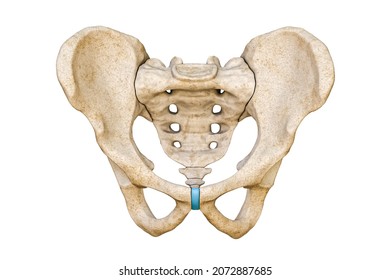
Synovial fluid
lubricate, nourish chondrocytes, shock absorber
Uniaxial
bone moves 1 plane or axis
Biaxial
bone moves 2 planes or axis
Multiaxial
bone moves in multiple planes or axes
Plane Joints
(planar/gliding joints)
flattened or slightly curved faces slide across 1 another
least mobile joint
uniaxial
Ex: intertarsal joint (ankle), intercarpal joint (wrist)

Hinge Joints
convex surface of 1 bone articulates with concave surface of another
uniaxial
Ex: elbow and knee joints

Pivot Joints
formed by articulating bone and rounded surface (ring) - most commonly by a ligament and another bone
uniaxial
Ex: annular ligament permitting a pivot action of radial head

Condylar Joints
not hinge
biaxial
Ex: metacarpophalangeal joints in digitals 2-5 (knuckles)

Saddle Joint
appears like a saddle
greater range of motion compared to condylar
biaxial
Ex: carpometacarpal joint

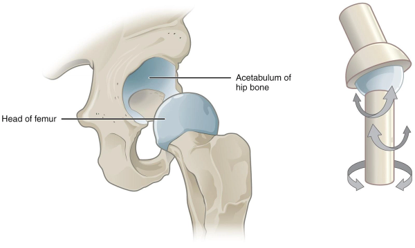
Ball and Socket Joints
Spherical articulating head of 1 bone fits into rounded “cup-like” structure of another bone
multi-axial
Ex: hip joints, shoulder joints
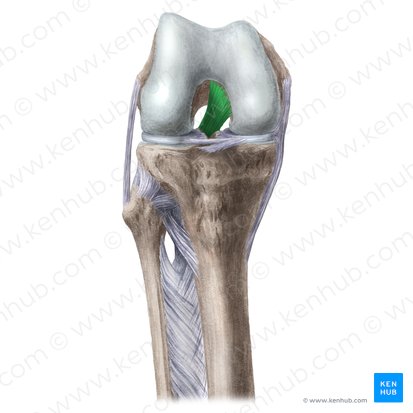
Anterior cruciate ligament
prevent hyperextension of the leg

Posterior cruciate ligament
prevents hyperflexion of the leg
Menisci
fibrocartilage pads on the condyles of the tibia to partially stabilize the joint and act to cushion the articular surfaces of femur and tibia

Flexion
decreases the angle btwn 2 bones

Extension
increases angle btwn 2 bones

Hyperextension
extension past the anatomical postition
Abduction
occur in frontal plane
moves limb away from median plane
Adduction
moves limb toward the median plane
Circumduction
moves distal end of a limb in a a circle
Pronation
rotates radius over ulna

Supination
rotates radius and ulna parallel

Opposition
brings tip of thumb in contact with any finger on the same hand
Dorsiflexion
moves superior surface of foot toward the shin (standing on heel)
Planter Flexion
moves superior surface of foot farther away from shin (pointing your toes)
Inversion
turns sole of foot medially
Eversion
turns sole of foot laterally
Elevation
moves mandible up (closing the mouth)
Depression
moves mandible down (opening the mouth)
Protraction
moves mandible forward
Retraction
moves mandible back