micro written exam 2
 Call Kai
 Learn
 Practice Test
 Spaced Repetition
 Match
 Flashcards
 Knowt Play
1/132

There's no tags or description
Looks like no tags are added yet.
133 Terms
Kirby–Bauer disk diffusion test
A test in which antibiotic disks are placed on a bacterial lawn to see how much they inhibit growth.
Bacterial lawn
An even layer of growth across the agar that makes clear zones easy to see.
Antibiotic
An antimicrobial substance originally produced by a microorganism. ex β-lactams, aminoglycosides, macrolides, and quinolones.
Susceptibility cutoff
The zone-size threshold used to interpret a result for a particular organism and antibiotic. Different drugs diffuse differently, so compare each zone with its own cutoff.
Bactericidal
Kills bacteria, causing viable counts to fall. Cells can remain intact, so turbidity may stay the same.
Bacteriolytic
Kills bacteria by breaking cells open. Both viable counts and turbidity can decrease.
Membrane filtration
Passing a known volume of water through a filter that traps bacteria, then placing the filter on agar and counting the colonies that grow.
R2A agar
medium is not selective but allows the development of slow-growing heterotrophic water bacteria.
gives you an idea of the bacterial content of “clean water"
EMB agar
The selective and differential medium used to look for coliforms.
selective for gram negative species and diagnostic for most coliforms.
The dyes inhibit many Gram-positive bacteria, and lactose fermentation helps distinguish colonies.
CFU per 100 mL
For undiluted water, divide colony count by the volume filtered in mL and multiply by 100. Three colonies from 5 mL = 60 CFU/100 mL.
Detection limit
The concentration that would produce one detectable colony in the volume tested. If you filtered 5 mL and saw no colonies, report <20 CFU/100 mL.
Resident bacteria
Bacteria that colonize the skin and are regularly recovered from it.
Transient species
contact with contaminating objects
found intermittently
can’t colonize skin
resident species fight transient species
Forearm versus forehead
The forearm represents a dry skin site. The forehead represents an oily skin site.
Sebum
An oily skin secretion that supplies lipids and other compounds that some skin microbes can use.
HBA (heated blood agar)
non-selective heated blood agar
incubated aerobically and allows for growth of species of micrococcacae
GE12:RCMF
incubated anaerobically
selects for species of propionibacterium/cutibacterium
Culturable population
The organisms that grew on the medium under the incubation conditions used. Changing the medium or oxygen level can change which organisms you recover from the same skin site.
Coliform
a Gram-negative, non-spore-forming, facultatively anaerobic rod that ferments lactose and produces gas.
many reside in the mammalian digestive tract and are indicative of fecal or sewage contamination in our waters.
Most probable number (MPN)
A statistical estimate of viable organisms based on the pattern of positive broth tubes across sample volumes or dilutions.
Durham tube
The small inverted tube that traps gas produced during fermentation. In these tests, check for gas when scoring a tube.
LTB
The presumptive coliform broth.
includes lactose
not selective
BGLB
The broth used to confirm total coliforms.
includes lactose
It inhibits many non-coliform bacteria.
Gas and turbidity is the key positive readout.
EC broth
Used to test for fecal coliforms.
includes lactose
incubate at elevated temperature 45 degrees celcisus
MPN index
The pattern of positives used to look up the estimate. 5–3–1 means five positives at the first selected dilution, three at the next, and one at the third. Keep the pattern in order for the lookup.
Gram stain
The first test in the P1 manual. Gram-positive cells appear purple, and Gram-negative cells appear pink. Use the result to narrow your unknown.
Catalase test
aerobic and aerotolerant bacteria can produce toxic oxygen by products (hydrogen peroxide)
to detoxify these compounds, cells produce the enzyme catalase
it decomposes hydrogen peroxide to water and oxygen
cytochrome oxidase
bacteria that perform aerobic respiration may possess the electron carrier molecule cytochrome oxidase, which catalyzes the terminal reduction of O2 to H2O. many species have an alternative enzyme in the electron transport chain meaning they are cytochrome negative.
Indole test
bacteria that possess the enzyme trytophanase hydrolyze tryptophan to pyruvic acid, ammonia and indole.
indole production in a growth medium can be detected by the addition of dimethylamino-benzaldehyde, which reactions wit indole to produce a red-colored compound
detects for indole
postive = red-colored compound
H₂S
Gelatin hydrolysis
gelatin is a protein that can be hydrolyzed to amino acids by organisms that possess the extracellular enzyme gelatinase
a gelatin-solidified medium is liquefied by such bacteria
Voges–Proskauer (VP) test
a number of acidic products are generated from the fermentation of carbohydrates by various bacteria. some convert their acids to the dialchol 2,3-butanediol. an intermediate in butanediol production is the compound acetone, which can be detected with a simple test from a broth in which cells ferment glucose
Ornithine decarboxylase test
this procedure test for the presence of one of the decarboxylase enzymes that is required for the breakdown of amino acids to amines and carbon dioxide. alkaline byproducts are detectable in a growth medium that contained a colored PH indicator
Acid from lactose or mannitol
the ability of bacteria to ferment different carbohydrates
fermentation products or intermediates include gases/acids
detection of lower pH
Nitrification
The aerobic oxidation of ammonia/ammonium → nitrite → nitrate.
Nitrosifier
An ammonia oxidizer such as Nitrosomonas. It produces nitrite.
Nitrite oxidizer
An organism such as Nitrobacter. It consumes nitrite and produces nitrate.
Denitrification
Reduction of nitrate → nitrite → NO → N₂O → N₂, typically under low-oxygen conditions. In EN1, look for gas and growth in the nitrate enrichment.
Prodigiosin
The red pigment made by wild-type Serratia marcescens. Visible red pigment is the readout for rescue
Cross-feeding
One mutant releases a chemical intermediate that diffuses to a neighboring mutant and lets it finish making the pigment.
Single rescue
Only one mutant turns red. In a linear pathway, the rescued mutant is blocked upstream and the rescuer is blocked downstream.
Double rescue
Both mutants turn red near each other. In the lab model, this fits mutants blocked on different branches of a convergent pathway.
Cyanobacteria
Oxygenic photoautotrophs. They use light for energy, CO₂ for carbon, and water as an electron donor, releasing O₂.
BG-11
The liquid mineral medium used for the cyanobacterial enrichment. It supplies inorganic nutrients and contains no added organic carbon. no sugar added.
EN3: Purple non-sulfur bacteria
physiologically diverse groups of phototrophs with purple hues
purple is due to compliment of carotenoid pigments and unique to chlorophyll: bacteriochlorophyll a and b
they can grow in light:
photoautotrophs (carbon from CO2)
photoheterotrophs (carbon from organics)
they can grow in the dark
aerobically (via respiration)
anaerobically (via fermentation or anaerobic respiration)
Photoheterotroph
An organism that uses light for energy and an organic compound for carbon. The EN3 setup is the course example.
Anaerobic conditions in EN3
Suppress aerobic respiration, helping the combination of light and malate select for the desired phototrophs.
DCMU
Inhibits photosystem II. It was added to EN3 to suppress competing oxygenic phototrophs such as cyanobacteria.
what are the two purposes of light in metabolism
energy generation used primarily in CO2 fixation
reductant (electron donor) generation for CO2 fixation
what redox state is sugar compared to CO2?
reduced
what is the purple due to
complement of carotenoid pigments and unique chlorophyll: bacteriochlorophyll A and B
g-9 is a carotenoid-less mutant. what color is bacteriochlorophyll a?
blue
bacteriochlorophyll has a ___ redox potential than O2/H2O couple
LOWER. that means h2o cannot serve as the electron donor
for autotrophic growth
an electron source with a lower potential than bacteriochlorophyll is needed. H2 and low levels of H2S can serve as a electron source
photoautotrophy for purple nonsulfer bacteria
photoautotrophy is costly for PNS: chemical, not light, energy used to make reductant
it may provide an ecological advantage
use variety of organics for carbon (alcohol, fatty acids, sugars, aromatics)
light for energy
enrichment conditions for purple non-sulfur bacteria
light
anaerobic (prevents respiration)
non-fermentable organic carbon
citrate (some can use)
malate (most can use — we add this)
benzoate or cinnamate (only rhodopseudomonas palutris)
DCMU (inhibits PSII [p680 Center] of cyanobacteria)
an additive that will inhibit photosynthesis II and in particular that p600 reaction center signing cyanbacteria by preventing photosynthesis from working (kill all cyanobacteria)
what proportion of the cells in and on your body are prokaryotic?
90%
what layers of the skin are actually consider the skin layer
dermis and epidermis
where do bacteria prefer to live in regard to locations of the skin?
just under the epidermis near the ducts or glands
want oxygen
love bacteria - sebum, sweat
more protected than top layer
where do your resident species come from?
from mom at birth — bacteria stays with you for life from your mother from birth
what can you resident microflora be affected by?
weather
what can your transient species be affected by
personal hygiene
bacteria love: sweat
urea
amino acids
lactic acids
salts
bacteria love: sebum
lipids, fatty acids, wax glycerol, hydrocarbons
bacteria density on dry skin
10² cells/cm
bacteria density on oily skin
10^6 cells/cm
micrococcaceae
gram-positive, aerobic micrococcus, staphylococcus
propionibacterium (known as cutibacterium)
gram-positive, anaerobic, make propionic acid
why you form zits
what method for enumerating will you not use if an organism don’t grow well (or at all) on solid media?
plate counts and membrane filtration
what method do you not use if the sample is too diluted
plate count and microscopic count
A small plated sample may contain too few cells to give a reliable count. Under the microscope, you may see too few cells per field.
what method do you not use if the sample is too concentrated
membrane filtration
A concentrated sample can clog the membrane or leave so many colonies that they merge. For plate counts and direct microscopic counts, you can dilute the sample before counting
what method are you not able to distinguish from the dead and alive cells
direct microscope counts
In a standard direct count, an intact dead cell can look like a living one. Plate counts and membrane filtration measure colony-forming units (CFUs), so the cells must be able to grow under the test conditions.
most probable number (MPN) methods
liquid-based protocol for live cells
can be adapted to enumerate wide range of species
can work with concentrated or dilute samples
sensitivity/recovery better for low population densities
steps for MPN
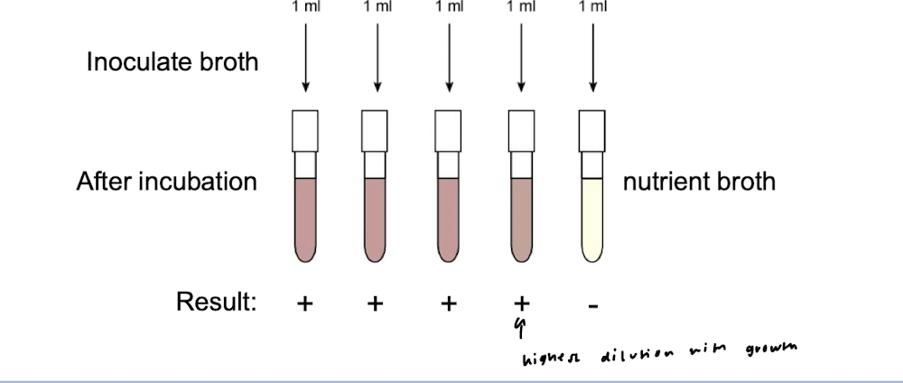
perform a volume series (membrane filtration or serial dilution)
“dilution to extinction” the original sample i.e dilute to point at which there will be no detectable organisms
test each dilution for the presence of targeted organisms by inoculating liquid medium with an aliquot from each dilution
compare the results to a probability distribution, which yields a “most probable number”
what concentration do you report?
< 1 × 10^4 cells/ml
“1” really means 0.1-5.6 if we are to be 95% confident

what is the combinations of positives
5-3-1

what is the dilution between MPN index volumes and the volumes in our sample?
1000x
rule 1 for MPN
choose the highest dilutions in which all 5 replicates are positive

5-3-1

3-1-0
rule 2 for MPN
choose the dilution with the highest number of positives and the next two higher dilutions
rule 3 for MPN
after choosing the three highest dilutions, all remaining positives in higher dilutions are counted and added to the third number of positives

5-4-2
steps for acids for lactose or mannitol
heavily inoculate a tube of BP (bromocresol purple) plus carbohydrate broth (either lactose or mannitol with your culture
incubate for 37 degrees Celsius without shaking
check for acid productions - change from purple to yellow is positive
check up to 6 days
catalase steps
need fresh (~24 hr) culture for accurate test results
obtain a bottle of 3% hydrogen peroxide from refrigerator
place a drop on a glass slide
emulsify a loop full of cells in the drop
production of bubbles=positive, no bubbles = negative
gelatin hydrolazye steps
1) using a microbial needle, stab a nutrient gelatin tube once with heavy incoculum of your organism (do not touch side of the tube)
2) incubate at room temp
3) exam daily for liquefaction of the gelatin
4) for weak hydrolyzes, liquefaction apparent only at surface of medium
Hydrogen Sulfide production
reduction from sulfur to hydrogen sulfide (H2S) occurs anaerobically in two different ways, depending upon the metabolism of the organism.
enzyme cysteinę desulfurase catalyzes the hydrolysis of cystenin to pyretic acid with the production of H2S and Ammonia.
the enzyme thiosulfate reductase catalyzes the reduction of sulfur as the terminal step in anaerobic respiration in the group of bacteria known as sulfidogens,
when H2S combines with ferrous ions, a black precipitate of ferric sulfide forms
steps of H2S test
using a microbiological needle stage a tube of SIM agar
insert the needle 2/3 of the depth of the agar
incubate at 37 degrees celsius without shaking for 2 days
blackening of the medium indicates H2S production
indole test
bacteria that posses the enzyme tryptophanase hydrolyze tryptophan to pyruvic acid, ammonia and indole. indole production in a growth medium can be detected by the add-on of dimethylamino-benzaldehyde which reacts with indole to produce a red-colored compount
steps for indole
1) use same medium from H2S test
2)add a few drops of kovacs reagent using a p1000 micropippette
3) the appearance of pink or red color in the reagent layer within several seconds indicates the presence of indole
4)when finished close the kovacs reagent tube and dispose of in the designated beaker in the hood
Ornithine decarboxylase steps
1) lightly inoculate a tube of liquid orthithine decarboxylase medium
2) using a p1000 pipette, overlay the medium with 0.5 ml of sterile mineral oil
3)incubate at 37 celcius and observe periodically for up to 3 days
4)for this test, the solution starts as a bronze purplish color
turns a strong purple is positive
turns yellow is negative
oxidase test
do this test on the bench top (no metal!)
1) obtain a tube of the powdered reagent from the refrigerator and suspend the reagent in a 5ml of water (dH2O).
2) smear a visible paste of cells from a fresh (<48hr) culture onto a piece of bibulous paper with a toothpick (NOT A LOOP)
3)place one drop of oxidase reagent on the smear using the p1000 and observe color change
4)dark blue within 10 seconds means positive, don’t wait for it to turn blue afterwards
steps for Voges-proskauer reaction
1) inoculate a tube of VP broth and incubate a 37 degrees celcius
2) after 6-7days of incubation transfer a 2ml aliquot to an empty non-sterile, spec tube, taking care not to disrupt any pallet of cells.
3) reagent A and B come in a closed glass vial encased by a plastic dropper tube
Access the liquid by breaking/cracking the vial as you would a glow stick
4) add 18 drop (0.6ml) reagent A and agitate the tube for one minute to oxygenate the medium
5) to the same tube add 6 drops (0.2ml) of reagent B and mix briefly
6)place the tube in a rack and do not move for at least 20 minutes
positive result will turn pink or red
if no red is detected let sit for an hour to see if its negative (yellow or copper color)
dispose of reagent an and b tubes in the designated beaker in the hood
do not bleach the reaction you prepared
hemolysis test
some pathogens that invade tissue produce the protein toxin hemolysin to lyse red blood cells to either release nutrients for their own growth or combat the immune system. bacteria that secrete hemolysis are defined by the amount of hemolysis they produce.
steps for hemolysis
1)to view plates for hemolysis, hold the plate up against your shelf light
complete lysis = clear halo = beta hemolysis
incomplete hemolysis green-ish halo is alpha hemolysis
no lysis (no halo) is gamma hemolysis
coagulase test
some pathogens produce the enzyme coagulase as a defensive means to ward off the host’s immune system. the enzyme works in conjunction with components of the host’s serum to produce fibrin barriers around bacterial cells this protecting them from immune attack.
steps for coagulase test
1) obtain a tube of coagulase plasma from the freezer and thaw it in your hands
2)place a loophole of cells in the plasma and gently mix
3) incubate at 37 celsius no shaking for up to 24 hrs.
4) check for coagulation by 24 hrs (increase viscosity or lumpiness)
do not check after 24 hrs
clumps of bacteria at the bottom of tube is not a positive result
L-pyrrolidonyl arylamidase (PYR) test
Group A streptococci, enterococci and some micrococcaceae possess this enzyme, which can be detected with a simple disk test.
steps for L-pyrrolidonyl arylamidase (PYR) test
PYR reagent are in a small box in the refrigerator. You need a fresh 24hr culture to get an accurate PYR test result.
place PYR disk on a class slide or on top of a petridish
add a sterile loopful of distilled water to the disk
heavily inoculate the disk with a paste from culture
wait 2 minutes and then add a drop of a p-dimethylaminocinnamaldehyde solution (color developer) to the disk
pink to red within 1 minute is a positive result
orange or no color is negative
antibiotic sensitivity test
sensitivity or resistance to a variety of antibiotics can sometimes be diagnostic of certain bacterial groups; these test are particular useful in the gram positive cocci. a paper disk impregnated with the antibiotic placed on a solid medium on which a heavy inoculum of test bacteria has been spread. the bacteria will grow to form a lan on the medium, but if the culture is sensitive to the antibiotic, a zone of clearing will form around the disk where the anitbiotic has diffused into the medium