Exercise Physiology Exam 2: Chapter 11
 Call Kai
 Learn
Learn Practice Test
 Spaced Repetition
 Match
Match Flashcards
 Knowt Play
1/57

There's no tags or description
Looks like no tags are added yet.
Name | Mastery | Learn | Test | Matching | Spaced | Call with Kai | Chat |
|---|
No analytics yet
Send a link to your students to track their progress
58 Terms
What does Q represent, and what is it equal to?
Q = cardiac output = HR x SV
how much blood moves
What is CaO2?
arterial oxygen content, the total amount of O2 carried in a given volume of arterial blood (bound to hemoglobin and dissolved in plasma)
what is a-VO2 diff?
arterial venous oxygen difference
the difference in the amount of blood between arterial blood and venous blood
how much oxygen your working muscles and tissues extract from the bloodstream
over a minute, which ventricle pumps more blood?
they pump the same amount
the left ventricle is larger because it pumps blood further but its still the same volume of blood (left = systemic)
arteries vs. veins function
arteries carry blood away from the heart, veins return it
components of the pulmonary circuit
right ventricle, lungs, left atrium
components of the systemic circuit
left ventricle, body, right atrium
Path of blood
enters through the superior or inferior vena cava
into the right atrium- through the tricuspid valve
right ventricle- pulmonary semilunar valve - pulmonary arteries- lungs (pulmonary capillaries) -pulmonary veins (right and left)
left atrium - bicuspid valve
left ventricle - aortic semilunar valve - aorta -arteries -systemic capillaries - veins
superior/inferior vena cava
What is VO2 max equal to?
VO2max = cardiac output x stroke volume
How does the heart contract as one unit?
Excitation passes cell to cell through gap junctions, so the atria contract as one unit, and the ventricles as another: two syncytia create a functional syncytium
cardiac and skeletal muscles are both striated (use actin and myosin), adjacent cells are joined end to end by intercalated discs. Discs carry gap junctions, which pass electrical activity cell to cell
A donor heart with every nerve cut in a supportive environment. What does it do?
It beats faster than resting HR
because resting HR sits below the SA node’s intrinsic rate because vagal tone predominates at rest, if you remove those nerves, the heart speeds up
resting HR of 75 is a break, not an engine speed
pacemaker cells depolarize spontaneously and the fastest sets the rate
SA node is the fastest in the right atrium, AV node is about 40 to 60 per minute, and takes over if the SA node fails
vagal tone holds resting rate at about 75, below the intrinsic rate
a resting rate of 40 does not itself tell you an athelete has a failing SA node
What does the P wave represent?
depolarization and contraction of atria
What does the QRS complex represent?
the ventricles depolarize, and then contract
What does the T wave reprsent
the ventricles repolarize and then relax
atrial repolarization is buried under the QRS and can surface in exercise
The whole conduction system has fired before the QRS appears
the U wave is inconsistent, and commoner in slower cycles
from rest to hard exercise: the beat goes from 0.8s (75bpm) to 0.33s (180bpm). What happens to filling time?
filling time falls much further to about a quarter of what it once was.
What is the first thing to be shortened when you speed up the heart?
filling time is shortened because diastole is shortened more than systole is
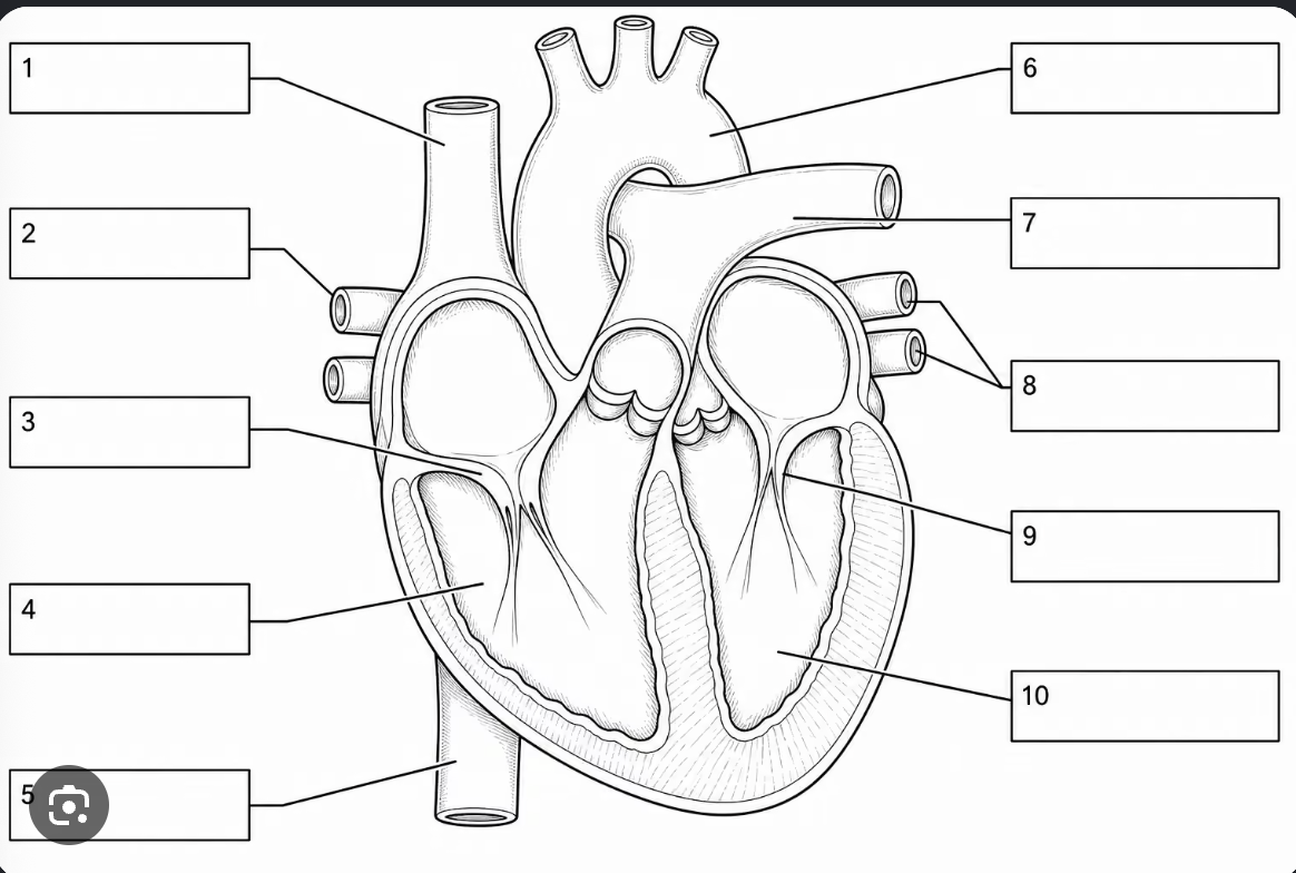
Label the parts of the heart
superior vena cava
right pulmonary veins
tricuspid valve
right ventricle
inferior vena cava
aorta
left pulmonary artery
left pulmonary veins
bicuspid valve
left ventricle
How to read an ECG strip
determine HR
determine whether the rhythm is regular
identify P wave, QRS complex, and T wave
regular rhythm: HR = 60/( R -R interval in seconds)
The cardiac cycle
ventricular filling and ejection
diastole: ventricles relax and fill
systole: the ventricles contract and eject blood
pressure opens valves and valves set volumes
What is the effect of speeding up HR on cardiac cycle?
cuts filling time first because it shortens diastole more than systole
for EDV to hold up, filling rate must rise to compensate
End diastolic volume
the volume in the ventricle at the END of diastole, about 130 mL
End systolic volume
the volume left at the end of systole, about 60mL
Stroke volume
EDV - ESV about 70mL
ejection fraction
SV/EDV, the fraction of what was there that left
Cardiac Output
Q = HR x SV about 5L/min at rest, near the whole blood volume every minute
What three things set SV
Preload, contractility, afterload
only preload and contractility explain stroke volume differences between people
What is preload?
volume of blood returned to the heart (represented by EDV)
stretch is the mechanism, not the definition - increases stroke volume
raised by venous return
what is Contractility?
the force of contraction at a given fill
set by sympathetic nerves and catecholamines
acts independently of filling volume
what is afterload?
the resistance the ventricle ejects against
set mainly by aortic pressure
does not explain differences in SV
what is the Frank Starling mechanism
greater ventricular filling increases cardiac muscle stretch, which increases contraction force and stroke volume
only increases up to a point
How do your leg muscles act as a pump?
contraction of leg muscles compresses veins, and empties them toward the herat because they have one way valves
becomes important when standing because gravity promotes blood pooling in the legs
if you reduce a vessels radius to 1/2, what happens to flow through it?
it falls to 1/16
Poiseuille’s law
R = 1/r^4
Resistance is equal to 1/radius^4
Lining of blood vessels
3 layers- adventitia, tunica media, tunica intima
media- smooth muscle, sets diameter
intima- once cell layer - endothelium
capillaries are endothelium only
endothelium moves materials out and prevents clots, releases factors that dilate and constrict the media
Where is blood velocity the slowest?
in the capillaries
as total cross-sectional area increases, average blood velocity decreases
slow capillary flow facilitates gas and nutrient exchange
What is hematocrit
the fraction of blood volume occupied by red blood cells
men: 43-48%
women: 38-45%
red blood cells carry oxygen through Hb
Plasma
55% of blood, and 90% of it is water
Why does blood volume matter?
it influences venous return, ventricular filling, and stroke volume
How do you calculate pulse pressure
SBP - DBP
represents the force you heart generates with each contraction
at rest DBP is one third of pulse pressure
Mean arterial pressure
MAP = DBP + 1/3 (Pulse pressure)
cardiac output equation in terms of Resistance to flow
Q = MAP / TPR so MAP = Q x TPR
flow = pressure/resistance
total peripheral resistance
opposes flow, mostly friction on vessel walls
Three cardio centers in the medulla
Cardioaccelerator: sympathetic nerves, raises rate and force
cardioinhibitor, the vagal nucleus: vagus nerve (PNS), lowers HR and force
vasomotor: arterial smooth muscle, SYMPATHETIC only, vasodilates or constricts
parasympathetic has little direct control over systemic arterioles
Sympathetic nervous system on cardiovascular system
heart: raises rate and force
vessels: constricts most arterioles
skin is the one exception
also drives the adrenal medulla
Parasympathetic nervous system on cardiovascular system
Heart: lowers rate and force via vagus nerve
vessels: essentially none
opposite of sympathetic only for the heart
dilation comes from sympathetic withdrawl
baroreflex
detect arterial stretch in the carotid sinus and aortic arch
when blood pressure increases:
baroreceptor firing increases, medulla recieves the signal, PNS activity increases, SNS decreases, HR and vascular resistance decrease
during exercise, the baroreflex resets to regulate pressure around a higher operating point
How does sympathetic drive raise SV without more filling?
Frank-Starling: more EDV, more SV
contractility: more force at the SAME end-diastolic volume
sympathetic nerves and circulating catecholamines raise contractility
in exercise both levers pull at once
Neural vs. Hormonal control
Neural control acts in seconds
hormonal control operates over longer periods of times
epi and norepi do what nerves do but slower
aldosterone: retain sodium, and water follows
ADH: retain water, and constrict vessels
more volume = more venous return, so more stroke volume
Heart rate variability
varies from person to person
variation in the time intervals between normal heartbeats - never like a perfect metronome —> index of autonomic balance
low variability predicts cardiovascular events and death in the general population- means its too much like metronome and not adapting to changing stimulus
Why is cardiac output almost never measured directly
It would require an arterial and mixed venous blood sample
Q = VO2/a-vO2diff
Doppler echoL SV from aortic area x velocity - doesn’t require steady state, so it works at maximal exercise but very expensive and hard to do during exercise
Everyday: measure HR and SV then Q = HR x SV
Why is VO2 max a cardiovascular measurment?
O2 delivery is limited by the cardiovascular system, so the highest O2 uptake a person can reach is a measure of cardiovascular capacity
What field tests estimate VO2max
one mile run or walk
20 meter shuttle run PACER
rockport walking test
walking tests suit older, heavier, or sedentary individuals
even an estimate improves health-risk classification
blood pressure at rest vs exercise
at rest: DBP2 (fifth Korotkoff sound) - sound disappears through stethescope
exercise: DBP1 (fourth korotkoff sound) - sound is muffled - recommended for children, adults exercising and if DBP2 is lower than 40
Why does the heart recieve most of its blood during diastole?
the myocardium contract forcefully during systole, so coronary blood vessels are compressed- especially those supplying the LV
Why can’t the myoxardium increase its O2 extraction during exercise?
the heart already extracts 60-70% of delivered O2 at rest, so there is little room for increase a-vO2 lever is unavailable
meet greater O2 demand by increasing coronary blood flow - the lever
harder contraction forces more in
Rate pressure product
Rate pressure product (RPP) estimates myocardial O2 use - dynamic and static
RPP = HR x SBP
ST depression
occurs when myocardial O2 demand exceeds supply
not enough O2 causes ischemia, which changes ventricular repolarization - downward displacement of ST on ECG
demand rises with HR and SBP so with RPP