Exam Three Parasitology
 Call Kai
 Learn
 Practice Test
 Spaced Repetition
 Match
 Flashcards
 Knowt Play
1/91

There's no tags or description
Looks like no tags are added yet.
92 Terms
Protozoa
single celled eukaryotic organisms that are not plants, animals, or fungi
Protists (formerly a kingdom)
does not form a monophyletic groups with protozoa
SAR
Plasmodium, Toxoplasma, Cryptosporidium, Blastocystis, Balantidium, Babesia
Discobids
Trypanosoma, Leishmania
Metamonds
Giardia, Trichomonas, Dientamoeba
Amoebozoa
Entamoeba, Acanthamoeba
Protozoa Classification
The protozoa that are infectious to humans can be classified into four groups based on their mode of movement.
sarcodina
amoeba- e.g. Entamoeba
Mastigophora
Flagellates e.g. Giardia, Leishmania
Ciliophora
Ciliates e.g. Balantidium
SPorozoa
Organisms whose adult stage is not motile e.g. Plasmodium, Cryptosporidium
Alveolata
Most have the presence of sacs of fluid under the cell membrane
Apicomplexa- have apical complex
Ciliates- have cilia
Excavata
most have flagella and excavated feeding structure on one side
Amoebozoa
most have amoeboid body structure
Trophozoite
feeding and reproductive stage in the life cycle of certain protozoa
Cyst
Resting or dormant stage of a microorganism
May have protective element if found in the environment
cysts can also form in tissues
Apicoplast
plastid (like chloroplasts and mitochondria)
Apical complex
Rhoptries and micronemes- secretory organelles
Apical polar ring- microtubule organization site
Conoid- filament structure.
Morphology- Apicomplexans
facilitates entry into host cells
Gliding motility site
New daughter cell formation
feeding site in micropredator myzozoa
Medically important apicomplexans include
Toxoplasma gondii
Plasmodium spp.
Cryptosporidium spp.
Cyclospora
Babesia
Eimeria
Sporozoites
motile infective form
merozoites
lives within infected host cells
gametocytes
sex cells
Toxoplasma Tachyzoites
extracellular parasite, rapidly dividing`
Toxoplasma Bradyzoite
Dormant encysted stage
Kinetoplastids
a network of circular DNA (called kDNA) inside a large mitochondria that contains many copies of the mitochondrial genome
Kinetoplastid parasites
Trypanosoma
Leishmania
Trypomastigote
main infective form, found in both mammal and insect host
Epimastigote
replicating form in insect host
Promastigote
motile, elongated, extracellular form in the life cycle of some kinetoplastids
Amastigote
Intracellular form
Prototheca spp.
Parasitic algae related to green algae but lack chloroplasts, opportunistic pathogen.
Hemiparasites
extracts water and minerals from the host but performs photosynthesis
Example- mistletoe
Holoparasites
Completely dependent on the host for all water, nutrients, and organic carbon (no photosynthesis).
Indirect parasites
Mycoheterotrophic, steals nutrients from tree/rhizome relationship or from parasitizing fungi.

Entamoeba histolytica
Geographic distribution: mostly in west coast africa like nigeria and ghana.
Life cycles: Mature cyst is ingested, excystation in the small intestine, trophozoites go to the large intestine.
Vector: fecal oral route
Symptoms: Stomach pain, nausea/vomiting, diarrhea, bloody stool, fever, liver abscesses.

Naegleria fowleri
Life stages: cyst form, trophozoite form, flagellated form.
Geographic distribution: Rare but does occur in some countries usually <10 cases
Hosts: Humans, cows and Tapirs
Life cycle: Starts as a cyst → trophozoite → Flagellated form → host nose into brain
Symptoms: fever, head pain, nausea, vomiting, eating aversion, convulsions and body shaking, neck stiffness, light sensitivity, mental disorientation, loss of consciousness.
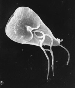
Giardia lamblia
Life stages: cyst (infective stage) and trophozoite ( growth and reproductive stage)
Geographic distribution: is spread worldwide but occurs mostly in warm climates in impoverished areas with poor sanitation and untreated water sources.
Hosts: humans, beavers, cows, cats, dogs and sheep
Life cycle: Ingestion of cysts via fecal oral route, excystation releasing two trophozoites, trophozoite stage, encystation moving to colon and turn into cysts, cysts are passed in the feces.
Symptoms: diarrhea, stomach pain, dehydration, nausea, gas, fever, reactive arthritis, IBS, recurring diarrhea, stunting.
Prevalence: 1.1 million in the USA, 5.2 cases per 100,000
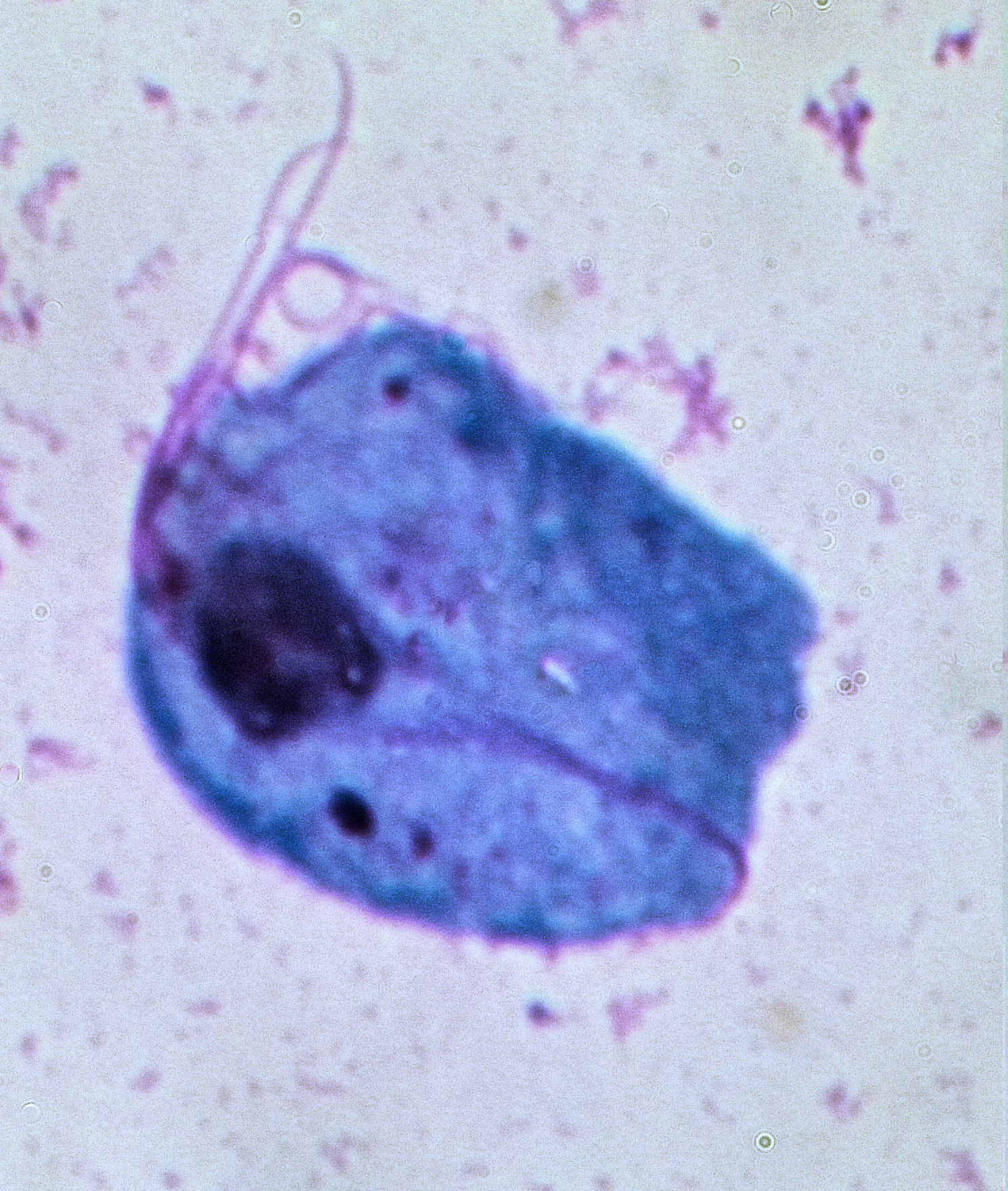
Trichomonas vaginalis
Life stages: exists as a trophozoite only. Oval and pear shaped with four flagella in front, fifth runs along the undulating membrane.
Geographic distribution: it is found worldwide with highest incidences in east and southern africa, as well as oceania and latin america.
Hosts: Humans with no vector and no other hosts.
Life cycle: none just resides in the vagina
Site of infection and symptoms: Vagina or urethra and prostate, in women vaginal discharge, itching, redness and soreness, development of strawberry cervix, usually no symptoms; urethral discharge or pain when urinating. 70% of people have no symptoms.
Cases: 156 million new cases in 2020, about 4,133 new cases per 100,000 worldwide. ~5% of women and 0.6% of men were infected in 2016

Plasmodium falciparum
Life stages: marginal form, ring form, double dotted rings, ring form, young trophozoite, trophozoite, early schizont, schizont, mature schizont, female and male gametocyte.
Geographic distribution: Found in most coutnries south of the equator besides aussy
Vector: Anopheles mosquitos
Hosts: humans
Life cycle: complex but involves the parasite taking over and using red blood cells
Symptoms: Flu like symptoms, chills, headache, muscle aches, and tiredness, nausea, vomiting, and diarrhea, anemia, jaundice, kidney failure, seizures, mental confusion, coma, death
Statistics: most deadly form of malaria, most prevelent in africa, ~282 million malaria cases and 610,000 malaria deaths in 80 countries
Metazoans
further classified by whether they have specialized tissues
Eumetazoans
animals having the body composed of cells differentiated into tissues and organs (usually a digestive cavity lined with specialized cells)
Diploblastic
two cellular body layerst
triploblasts
three cellular bofy layers
Coelom
fluid filled cavity that contains internal organs, lined with mesoderm
Pseudocoelomate
fluid-filled body cavity separating the gut of the organism from the body wall, but it is not lined by mesoderm
acoelomate
lacks coelom
Plathyhelminthes- Acoelomates
flatworms, trematodes, and cestodes
Pseudocoelomates
phylum acanthocephala and nematoda.

Toxoplasma gondii
Life stages: Includes tachyzoite, bradyzoite, merozoite, macrogamete, microgamete, zygote, unsporulated oocyst, sporulated oocyst and sporozoite
Global distribution: Found worldwide most common in Africa, 17.5% in North America,
Vectors: does not need a biological vector, transmitted through contaminated food, water, soil and infected animal tissues.
Definitive hosts: Cats, African Lions, Jaguars.
Intermediate hosts: Humans, house mouse, domestic chicken
Life cycle: Cats eat mice or birds who have tissue cysts. Cats will then poop out fecal eggs which can be eaten by sheep or pigs and then eaten by humans, or the poop can infect food or water which is then not properly washed and eaten by humans.
Site of infection: the GI tract, Brain, Eyes, Skeletal and cardiac muscles, blood and lymph nodes.
Symptoms: Usually asymptomatic, swollen lymph nodes, fever, fatigue, sore throat, body aches, malaise, eye pain, blurred vision, eye redness
Prevelence: about 50% of the worlds population is infected with Toxoplasmosis. Regional rates range from 11% in the USA to 60-90% in central/south americas. 190,100 cases per year of congenital toxoplasmosis. more than 3,500 newborns in the US each year.

Leishmania donovani
Life stages: Promastigote and amastigote
Geographical distributions: mostly in eastern africa, the middle east and asia.
Vectors and intermediate host: sandflies and humans
Life cycle: digenetic (requires two different hosts to complete its life cycle), Anthroponotic (transmitted from person to person without a non-human obligatory host.
Symptoms: Splenomegaly, hepatomegaly, lymphadenopathy, anemia, leukopenia, weight loss, fever, Kala-Azar, Death, Post Kala-Azar Dermal Leishmaniasis.
Prevalence: ~500,000, ~50,000 deaths annually

Trypanosoma Cruzi
Life stages: Amastigote, epimastigote, Trypomastigote which form metacyclic trypomastigotes as well.
Geographic distribution: uncommon in the USA, Widespread across southern states though all the way down to Argentina
Vector: Triatoma infestans “kissing bug” → feed on blood and deposit fecal matter to infect the humans
Host: humans, armadillos, possums, racoons, woodrats, other rodents, and domestic dogs.
Life cycle: Starts by a kissing bug taking a meal, then metacyclic trypomastigotes penetrate cells at bite wound sites, amastigotes multiply by binary fission in cells, amastigotes then transform into trypomastigotes then burst out of the cell and a kissing bug takes another meal and transforms into epimastigotes in the midgut of the kissing bug and it is multiplied in the midgut then forming metacyclic trypomatigotes which then restart the cycle.
Site of infection: enters through bite wound
Symptoms: skin lesions, purplish swelling of the eyelid, Fever, headaches, enlarged lymph glands, muscle pain, Dyspnea, abdominal or chest pain. Chronic Phase: Development of cardiac disorders and around a 10 percent chance of experiencing digestive neurological, or a mixture of disorders.
Statistics: ~288,000 Latin American Adults were infected with T. cruzi in the United States. Around 57,000 adults are estimated to have Chagas-related heart disease.

Trypanosoma brucei
Life stages: 1. metacyclic trypomastigote, procyclic trypomastigote, epimastigote, metacyclic trypomastigote.
Geographic distribution: mainly in Sub-Saharan Africa, West and Central Africa have the T. b. gambiense subspecies East and Southern Africa have the T. b. rhodesiense subspecies.
Vector: Glossina spp. “Tsetse fly”
Hosts: Humans and Cows
Life cycle: Starts when Tsetse fly takes a blood meal injecting the metacyclic trypomastigote → the metacyclic trypomastigote transforms into bloodstream trypomastigotes which are carried to other sites → Trypomastigotes multiply by binary fission in various body fluids. → Circulating trypomastigotes in blood during acute phase usually undetectable in latent phase → Tsetse fly will take another blood meal → bloodstream trypomastigotes transform into procyclic trypomastigotes in the vector midgut, and multiply by binary fission. → Procyclic trypomastigote leave the midgut and transform into epimastigotes → epimastigotes multiply in the salivary gland and transform into metacyclic trypomastigotes.
Symptoms: fever, headache, muscle and joint pain, swollen lymph nodes, itching.
Late stage symptoms: sleep disturbances, confusion, behavioral changes, poor coordination.
Prevalence: 471 cases reported in 2025, 97.5% decrease from 1999 to 2025, Incidence: new reported cases each year are now <1000 due to better screening, treatment, and tsetse fly control.

Cyclospora cayetanensis
Life Stages: Oocysts, sporozoites, schizonts, zygote
Geographic distribution: found worldwide, most common in tropical and subtropical regions, outbreak in USA 2026
Host: humans are the definitive host passed via fecal oral route
Life Cycle: Sporulated oocyst sporulation in the environment→ contaminated water or food is ingested → unsporulated oocyst is passed in feces → cycle continues
Site of infection: small intestine mucosal cells
Symptoms: watery diarrhea, headache, fever, loss of appetite, bloating, and fatigue
Statistics: 20,996 confirmed USA cases, 2,218 around the world, 49 states reported cases.
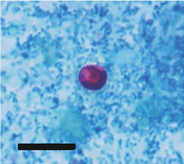
Cryptosporidium hominis
Life stages: oocysts (infectious stage secreted by infectious host in feces) → sporozoite (excystation occurs and the oocyst releases four sporozoites in host SI)→ Trophozoite (asexual active feeding form) → Meront (sexual stage, type I produces trophozoites, type II leads to new oocyst formation)
Life cycle: Thick walled oocyst exits host → contamination of water and food with oocysts → thick-walled oocyst ingested by host → cycle continues.
Geographic distribution: relatively common worldwide, however more prevalent in areas with poor WASH.
Vector and host- anthroponotic parasite.
Symptoms: Diarrhea, abdominal pain, fatigue, nausea, vomiting, fever.

Fasciola hepatica
Life Stages: Egg, miracidium, cercaria, metacercariae, adult worm
Geographic Distribution: high altitude and temperate regions, found in continents, mostly where sheeps and cattles are raised.
Intermediate hosts- snails
Definitive hosts: sheep, cattle, humans and goats
Life cycle: embryonated eggs are passed in feces → eggs become embryonated in water → miracidia hatch from eggs, seek out snails → in snails sporocysts to rediae to cercariae → free swimming cercariae encyst on aquatic vegetation → metacercariae on vegetation ingested by definitive host → immature flukes excyst in duodenum, penetrate intestinal wall and migrate through liver parenchyma to biliary ducts → adult flukes in hepatic biliary ducts
Symptoms: fever nasuea, hepatomegaly, abdominal pain, anemia, eosinophilia, rash, can be asymptomatic, cholelithiasis, obstructive jaundice, pancreatitis
Prevalence: occurs in all continents, 2.4 million affected worldwide, 200,000 illnesses and 7,000 deaths annually
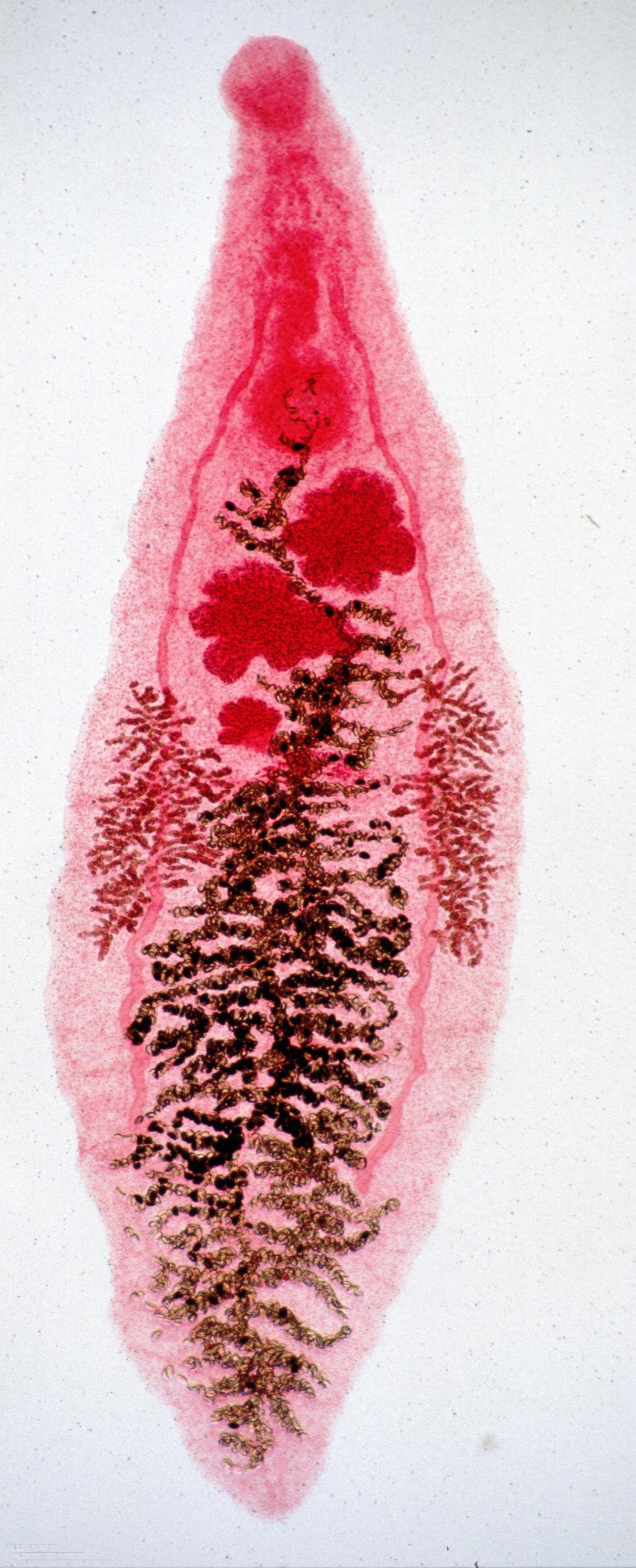
Dirocoelium dendriticum
Life stages: egg → miracidium → sporocyst → cercaria → metacercaria → adult
Geographic distribution: Predominantly in Europe, Asia, and North Africa, uncommon in North America.
Intermediate hosts: Terrestrial snails, ants
Definitive host: ruminants
Accidental host: humans
Life cycle: ruminants release embryonated eggs in feces → snails consume the eggs ( Miracidia → sporocysts → cercariae → slime ball) → ants consume slime ball (cercariae → metacercariae) → Ruminants consume ants (metacercariae → adults)
Symptoms: Snails: tissue loss in infected regions
Ants: altered behavior, tetany of mandibular muscles
Ruminants: hepatic fibrosis, biliary dysfunction, inflammation
Humans: abdominal comfort, weight loss, chronic diarrhea
Incidence and prevalence: Not good statistics on this.
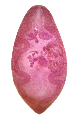
Paragonimus westermani
Life stages: Egg → miracidium → sporocyst/rediae → cercaria → metacercaria → flukes
Geographic distribution: in Eastern Asia and pacific islands of asia, some parts of the philippines and india.
Intermediate host: freshwater snails and freshwater crustaceans
Definitive host: humans.
Life cycle: embryonated egg in source of freshwater → the fertilized egg develops an embryo → miracidia hatch from the egg and penetrate a snail → Miracidia asexually reproduce going through three developmental stages within the snail → cercariae leave the snail now in their free swimming form invase a crustacean where they encyst into metacercariae → the metacercariae remain encysted until raw crustacean is consumed by host → metacercariae excyst in duodenum releasing juvenile flukes → adults lay eggs that are released into environment via coughing or swalling which are then excreted via feces.
Infection and symptoms: migration stage: diarrhea, abdominal pain, fever, cough, and some pulmonary abnormalities. Symptoms closely mimic symptoms of tuberculosis. can migrate from lungs to extrapulmonary paragonimiasis, cerebral paragonimiasis being the most serious outcome.

Schistosoma mansoni
Life stages: egg → miracidia → sporocysts → cercaria → schistosomule → adult male and female
Geographic distribution: found primarily across sub-Saharan Africa and some South American countries and the Caribbean, with sporadic reports in the Arabian Peninsula.
Definitive host: humans.
Intermediate host: freshwater snails
Reservoir host: rodents and primates
Life cycle: Eggs leave the body and hatch in water → miracidium infects a snail and two generations of sporocysts, whihc produce lots of cercariae → cercariae are released into the water and penetrate the skin of a person in the water → the schistosomula travel through the bloodstream and mature into adults in the liver → adults settle and mate, females deposit eggs in tiny veins.
Site of infection: mesenteric venules of large intestine. Eggs trapped in the liver spleen, intestine, bladder, and occasionally the CNS.
Symptoms: Often asymptomatic, itchy rash at the skin entry site, acute schistosomiasis, fever, cough, abdominal pain, diarrhea, hepatosplenomegaly, eosinophilia.
Prevalence: about 151 million people infected worldwide 84% in Africa

Clonorchis sinensis
Life stages: Eggs (release miracidia), Miracidia go through development stages, cercariae encyst to metacercariae, metacercariae excyst in duodenum and mature here
Geographic distribution: endemic in east Asia
Intermediate hosts: freshwater snails and fish
Definitive hosts: humans and other animals that eat fish
Life cycle: Eggs are discharged in stool and biliary ducts, snail ingests the eggs, eggs release mircadia, cercariae are released from the intermediate host and are ingested by fish, encystation occurs to become metacercariae, ingested by humans, cycle continues.
Symptoms: Indigestion, nausea, constipation, fever, jaundice, abdominal pain.
Statistics: prevalence in fish species: 18.36%, average detection rate in humans was 4%

Diphyllobothrium latum
Geographic distribution: North and south poles in the north and very south
intermediate and paratenic Hosts: Perch, pike, crustaceans
Definitive hosts: humans, bears, canids, felines, mustelids
Life cycle: unembryonated eggs passed in feces of definitive host, eggs embryonate in water, coracidia hatch from eggs and are ingested by the first intermediate host crusteceans, procercoid larvae develop in body cavity of crustaceans, infected crustacean ingested by second intermediate host fish, predator fish eat small fish and invades tissue, definitive host ingests plerocercoids in infected fish and cycle continues by eggs being released into water.
Site of infection: intestines, gallbladder, bile ducts
Symptoms: diarrhea, vomiting, abdominal discomfort, weight loss, vitamin B12 deficiency.
Taenia solium
Life Stages: Egg → oncosphere → cysticercus → adult → gravid adult →
Geographic distribution: worldwide mostly in impoverished areas that have a close relationship with pork
Intermediate host: pigs
Definitive host: human
Life cycle: 1. ingestion of eggs → 2. Oncosphere eggs hatch and larvae penetrate intestinal mucosa, travel through the bloodstream and settle in tissue → 3. cysticercus become cysts in muscles, brain, eye.
OR
Ingestion of cysts → scolex attachment to the intestinal wall → growth of proglottids → reproduce → transmission.
Symptoms: Taeniasis: loss of appetite, weight loss, upset stomach, abdominal pain.
Cysticercosis: lumps under skin, swelling or detachment of retina, blurry vision, difficulty balancing, hydrocephalus, seizures.

Echinococcus granulosus
Life stages: Embryonated egg → oncosphere → hydatid cyst → protoscolex → adult tapeworm
Geographic distribution: Mostly in Russia, North western asia, middle east and north eastern africa.
Intermediate host: humans and sheep
Definitive host: dogs
Life cycle: Adult worm in dog small intestine → embryonated eggs in feces → oncosphere hatches and penetrates small intestine → hydatid cyst in various organs → protoscolex from cyst → scolex attaches to intestine.
Site of infection: usually liver and lungs
Symptoms: abdominal pain, nausea, vomiting, chronic cough, chest pain, shortness of breath, loss of appetite, weight loss, weakness.
Prevalence: ~148,500 new cases and 1,364 deaths

Trichuris trichiura
Life stages: embryonated egg, embryonated egg, adult
Geographic distribution: worldwide distribution, highest prevalence in Africa, South America, East and Southeast Asia.
Life cycle: infection occurs through ingestion of embryonated eggs usually from water or soil contamination, eggs hatch in small intestine, adult whipworms live in and attach to cecum or ascending colon
Definitive host: humans
Other hosts: non-human primates
Symptoms: Rectal bleeding, abdominal pain, weight loss, rectal prolapse, mucus discharge, and diarrhea.
Estimated cases: 450 million- 1 billion

Toxocara canis
Life cycle: Egg→ embryonated egg → larvae hatch in intestine → eggs
Geographic distribution: Global infection rates
Definitive host: dogs wolves coytes foxes
Accidental hosts: small mammals, birds, live stock, and borat
Site of infection in humans: lungs heart and muscles
Symptoms in humans: fever, coughing, wheezing, abdominal pain, and enlarged liver, blurred vision, eye inflammation, possible vision loss, headaches, seizures, and neurological changes, or asymptomatic
Site of infection in dogs: small intestine
Symptoms in dogs: diarrhea, vomitting, poor apetite, and abdominal discomfort.
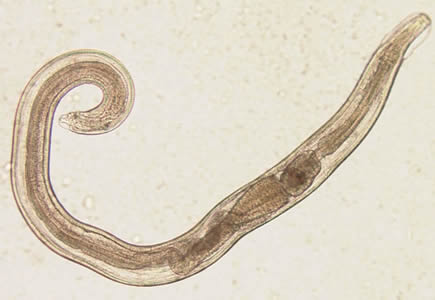
Enterobius vermicularis
Life stages: egg → larval form → adult form
Geographic distribution: occurs worldwide
Host: humans
Life cycle: eggs are ingested → larvae hatch and move to colon → adult females travel to perianal region and lay eggs —> butt is scratched→ eggs ingested.
Symptoms: asymptomatic, itching around the anus, teeth grinding, enuresis, insomnia, anorexia, irritability, abdominal pain.
Prevalence: ~12.9% prevalence rate in children.
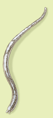
Trichinella spiralis
Life stages: egg → Larvae → encysted larvae → adult
Geographic distribution: found worldwide
Hosts: All carnivorous and omnivorous mammals and humans
Life cycle: encysted larvae is eaten by humans, the larvae is released in small intestine, adults form in small intestine, larvae deposit in mucosa and encyst in striated muscle.
Symptoms: diarrhea, abdominal pain, vomiting, swelling around eyes or face, pink eye, fever, muscle pain, splinter hemorrhages, rashes, weakness, and peripheral eosinophilia, myocarditis, pneumonitis, CNS involvement.

Necator americanus
Life stages: Egg stage → Rhabditiform larvae → Filariform Larvae → adult hookworm
Geographic distribution: Worldwide distribution.
Definitive host: humans
Life cycle: Eggs→ hatch into Rhabditiform larvae → filariform larvae → filariform larvae infect host via direct skin penetration → Adult hookworms infect intestine → cycle repeats as eggs are defecated.
Site of infection: small intestine
Symptoms: abdominal pain, nausea, anorexia, iron deficiency anemia, dermal reaction, rash.
Statistics: 406-480 million people.
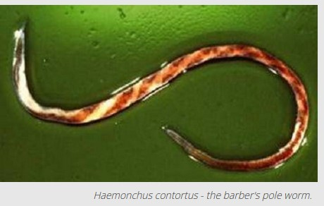
Haemonchus contortus
Geographic distribution: Africa, parts of europe and south america
Hosts: Sheep, cow, goats and humans
life cycle: eggs → larvae stage 1 → larval stage 2 & 3 → ingested laraval 3 will exsheath and become xL3 → xL3 grows into larval stage 4 → L4 will feed on the blood of its host and mature into an adult.
Site of infection: the abomasum of small ruminants
Symptoms: Anemia, edema, diarrhea, potential to cause death. CONSUMES BLOOD
Prevelence: 37% worldwide, highest rates seen in Europe, lowest in oceania.
Dirofilaria immitis
Life stages: Larvae → adult worms → microfilariae
Geographic distribution: found everywhere
Vector: mosquitos
Dead-end host: humans
Definitive host: dogs
Life cycle: Mosquito takes blood meal → into dogs or humans → adults move into subcutaneous tissue → adults produce microfilariae that typically are found in peripheral blood → mosquito takes blood meal → microfilariae penetrate malpighian tubes → L1 larvae → L3 larvae → migrate to head and mosquito proboscis
Symptoms: Coughing, low exercise tolerance, lethargy, difficulty breathing, loss of interest in food, discolored gums.

Dracunculus medinensis
Life stages: Larval stage → L2 → L3 (infective) → L4 → adult male and female.
Geographic distribution: Rural communities in low-income countries → widespread across africa, middle east and india
Definitive host: humans
Intermediate host: copepods
Vector: copepods
Life cycle: humans drink unfiltered water containing copepods → copepods die in digestive system releasing larvae → larvae penetrate the stomach and intestinal walls → larvae mature into adult worms and mate → one year later the emale causes a blister or ulcer on lower leg and emerge when the affected area touches water → the female releases larvae into the water → copepods ingest the larvae which develop into infective L3 larvae after approc two weeks and two molts → humans become infected again.
Site of infection: stomach and intestinal walls, abdominal cavity and retroperitoneal space. subcutaneous tissues.
Symptoms: painful blister or ulver develops when the adult female worm emerges through the skin. intense itching and burning pain around the affected areas. Skin redness and inflammation. Possible allergic reactions. Secondary bacterial infection. Joint inflammation, pain and arthritis.
2024: 13 human cases reported. close to eradication.

Baylisascaris procyonis
Life stages: unembryonated egg → embryonated egg → larvae
Geographic distribution: Where racoons are
Definitive hosts: racoons and dogs
Paratenic hosts: Humans, birds, mice, squirrels
Life cycle: Eggs passed in feces → emrbyonated eggs with larvae ingested → enter the definitve or paratenic host→ adults mature in small intestine in raccons and the cycle continues.
Site of infection: Racoon GI tracts, Paratenic host: eyes, CNS, and visceral organs
Symptoms: nausea, vomiting, fatigue, loss of appeite, fever,neural larva migrans, ocular larva migrans, visceral organ inflammation
Prevelence: not common in humans but common in racoons

Onchocerca volvulus
Life stages L1 → L2 → L3 → microfilariae → filarial larvae → adult worms
Geographic distribution: affects 37 million people, 99% of cases in Africa, caused 987,000 disability adjusted life years.
Vector: Female blackfly
Definitive host: Humans
Life cycle: blackfly takes blood meal → microfilariae develop maturing into L1-L3 in midgut of fly → L3 larvae migrate to blackfly mouth parts, so when the blackfly feeds the L3 larvae are transmitted to host through the wound → L3 larvae develop into adults in subcutaneous tissue → the mature adults encase into fibrous nodules called onchocercomas → through sexual reporduction in nodules, thousands of microfilariae are formed → microfilariae migrate through tissue, potnetially infecting ocular tissue, sputum, and can be found in blood and urine → another blackgly takes blood meal.
Symptoms: Over years of chronic infection, extended fibrosis and ketatosis develop with pigmentary changes and lymphatic dilation. Onchocercomata: lumps that present beneath the skin due to the formation of fibrous nodules around adult worms. the nodules are usually palpable at bony areas like the pelvic girdle, ribs, knees, tops of hands, and skull, Ocular inflammation and blindness.

Ancylostoma duodenale
Life stages: Eggs → Rhabditiform larvae → filariform larvae → adult hookworm
Geographic distribution: Favors tropical and subtropical regions between 30 degrees north and south of the equator.
Hosts: Humans
Life cycle: eggs passed down through human feces → eggs hatch into rhabditiform larvae → transforms into the filariform infective stage → filariform penetrates the skin → enters the jejunum and matures into adult worms, attaching to the intestinal walll → adults may survive for years and produce eggs that are passed down into the feces.
Site of infection: Feet and intestines
Symptoms: Skin rashes, fevers, coughing or wheezing, abdominal pain, loss of appetites, diarrhea, weight loss, iron deficiency anemia
number of cases: An estimated 72.5 million people
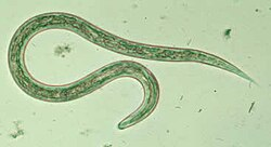
Loa loa
Life stages: Microfilaria → L1 → L2 → L3 → Adult worm
Geographic distribution: Found only in central western africa
Intermediate host and vector: Chrysops silacea and chrysops dimidata
Definitive host: Humans
Life cycle: 1. infected fly bite giving L3 → 2. L3 migrate to subcut tissue and develop into mature worm → 3. reproduction → infected human blood and taken up by fly → development in the fly → larvae become infectious.
Symptoms: asymptomatic, calabar swelling, eye worms, visible crawling under skin, itching, muscle or joint pain, fatigue, inflammation of the lungs, fluid in the lungs, scarring of heart, swelling of Lymph Nodes.
Prevalence: Affects 23.6 million people in Central and West Africa in medium and high transmission regions.
Acoelomates
includes trematodes and cestode (flukes and tapeworms)
Pseudocoelomates
nematodes.
Platyhelminthes
flatworms
flattened body
acoelomates
bilaterally symmetrical
sucker often present
many are hermaphroditic
mouth end with blind ending sack or branching tubules (except tapeworms)
trematodes
flukes
complex lifestyles with 1 or 2 intermediate hosts, usually snails
often have cercariae that can breach through the skin and most are water dependent
Cercaria
larval form of trematodes if aquatic, swimming form.
Cestodes
tapeworms
Lack mouth and gut, body surface has tegument that can absorb nutrient and release waste
Scolex: head region contains hooks and suckers that aid in attachment
Neck - forms new proglottids
usually are hermaphroditic
Proglottids contain eggs and detach from adult worm and migrate once defecated out.
Nematodes
Estimated 1 million species, 300,000 characterized, very abundant, diverse, found in many different habitats.
Parasitic nematodes include- ROundworms, whipworms, hookworms.
Pseudocoelomates
Caenorhabditis elegans non-parasitic, model organism
Male/female sexual reproduction.
Nematode life cycle
have multiple larval stages and produce a whole lot of eggs.
Amoebae and ciliates
Entamoeba histolytica, Naegleria fowleri, Balantidium coli
Flagellates
Giardia lamblia, Trichomonas vaginalis,
Apicomplexans
Cryptosporidium hominis, cyclospora cayetanensis, Toxoplasma gondii, Plasmodium falciparum.
Kinetoplastids
Trypanosoma brucei, trypanosoma cruzi, Leishmania donovani
Cestode profiles (the 3 tapeworms)
Diphyllobothrium latum, Taenia solium, Echinococcus granulosus
Intestinal Nematodes: Fecal-oral egg ingestion
Ascaris lumbricoides, Trichuris trichiura, Enterobius vermicularis.
Intestinal Nematodes: Skin-penetrating Soil Larvae
Ancylostoma duodenale, Necator americanus, Strongyloides stercoralis,
zoonotic and vet GI/Tissue Nematodes
Trichinella spiralis, Toxocara canis, Baylisascaris procyonis, Haemonchus contortus,
Filarial and Tissue Nematodes
Wuchereria bancrofti, Loa loa, Onchocerca volvulus, Dirofilaria immitis, Dracunculus medinensis.