Anatomy and Physiology Quiz 3
 Call Kai
 Learn
 Practice Test
 Spaced Repetition
 Match
 Flashcards
 Knowt Play
1/111

There's no tags or description
Looks like no tags are added yet.
112 Terms
What are the two subdivisions of Efferent neurons?
somatic and aautonomic
somatic
controls skeletal muscles, voluntary
autonomic
smooth muscle, cardiac muscle, glands, and adipose tissue; involuntary
what are the two subdivisions of the autonomic division?
sympathetic branch and parasympathetic branch; homeostasis is the balance between the two
sympathtic
fight or flight
parasympathetic
rest and digest
autonomic pathways
CNS, preganglionic neuron, autonomic ganglion, postganglionic neuron, target tissue; can be modulated

the neuroeffector junction
synapse between postganglionic autonomic neuron and target cell
Somatic motor pathway
1 neuron, myelinated, very long, always excitatory

Neuromuscular Junction
synapse between somatic motor neuron and skeletal muscle fiber; acetylcholinesterase in synaptic cleft

What are the three types of muscles?
skeletal, cardiac, and smooth
skeletal muscle
large fibers(cells), multinucleate cells that are striated/striped
attatched to bones by tendons and surrounded by connective tissue
cardiac muscle
fibers are striated but smaller, branched, uninucleate. Joined together by junctions called intercalated disks
smooth muscle
fibers are small and lack striations
satellite cells
stem cells, differentiate into muscle for growth or repair
muscle fibers
or cells, bundles into fascicles surrounded by connective tissue sheath
structure of the skeletal muscle
muscle, fascicle, fiber, myofibril
Sarcolemma
muscle cell membrane
sarcoplasm
muscle cell cytoplasm
sarcoplasmic reticulum
modified endoplasmic reticulum
thin filament
actin with regulatory proteins

thick filament
myosin, heavy chains, motor domain, myosin head movement

regulatory proteins
on Actin, tropomyosin and troponin
accessoy proteins
titin (stabilizes myosin and returns to length after contraction) and nebulin (inelastic, aligns actin)
T-tubules
extensions of sarcolemma that associate with the ends of the sarcoplasmic reticulum
Sarcomere
junction of actin and myosin, stuck to other sarcomeres
what are the 4 sections of a sarcomere?
I band, H zone, M line, A band

I band
actin only, contains the z disk (where the sarcomeres are linked)
H zone
myosin only, contains the M line
M line
myosin linked with accessory proteins
A band
edges of the sarcomere, actin and myosin overlap
Muscle tension
force created by muscle
Load
weight or force opposing contraction
what steps lead up to a skeletal muscle contraction?
events at neuromuscular junction
excitation-contraction coupling
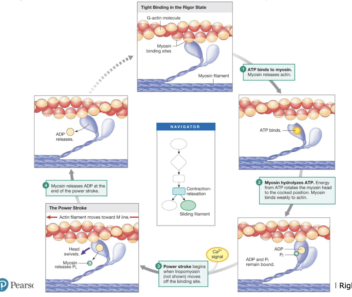
Contraction-relaxation cycle

Steps of the Contraction Cycle
ATP binds, myosin releases actin, myosin hydrolyzes ATP, Power stroke, Myosin releases ADP, Myosin stuck bound until given ATP
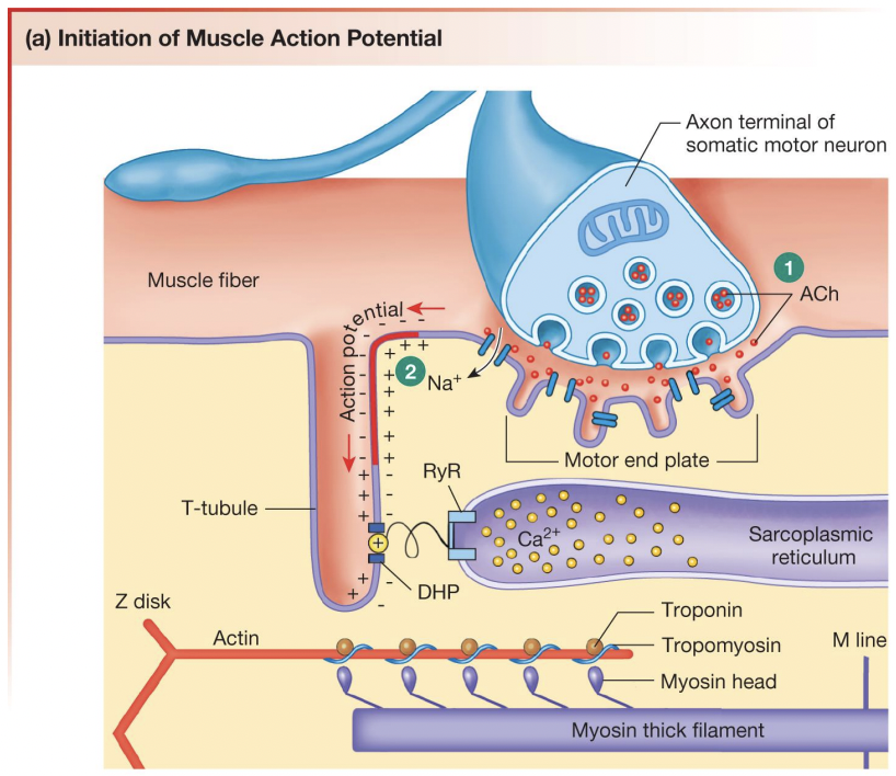
Acetylcholine (ACh)
released from somatic motor neuron and initiates action potential in muscle fiber,
How does ACh initiate contraction?
ACh is released from somatic motor neuron, ACh binds to sarcolemma, Na+ enter and K+ exits cell, depolarization, end-plate potential (EPP), muscle action potential, releases sarcoplasmic reticulum, calcium combines with troponin to initiate contraction
Summation
stronger contraction when the muscle does not relax completely between action potentials
Tetanus
maximal contraction
What causes decreased tension in resting muscles?
too much overlap between thick and thin filaments

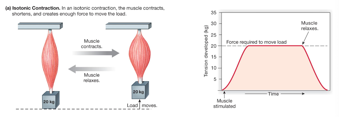
Isotonic contractions
concentric action (shortening action) and eccentric action (lengthening action)
Isometric contractions
series elastic elements
sarcomeres shorten while elastic elements stretch

smooth muscle
in an organ, they contract and relax slowly, use less energy to generate and maintain a given amount of force. Vascular, gastrointestinal, urinary, respiratory, reproductive, ocular
Caridiac muscle
striated, sarcomere structure, short fibers, single nucleus, gap junctions, sympathetic and parasympathetic control
Compare fibers of skeletal, smooth, and cardiac
skeletal: sarcomeres
smooth: no sarcomeres
cardiac: sarcomeres
Compare location of skeletal, smooth, and cardiac
skeletal: attatched to bones, a few sphincters
smooth: forms the walls of hollow organs and tubes, some sphincters
cardiac: heart muscle
Compare tissue morphology of skeletal, smooth, and cardiac
skeletal: multinucleate, large, cylindrical fibers
smooth: uninucleate, small spindle-shaped fibers
cardiac: uninucleate, shorter branching fibers
Compare what each muscle controls with the skeletal, smooth, and cardiac
skeletal: Ca2+ and tropinin, fibers independent from one another, Ca2+ from SR
smooth: Ca2+ and calmodulin, some fibers electrically linked via gap junctions, Ca2+ from ECF and SR
cardiac: Ca2+ and troponin, fivers electrically linked via gap junctions, Ca2+ from ECF and SR
Autonomic reflexes
visceral reflexes, some are spinal reflexes with brain modulation, some are integrated in the brain.
polysynaptic, with at least one synapse in the CNS
Skeletal muscle reflexes
proprioceptors send input to CNS and then somatic motor neurons carry output signal (alpha motor neurons), and effectors called extrafusal muscle fibers contract muscle fibers
Golgi tendon organs
free nerve endings wound inbetween collagen fibers in junction of tendons and muscle fibers, send info to CNS

Muscle spindles
stretch receptors encode signals about muscle length and changes in muscle length
myotatic unit
collection of neural pathways controlling a single joint
reciprocal inhibition
antagonistic muscles must relax as the prime mover muscles contract. sensory fibers branch upon arriving at the spinal cord
flexion reflexes
reflex to pull away from painful stimuli
3 classifications of movement
reflex, voluntary, or rhythmic
Integrated, coordinated responses require?
input from multiple regions of the brain
Where are reflex movement integrated?
least complex, integrated in spinal cord or brain
voluntary reflexes
most complex, integrated in cerebral cortex
rhythmic movements
intermediate complexity, integrated in spinal cord with higher center input required, Central pattern generators (CPGs)
3 levels of movement through CNS integration
spinal cord: reflexes and central pattern generators
Brain stem and cerebellum: posture and eye/hand movement
Cerebral cortex and basal ganglia: voluntary movements
the corticospinal tract
interneurons run from motor cortex to synapses with somatic motor neurons

Pakinson’s disease and basal ganglia function
abnormal movements, speech, cognitive changes, dopamine release reduction
Visceral Muscle Movement
smooth and cardiac, spontaneous depolarizing fibers called pacemakers
Cardiovascular system includes
heart, blood, vessels and capillaries
What is transported through the body in cardiovascular system?
nutrients, water, gases, hormones, immune cells, antibodies
What is eliminated by cardiovascular system?
CO2, heat, metabolic waste
Types of blood vessels
arteries, veins, capillaries, portal system (joins two capillary beds in series)
Parts of the heart
septum divides halves (left, right), atrium (in), ventricle (out)
Pulmonary circulation
short circuit, heart and lungs
arteries (O2 poor) and veins (O2 rich)
Systemic Circulation
heart and rest of the body,
aorta (O2 rich)
superior and inferior vena cava (O2 poor)
Blood flow motion components
dynamic (kinetic energy) and later (hydrostatic pressure)
Factors of blood flow
pressure, distance/length, radius, resistance, viscosity
Poiseuille’s law
resistance proportional to length and viscosity
resistance is inversely proportional to radius^4
velocity of flow
v, velocity = q, flow rate / a, cross-sectional area of tube
Mean arterial pressure (MAP)
pressure reserved in arteries during heart relaxation
MAP =~ cardiac output (CO) * peripheral resistance (PR)
What are the four chambers of the heart?
right and left atrium, right and left ventricle
What is the heart mostly made of?
myocardium, encased in pericardium
path of deoxygenated blood
vena cava → right atrium →right ventricle → pulmonary trunk
path of oxygenated blood
pulmonary veins → left atrium → left ventricle → aorta
atrioventricular valves
between atria and ventricles (tricuspid and bicuspid valve)
semilunar valves
between ventricles and arteries (aortic and pulmonary valve)
coronary circulation
supplies blood to the heart
autorhythmic cells
pacemakers, signal contraction, few contractile fibers
Contractile cells
striated fibers organized into sarcomeres
cardiac muscle compared to skeletal muscle
single nucleus per fiber
branch and join cells through intercalated disks
gap junctions
sarcoplasmic reticulum is smaller
myocardial contractile cells action potentials
depolarization (Na channels open) → initial repolarization (Na channels close) → plateau (Ca channels open, K channels close) → rapid repolarization (Ca channels close, K channels open)→ resting membrane potential (-90mv)

Myocardial autorhythmic cells action potentials
pacemaker potential (-60mV, Na+ in) → action potential (Ca2+ in and then K+ out) → back to resting (-60mV, K+ close If open)

What happens to the action potential of a myocardial autorhythmic cell if tetrodoxin, which blocks voltage-gated Na channels, is applied to the cell?
nothing because myocardial autorhythmic cells don’t use Na channels
What are the waves of the Electeocardiogram (ECG)?
P wave: depolarization of the atria
QRS complex: wave of ventricular depolarization and atrial repolarization
T wave: repolarization of the ventricle

What are the two segments of ECG waves?
P-R segment: AV nodal delay
T-P segment: ventricular atrial relaxation
What is the heart rate?
the time between two P waves or two Q waves
tachycardia
faster than normal heart rate
bradycardia
slower than normal heart rate
Diastole
cardiac muscle relaxes
Systole
cardiac muscle contracts
What are the five phases of the cardia cycle?
Heart at rest: Atrial and ventricular diastole
Ventricular filling: atrial systole
Early ventricular contraction (first sound)
Heart pumps: ventricular ejection
Ventricular relaxation (second sound)

Stroke Volume (SV)
End diastolic volume (EDV) - end systolic volume (ESV)
volume of blood before contraction minus after contraction
Cardiac output
volume of blood pumped by one ventricle in a given period of time
CO = HR * SV
average = 5 L/min
What are the three autonomic division controls?
parasympathetic control, sympathetic control, and tonic control