Respiratory system anatomy
 Call Kai
 Learn
 Practice Test
 Spaced Repetition
 Match
 Flashcards
 Knowt Play
1/190

There's no tags or description
Looks like no tags are added yet.
191 Terms
nasal cavity, oral cavity, pharynx
What parts of the respiratory system are shared between multiple body systems/part of the UPPER respiratory system? (3)
conducting airway
Part of the airway that GETS oxygen TO sites of gas exchange and does NOT participate in gas exchange
respiratory airway
Part of the airway that participates in gas exchange
filter, warm, moisten
The nasal hair and mucosa in the nose do these 3 things to make sure that the air does not cause damage to the lungs upon inspiration
Cartilage, smooth muscle, elastic fibers
Main components of airway wall: (3)
Cartilage
Structural component of the airway wall that supports the lumen and prevents collapsing
Smooth muscle
Structural component of the airway wall that changes the diameter of the airway
Elastic fibers
Structural component of the airway wall that allows for recoil of the lungs
Goblet cells, glands, cartilage
As we move down the respiratory tract, these components gradually decrease in relative abundance: (3)
Elastic, smooth muscle
As we move down the respiratory tract, these components gradually increase in relative abundance: (2)
Maintain, close airway
What are the two main functions of the larynx
Larynx
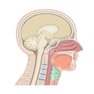
What part of the respiratory tract is highlighted in green?
Larynx
Which part of the respiratory tract are the vocal cords found in?
False vocal cords
What is denoted by #1?
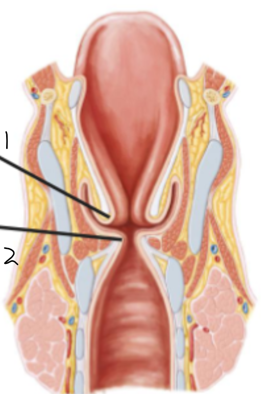
True vocal cords
What is denoted by #2?
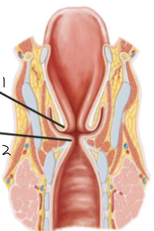
True vocal cords
Vocal folds that vibrate to produce sound; located in the larynx inferior to the false vocal cords
Attached to the thyroid and arytenoid cartilages
Stratified squamous
The true vocal folds are lined with this kind of epithelium
Pseudostratified columnar, cuboidal
As we progress down the respiratory tract, the epithelium starts as _______________ and becomes __________
Cricoid cartilage, larynx
What forms the only complete ring around the airway? Where is it located?
Skeletal
What kind of muscle lines the larynx?
Smooth
What kind of muscle lines tha trachea?
Thyroid, arytenoid cartilage
What are the true vocal folds attached to? (2)
Cricothyroid membrane
What location is usually used for emergency airways?
Inferior
The cricothyroid membrane is located ______ to the vocal cords
Arytenoid cartilage
Cartilage on the posterior side of the cricoid; moves the vocal folds to allow for breathing or vibration
Breathing
Arytenoid cartilages pulling the vocal folds open allows for:
Talking, singing
Arytenoid cartilages pulling the vocal folds closed allows for:
short, tall
The cricoid cartilage is ________ in the front, ________ in the back
Thyroid cartilage
_________________ is superior to the cricoid cartilage in the larynx
Thyroid cartilage
The hyoid bone is superior to the _________ in the larynx
Cartilage
The epiglottis is made of:
Hyoid bone
Inferior to the epiglottis
Cricoid
What structure is highlighted here?

Arytenoids
What structure is highlighted here?
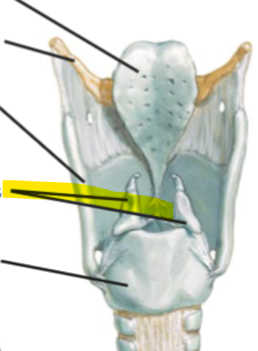
Thyroid cartilage
What structure is highlighted here?
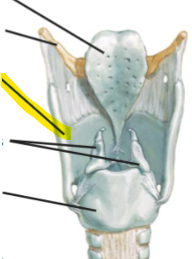
Hyoid bone
What structure is highlighted here?

Epiglottis
What structure is highlighted here?

Epiglottis, hyoid bone, thyroid cartilage, cricothyroid membrane, arytenoids, cricoid
Name the structures in the larynx from top to bottom: (6)
Anterior, posterior tilting of thyroid
Muscles change pitch of our voices through _____________
Trachea
Starts just inferior to the larynx, contained in the neck and thorax and branches into 2 main bronchi
Carina
Cartilage at the split of the trachea into the two bronchi
Sternal angle
The carina is located at the:
C-shaped hyaline
Type of cartilage that is characteristic of the trachea:
Esophageal expansion
The smooth muscle in the posterior of the trachea allow for:
Carina

What structure is highlighted in green?
Superior, inferior
What are the lobes of the left lung called?
Superior, middle, inferior
What are the lobes of the right lung called?
Oblique
What kind of fissure divides the superior and inferior lobes of the left lung?
Oblique
What kind of fissure divides the middle and inferior lobes of the right lung?
Horizontal
What kind of fissure divides the superior and middle lobes of the right lung?
Lingula, cardiac notch, aortic arch imprint
Left lung is characterized by these three structures:
Lingula

What is highlighted here?
Cardiac notch
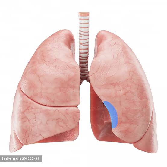
What is highlighted here?
Middle right
Which lobe cannot be seen from the posterior view of the lungs?
Left superior
Which lung lobe is in yellow?

Left inferior
Which lung lobe is in pink?

Right superior
Which lung lobe is in green?
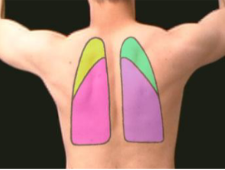
Right inferior
Which lung lobe is in purple?

Hilum
Location in the lung where vessels, nerves, all other structures enter and exit an organ like the lung
Bronchus, pulmonary artery, two pulmonary veins, lymph nodes, bronchial arteries
What structures are contained within the hilum of the lung? (5)
Bronchial arteries
Travel alongside bronchi and supply oxygen and nutrients to the lung parenchyma; branches off of the descending aorta
Descending aorta, systemic
The bronchial arteries branch off of the __________ and are a part of the __________ circulation
Oxygenated, deoxygenated
The bronchial arteries carry ________ blood and the pulmonary arteries carry __________
Hilum
What is this overall structure called?

Right
What side of the lung is this hilum on?

Left
What side of the lung is this hilum on?

Lung
There is one primary bronchi per _____
lobe
There is one secondary bronchi per _____
bronchopulmonary segment
There is one tertiary bronchi per _____
Segmental
Tertiary bronchi are also called:
Lobar
Secondary bronchi are also called:
Bronchopulmonary segments
Independent anatomical and functional units of the lung; a segment can be removed without damaging adjacent segments
Intersegmental, intrasegmental
Pulmonary veins are _____________ while pulmonary arteries are __________ in relation to bronchopulmonary segments
Tertiary bronchus, segmental pulmonary artery branch
One of each of these are located in each bronchopulmonary segment: (2)
Cartilage, submucosa
Bronchioles lack these structures in their walls: (2)
Both
Bronchioles are part of the conducting or respiratory airway?
Elastic
Alveoli have this kind of tissue that allows for passive expiration
Enlarged, elastic tissue, exhalation
Emphysema is characterized by ______ alveoli due to loss of _________ —> difficulty with _______
Less
Larger alveoli in emphysema patients —> ________ surface area for gas exchange
Visceral, parietal, fibrous
The lungs have these two layers in their pleura _______ but NOT ________
Mesothelium
Type of epithelium in the visceral and parietal pleura
costal
What part of the parietal pleura is shown in blue
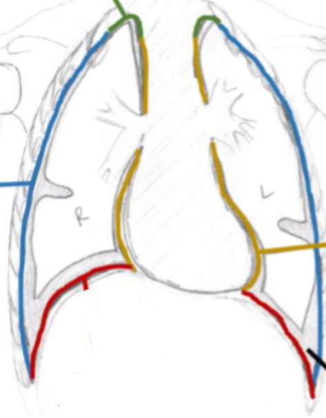
Cervical
What part of the parietal pleura is shown in GREEN

Mediastinal
What part of the parietal pleura is shown in yellow?

Diaphragmatic
What part of the pleura is shown in red?
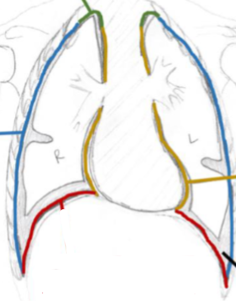
Costadiaphragmatic recess

What does #1 indicate?
Costal, cervical, diaphragmatic, mediastinal
What are the parts of the parietal pleura? (4)
Costodiaphragmatic recess
Edges of the pleural sac where the lungs will NOT reach
Two layers, parietal
In the pleural/costodiaphragmatic recesses, __________ of the ________ pleura will be touching
Potential space
The costadiaphragmatic recess is a ___________
Thoracoabdomial diaphragm
Primary respiratory muscle
Blood oxygen levels
Unconscious breathing via thoracoabdominal diaphragm is controlled by:
Central tendon, skeletal muscle
The thoracoabdominal diaphragm is primarily comprised of: (2)
somatomotor, phrenic, hemidiaphragm
The thoracoabdominal diaphragm has _________ innervation from the _______ nerve - one for each __________
Cervical 3-5
Phrenic nerves originate from these spinal levels: