Nervous System
 Call Kai
 Learn
 Practice Test
 Spaced Repetition
 Match
 Flashcards
 Knowt Play
1/65

There's no tags or description
Looks like no tags are added yet.
66 Terms
folding (gyri = ridges, sulci = grooves) are a way to pack more neurons into a limited skull space
more folding = more neurons = more complex processing capacity
Lissencephaly = "smooth brain" — normal in some small mammals, but a disease in humans (missing normal gyri/sulci).
~80% of the brain's neurons are in the cerebellum (heavily folded even in simpler species).
Humans: frontal lobe is proportionally largest → associated with reasoning/rationality vs. more "impulse-driven" responses in other species.
how does folding in the brain affect complex processing capacity
central sulcus - groove running from the top of the brain down the lateral surface (separates frontal from parietal lobe)
lateral (sylvian) fissure — runs horizontally along the lower lateral surface
preoccipital notch — small notch on the undersurface between cerebellum and rest of brain
parieto-occipital sulcus — imaginary line draw straight up from the preoccipital notch (better seen on medial view); separates parietal from occipital lobe
resulting lobes:
frontal lobe — everything anterior to central sulcus, superior to lateral fissure
parietal lobe — between central sulcus and parieto-occipital sulcus
occipital lobe — posterior to parieto-occipital sulcus
temporal lobe — inferior to lateral fissure
outline the landmarks used to define the lobes
primary motor cortex (precentral gyrus)
premotor/planning cortex (anterior to primary motor cortex)
frontal association cortex
what are the functional areas of the frontal lobe
located immediately anterior to central sulcus
contains pyrimdal cells — very large, triangular neurons that project directly down to spinal cord to activate muscles
job = execution of movement only (not planning/refinement)
organised as motor homonculus (body map) — somatopic organisation:
throat → most inferior/lateral
face, eyes, hand (large representation) → lateral
trunk, legs → medial surface
size of cortical representation is proportional to fineness of motor control needed (hence huge hand/face area vs small trunk area)
outline the primary motor cortex (precentral gyrus)
the “brains of the motor system” — plans movement before primary motor cortex executes it
Broca’s area: plans motor movements for speech
damage → Broca’s aphasia ( = non-fluent aphasia = motor aphasia — all synonyms)
comprehension intact, but speech is halting/effortful, poor fluency
front eye fields: plan eye-scanning movements (e.g. reading)
exner’s area: plans handwriting movements
outline the premotor/planning cortex (anterior to primary motor cortex)
large volume, functions less precisely defined
broad roles: intelligence, personality, behaviour, mood, cognitive function
outline the frontal association cortex
primary auditory cortex
Wernicke’s area (secondary auditory cortex)
arcuate fasciculus
temporal association cortex
what are the functional areas of the temporal lobe
buried in the lateral (Sylvian) fissure
decomposes sound into tone and timing only (tonotopic map — high vs low pitch mapped to different regions); doesn’t interpret meaning
outline the primary auditory cortex
surrounds primary auditory cortex
responsible for understanding/comprehension of spoken language
damage → Wernicke’s aphasia ( = sensory aphasia)
speech remains fluent but makes no sense/doesn’t answer the question asked
contrast directly with Broca’s aphasia (fluency differs; comprehension differs)
outline the Wernicke’s area (secondary auditory cortex)
white matter bundle connecting Wernicke’s area → Broca’s area (arch-shaped)
damage → conduction aphasia
comprehension is okay, but can’t produce an appropriate verbal response (can often still write an answer)
outline arcuate fasciculus
broad roles:
memory
mood
aggression
intelligence
outline the temporal association cortex
Type | Area damaged | Fluency | Comprehension | Alt. names |
|---|---|---|---|---|
Broca's aphasia | Broca's area | ❌ Non-fluent | ✅ Intact | Motor / non-fluent aphasia |
Wernicke's aphasia | Wernicke's area | ✅ Fluent | ❌ Impaired | Sensory aphasia |
Conduction aphasia | Arcuate fasciculus | ✅ Fluent (can understand) | ✅ Intact | Connection aphasia |
aphasia comparison table
postcentral gyrus — primary somatosensory cortex (immediately posterior to central sulcus)
supramarginal gyrus and angular gyrus (together = SMAGLA/AGLA on diagram; part of inferior partietal lobule)
parietal association cortex
what are the functional areas of the parietal lobe
receives sensory input: pain, temperature, discriminative touch (2-point discrimination), and proprioception (limb position sense)
also organised as a sensory homunculus — mirrors the motor homunculus layout
outline the role of the postcentral gyrus
support reading and writing
rough rule of thumb: supramarginal gyrus = more reading-related, angular gyrus = more writing-related, but overlap exists
outline the supramarginal gyrus and angular gyrus
spatial skills, 3D recognition (shapes, faces, concepts, abstract perception)
outline the role of the parietal association cortex
primary visual cortex located at the occipital pole (seen on medial view around the calcarine sulcus)
outline the role of the occipital lobe
non-verbal language (body language)
emotional expression/tone of language
spatial skills (3D)
conceptual understanding
artistic/musical skills
outline the role of the non-dominant (right) hemisphere of the brain
loss of non-verbal language
speech lacks emotion (flat prosody)
spatial disorientation
inability to recognise familiar objects
loss of musical appreciation
what are the effects of injury to the right hemisphere of the brain
primary visual cortex — at the very back of the brain (occipital pole/occipital protuberance)
secondary visual cortex — the rest of the occipital lobe; extends down into the inferior/bottom part of the temporal lobe
what makes up the occipital lobe
receives raw visual field input: right visual field → left hemisphere; left visual field → right hemisphere (info from both eyes crosses over)
organised as a visuotopic map of the visual field
extremely densely packed with neurons (huge amount of raw visual data to process)
only organises incoming information — does NOT interpret or add meaning to it
outline the role of the primary visual cortex
job = interpretation of what the primary visual cortex has organised (adds meaning — “what am I actually looking at”)
takes up far more cortical territory than the primary visual cortex → reflects how much more complex interpretation is than raw data collection
because it works from a learned model of "what normal looks like," it can be tricked — this is why magic tricks and acrobatics (e.g. Cirque du Soleil) feel so compelling: secondary visual cortex flags them as defying its expectations
primary visual cortex itself is very rarely "wrong" — the trickery happens at the interpretation stage
outline the role of the secondary visual cortex
cingulate gyrus — first major gyrus encountered on the medial surface
corpus callosum — large bundle of white matter connecting the left and right hemispheres
cerebellum — sits at the back, underneath the cerebrum
brainstem — made up of three parts
midbrain
pons
medulla
what new structures can be seen in the medial view of the brain
acts as a pathway that carries information from deep brain structures back out to the cortex so it can be acted on
outline the role of the cingulate gyrus
unique to higher primates (most other species don’t have one)
enables left-right coordination between hemispheres
outline the role of the corpus callosum
sits at the back, underneath the cerebrum
coordinates movement, balance, and posture
outline the role of the cerebellum
forebrain → cerebral cortex
midbrain → midbrain (stays small)
hindbrain → cerebellum + pons + medulla
outline the adult mapping of the three parts of the brain stem
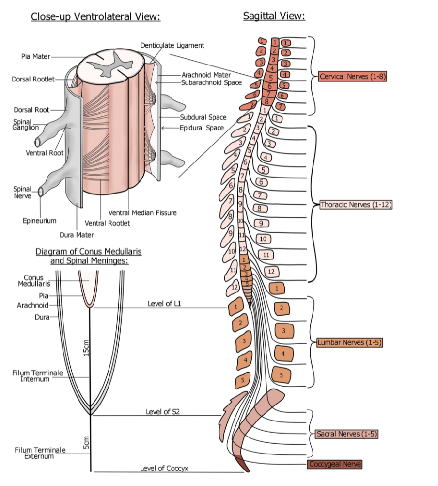
Region | # of nerve pairs | Notes |
|---|---|---|
Cervical | 8 | "Cervical" = narrowing (neck). 8 nerve pairs despite only 7 cervical vertebrae. |
Thoracic | 12 | One pair per thoracic vertebra. |
Lumbar | 5 | Spinal cord itself ends around vertebral level L1 — these nerves travel further down before exiting. |
Sacral | 5 | Individual nerve roots, not true spinal cord. |
Coccygeal | 1 | Single nerve at the very bottom. |
Total | 31 pairs | 8 + 12 + 5 + 5 + 1 |
outline the sagittal view
thickens at two points where extra limb innervation is needed
cervical enlargement — accommodates the large number of nerves serving the upper limbs
lumbar (thoracolumbar) enlargement — accommodates nerves serving the lower limbs
why does the spinal cord thicken at two points
Conus medullaris — the cone-shaped tapering point where the spinal cord proper ends (~level of L1)
Cauda equina ("horse's tail") — bundle of lumbar, sacral, and coccygeal nerve roots that continue down inside the vertebral canal after the cord has ended, before exiting at their respective vertebral levels (nicknamed "spaghetti fibres")
Filum terminale — a thread of pia mater extending from the tip of the conus medullaris down to anchor at the coccyx
Function: anchors the cord so it doesn't get painfully stretched every time you bend forward and back
Clinical note: rugby players can tear this structure — described as extremely painful; can be surgically reanchored
outline the end of the spinal cord

from innermost (closed to cord) to outermost:
Layer | Latin meaning | Key feature |
|---|---|---|
Pia mater | "soft mother" | Closely adhered directly to the spinal cord/brain surface — very hard to separate |
Arachnoid mater | "spider mother" | Named for its shimmering, spider-web-like appearance |
Dura mater | "tough mother" | Outermost, very tough — difficult to cut even with a scalpel |
meningitis = inflammation of these three layers
outline the meninges (the 3 protective layers)

Dorsal rootlets (back) + ventral rootlets (front) emerge along the cord → converge into a dorsal root and ventral root
Dorsal root + ventral root → one spinal nerve (per side, per level)
outline the anatomy at the single spinal segmental level
Dermatome = a map of the skin showing which spinal nerve level supplies sensation to each patch of skin (e.g. the thumb is supplied by C6, the pinky by C8)
Myotome = the equivalent map, but for muscles (motor output) rather than skin sensation
Clinical use: if someone comes in saying "I've lost feeling in my hand," you can use the dermatome map and test sensation systematically (e.g. with a pin or Q-tip) across the C6 → C7 → C8 regions of the hand/arm to work out exactly which spinal nerve level is likely affected (e.g. a pinched nerve in the spine).
outline dermatones and myotomes
receptors: Meissner’s corpuscles and Pacinian corpuscles — these sit just beneath the skin surface and are “encapsulated” (wrapped in their own structure)
fibre type: heavily myelinated (insulated) → fast conduction, roughly 50 metres/second
why so fast: these receptors are firing almost constantly — clothing touching your skin, everyday bumps and pressure — so it’s worth the body’s metabolic cost of building all that myelination insulation
route: receptor in skin → nerve fibre → cell body in dorsal root ganglion → enters the spinal cord via the dorsal root
outline the sensory pathway of discriminative touch and pressure
receptors: free nerve endings — unencapsulated, bare nerve endings in the skin
fibre type: unmyelinated → slow connection, roughly 1 metre/second
why so slow: real pain is felt relatively rarely for most people (often days apart), so it isn’t “worth” the expensive cost of myelinating these fibres
route: receptor in skin → nerve fibre → cell body in dorsal root ganglion → enters via the dorsal root → terminates directly in the back (posterior) part of the spinal cords’s grey matter
outline the sensory pathway of pain and temperature
| Pain & temperature (non-discriminative) | |
|---|---|---|
Receptor | Meissner's / Pacinian corpuscles (encapsulated) | Free nerve endings (bare) |
Myelination | Heavy | None |
Conduction speed | ~50 m/s | ~1 m/s |
How often it's firing | Constantly | Rarely |
both use pseudounipolar neurons — one single process leaves the cell body (which sits in the dorsal root ganglion) and splits into two branches: one running out to the receptor in the skin, and one running central into the spinal cord (“Ganglion” just means a cluster of nerve cell bodies grouped together)
compare the similarities and differences between discriminative and non-discriminative sensory pathways
dense clusters of cell bodies
located inside in the centre of the cord, outside (cortex) in the brain
what is grey matter and where is it located
the fibres/axons carrying information from A to B
located around the outside in the spinal cord, inside in the brain
what is white matter and where is it located
a funiculus — a large bundle/cable of white matter
there are three major ones per side of the spinal cord
Funiculus | Location | Job / carries |
|---|---|---|
Dorsal funiculus | Back of cord | Discriminative sensation (touch & pressure) |
Ventral funiculus | Front of cord | Pain & temperature |
Lateral funiculus | Side of cord | Motor columns |
outline the three funiculi
gracile fasciculus — medial (closer to the midline) → carries info from the lower limb (leg, thigh, foot)
cuneate fasciculus — lateral (further from midline) → carries info from the upper limb/trunk (arm, hand, fingers)
outline the division of the dorsal funiculus and its roles
Zone | Location | Function |
|---|---|---|
Substantia gelatinosa | Very back (tip of dorsal horn) | Jelly-like, unmyelinated — first stop for pain & temperature fibres |
Nucleus proprius | Just in front of substantia gelatinosa | Involved in proprioception — limb position sense |
(middle section — not named) | Central grey matter | Autonomic functions (e.g. balance) — not examined in detail |
Lower motor neuron (LMN) pool | Front (anterior horn) | Where lower motor neurons connect to upper motor neurons to produce movement |
outline the location and role of each key zone of grey matter
go straight up the cord in the gracile/cuneate fasciculus, or
detour briefly through the nucleus proprius, then rejoin the others heading up anyway
what are the two pathways a touch/pressure fibre coming in from the periphery can take
Imagine your entire forearm rests flat on a table. Thousands of receptors are all reporting essentially the same information at once. Sending every single one of those thousands of fibres all the way to the brain would need a spinal cord many times its actual size.
Instead, the nucleus proprius lets many similar fibres converge onto fewer neurons before they head upward — saving space and energy, and keeping the cord as small as it actually is (about the width of your pinky finger!).
why does touch info detour through the nucleus proprius
reflex — encapsulated receptor sends sensory fibre directly to the LMN (lower motor neuron) pool at the same spinal level, skipping the brain entirely
myotactic (monosynaptic) reflex — one synpase, very fast
e.g. knee-jerk test
withdrawal (polysynaptic) reflex — two synapses, involves the slower pain pathway
example: yanking your hand back from something hot
uses unmyelinated pain fibres and an extra synapse, therefore slower than the myotactic reflex
outline reflexes/reflex arc
cell body in dorsal root ganglion
enters dorsal root, synapses immediately in substantia gelatinosa
decussates immediately, right there at the spinal level, via the ventral (anterior) white commissure
travels up the opposite side as the sphinothalamic tract, in the ventral funiculus
synapses in the thalamus
travels up to the primary somatosensory cortex
key difference from DMCL: decussates immediately at the spinal cord level, not up in the medulla
outline the pain/temperature fibre pathway (sphinothalmic tract) — unmyelinated free nerve endings
cell body in dorsal root ganglion
travels up, same side, in gracile/cuneate fasciculus
synapses in the gracile/cuneate nucleus — in the lower medulla
decussates (crosses sides) via the internal acruate fibres
now called the medial lemniscus
synapses in the thalamus
travels via the internal capsule → primary somatosensory cortex
outline the touch and pressure fibre pathway (DCML system)
acts as a gatekeeper
constantly triages all incoming sensory information and decides moment to moment what’s important enough to actually pass onto the cortex
Example: a minor cut hurting less once a bigger pain occurs elsewhere — the thalamus has re-prioritised.
what is the role of the thalamus
| Non-discriminative (Spinothalamic) | |
|---|---|---|
Myelination | Heavily myelinated | Unmyelinated |
Conduction speed | ~50 m/s | ~1 m/s |
Function | Touch & pressure | Pain & temperature |
1st neuron terminates | Gracile/cuneate nucleus (lower medulla) | Substantia gelatinosa (spinal cord) |
Decussates | Internal arcuate fibres, in the medulla | Ventral/anterior white commissure, at the spinal cord level |
Named white matter tract after crossing | Medial lemniscus | Spinothalamic tract |
Final relay before cortex | Thalamus | Thalamus |
Destination | Primary somatosensory cortex | Primary somatosensory cortex |
Discriminative vs Non-discriminative sensory pathways
both discriminative and pain/temperature fibres from the same original (opposite) side have already crossed over by this point
a lesion here → loss of both touch/pressure and pain/temperature on the same side (opposite to the lesion)
this is called an associated sensory loss (both modalities lost together, same side)
outline the affects of lesions in the brain/brainstem
discriminative (touch/pressure) fibres haven’t crossed yet at this level → loss occurs on the same side as the lesion
pain/temperature fibres already crossed at the spinal level → loss occurs on the opposite side to the lesion
this is called a dissociated sensory loss (the two modalities are lost on different sides)
outline the affects of lesions in the spinal cord
Lesion location | Touch/pressure loss side | Pain/temp loss side | Type |
|---|---|---|---|
Brain/brainstem | Opposite to lesion | Opposite to lesion | Associated |
Spinal cord | Same side as lesion | Opposite to lesion | Dissociated |
lesion summary table
located in the primary motor cortex, arranged as the motor homunculus
cell body shape: pyrimdal (triangular) — hence also called pyramidal neurons, or Betz cells
huge compared to a typical neuron: up to ~160 micrometres wide
outline the location and structure of upper motor neurons
they have to carry an electrical signal an extremely long way — up to 1.2 metres, from the cortex down to as low as vertebral level
carrying a signal that far without it fading out requires a much bigger “battery” (capacitance), and that means a bigger cell
why are upper motor neurons so large
cell body in primary motor cortex, in the arm region of the homunculus
travels down through the internal capsule (completely bypasses the thalamus)
into the mid-brain, in an anterior (front) position — motor info always run toward the front
into the pons — here it temporarily breaks into many small bundles, weaving around the pons’ many nuclei, before regathering further down
into the lower medulla — the pyramidal decussation: ~85% of fibres cross to the opposite side here
after crossing, becomes the lateral corticospinal tract (in the lateral funiculus)
the remaining ~15% don’t cross here — they continue down the same side as the anterior corticospinal tract
at the relevant spinal segmental level (e.g. C5-C8 for the arm), the UMN terminates in the LMN pool (anterior/ventral horn grey matter)
lower motor neuron (LMN) — the second neuron — sends its fibre out through the ventral root to the muscle, causing contraction
outline the route a fibre takes down the pyramidal tract
15% (anterior corticospinal tract) → controls axial/trunk muscles — coarse, bulk movements (e.g. bending over to pick something up)
85% (lateral corticospinal tract) → controls fine manual dexterity (e.g. precise hand/finger movement)
Key insight: fine, precise movement requires a disproportionately larger share of neural resources than coarse trunk movement — small accurate movements are neurally "expensive."
why do 85% of fibres in the pyrimdal tract cross over but 15% don’t
About half the homunculus represents the face/head — but the face is above where the spinal cord starts, so it doesn't need spinal nerves
Instead, a side-branch called the corticobulbar tract carries this information out to the face
This is why the pons has a bulge on its front surface — extra nuclei needed to handle all this facial motor information
outline the corticobulbar tract
Neuron | Location | Path |
|---|---|---|
1st (Upper motor neuron) | Primary motor cortex | Internal capsule → midbrain (anterior) → pons (scatters and regathers) → pyramidal decussation (lower medulla, ~85% cross) → lateral or anterior corticospinal tract → terminates in LMN pool |
2nd (Lower motor neuron) | LMN pool (anterior/ventral horn) | Exits via ventral root → muscle |
upper and lower motor neuron summary table
| LMN lesion | |
|---|---|---|
Result | Spastic paralysis (spasticity / hypertonia — increased tone) | Flaccid paralysis (hypotonia — no tone) |
Why | Loss of central inhibition → local reflex loop runs unchecked → muscle tone increases | No nervous input reaches the muscle at all |
What you'd see | Arms held close to body (resting flexor tone); exaggerated knee-jerk reflex; resisted arm movement "overshoots" when released (person almost hits themselves) | Limb is limp when picked up and released — no resistance, no tone at all |
Bedside test | Ask patient to resist you pulling their arm, then let go — exaggerated overshoot = UMN lesion | Pick up hand, let go — completely limp = LMN lesion |
UMN vs LMN lesion comparison table
caudate nucleus + putamen — together called the striatum
globus pallidus — split into an external segment (GPe) and internal segment (GPi)
subthalamic nucleus — sits directly beneath the thalamus
substantia nigra — “black substance”, produces almost all of the brain’s dopamine
what five nuclei make up the basal ganglia
planning cortex (e.g. Broca’s area) → releases glutamate (excitatory) → activates the striatum
from the striatum, GABAergic (inhibitory) neurons branch three ways: to GPe, to GPi, and to the substantia nigra
striatum → GPi directly: uses GABA (inhibitory)
GPi → thalamus: uses GABA (inhibitory)
thalamus → planning cortex (back to where we started): uses glutamate (excitatory) — completing the loop
outline the direct pathway
planning cortex (e.g. Broca’s area) → releases glutamate (excitatory) → activates the striatum
from the striatum, GABAergic (inhibitory) neurons branch three ways: to GPe, to GPi, and to the substantia nigra
indirect pathway (striatum → GPe → subthalamic nucleus → GPi)
striatum → GPe: uses GABA (inhibitory)
GPE → subthalamic nucleus: uses GABA (inhibitory)
subthalamic nucleus → GPi: uses glutamate (excitatory)
GPi → thalamus: uses GABA (inhibitory)
thalamus → planning cortex (back to where we started): uses glutamate (excitatory) — completing the loop
outline the indirect pathway
direct pathway — more “conservative”, models what happens if the movement is under-done
indirect pathway — more “liberal” — models what happens if the movement is over-done (exaggerated)
the circuit effectively averages these two scenarios and sends the result via GPi → thalamus → back to the cortex, giving the planning cortex a refined sense of the right amount of movement
once refined, the planning cortex activates the primary motor cortex (UMN) → corticospinal tract → LMN → muscle
what do the direct and indirect pathways do
comes from the substantia nigra
primes the three GABAeric striatal output neurons (to GPe, GPi, and substantia nigra) so they’re ready to fire at the slightly excitatory input
analogy: a cup filled right to the brim — it only takes the smallest additional drop (glutamate) to make it overflow (fire)
what is the role of dopamine in the basal ganglia
the cerebellum feeds into the same glutamateric neuron that runs back up to the cortex, and does three jobs:
helps orient the head for visual focus/balance
general balance maintenance
holds a map of what the planned movement should look like, and compares it to what’s actually happening, if they don’t match, the cerebellum sends a stop signal, letting the basal ganglia re-plan and try again
outline the role of the cerebellum in the direct and indirect pathway
pathology: substantia nigra neurons die off (cause unclear, but linked to toxic protein accumulation) → dopamine depletion
consequence, traced through the circuit:
without dopamine “priming”, the three GABAergic striatal neurons are no longer ready to fire — much more glutamate is now needed to activate them
because they’re under-activated, less inhibition reaches the GPi
less inhibition on GPi → GPi neurons fire more (an inhibitory fibre that isn’t itself inhibited works harder)
more GABA released from GPi → thalamus is inhibited more than normal
thalamus sends less glutamate up to the cortex → hypo-excitability of the motor cortex → reduced movement (hypokinesia)
outline how Parkinson’s Disease occurs
Rigidity — increased tone (sitting stiffly, arms/abdomen taut)
Bradykinesia/hypokinesia — slowness, difficulty initiating movement (e.g. struggling to stand up from a chair, shuffling "freeze-then-go" gait)
Tremor at rest
Flat/reduced mood expression
what are the symptoms of Parkinson’s Disease