Heart
 Call Kai
 Learn
 Practice Test
 Spaced Repetition
 Match
 Flashcards
 Knowt Play
1/66

There's no tags or description
Looks like no tags are added yet.
67 Terms
Arteries
Blood vessels that transport oxygenated blood away from the heart.
Veins
Blood vessels that transport deoxygenated blood to the heart.
Pulmonary circulation
Transports deoxygenated blood from the right heart to the lungs and oxygenated blood back to the left heart.
Systemic circulation
Transports oxygenated blood from the left heart to body tissues and deoxygenated blood back to the right heart.

Fibrous pericardium
Outer dense connective tissue layer of the pericardium attached to the diaphragm and great vessels.

Myocardium
The thickest layer of the heart wall composed of cardiac muscle.

Endocardium
Internal epithelium and connective tissue lining the heart chambers.
Pericarditis
Inflammation of the pericardium causing excess fluid accumulation and limiting heart pumping.

Tricuspid valve
AV valve located between the right atrium and right ventricle.
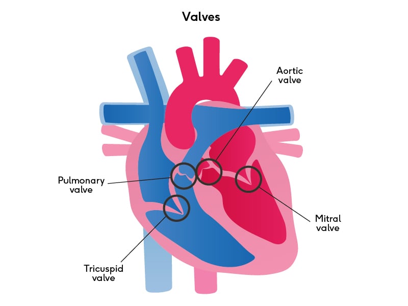
Bicuspid (mitral) valve
Valve located between the left atrium and left ventricle.

Chordae tendineae
Collagen fibers that anchor AV valves and prevent eversion during ventricular systole.

Sinoatrial (SA) node
The pacemaker of the heart that initiates muscle impulses for a heartbeat.

AV node
Delays electrical impulses on the floor of the right atrium to coordinate heart timing.

Purkinje fibers
Large specialized cells providing rapid impulse distribution to the ventricular myocardium.
Systole
Contraction phase of a heart chamber.
Diastole
Relaxation phase of a heart chamber allowing blood to flow in.
Ventricular hypertrophy
Thickening of the ventricular walls caused by increased workload and high blood pressure.
Cardiovascular Overview
Blood circulates throughout the body to maintain homeostasis, cells need oxygen, blood flow is unidirectional
Blood Pressure
Force of blood pushing against the vessel walls, established and maintained by heart
Pattern of Blood Flow
Right side of heart, lungs, left side of heart, systemic cells (rest of body), right side of heart
Mediastinum
The heart is behind the sternum in the mediastinum
Serous Pericardium
Double Layered, deep to fibrous pericardium
Parietal Layer
Superficial layer of serous pericardium
Visceral Layer (Epicardium)
Deep layer of serous pericardium

Pericardial Cavity
Filled with serous fluid, surrounds the heart
Base of the heart
Posterior giant artery
Apex of the heart
Pointy left side of the heart
Atria
Thin walled chambers, superiorly located, receive blood returning to the heart
Ventricles
Inferior chambers

Right Atrium
Blood from the systemic circulation, to right atrium is the superior vena cava and inferior vena cava

Left Atrium
Blood from the pulmonary circulation, to the left atrium is the pulmonary veins

Right Ventricle
Blood to pulmonary, from the right ventricle is the pulmonary trunk and the pulmonary arteries

Left Ventricle
Blood to systemic circulation, from the left ventricle is the ascending aorta

Coronary Atrioventricular Sulcus
Separates atria and ventricles, can be seen outside of heart

Anterior Interventricular Sulcus
Separates ventricles, outside anterior view
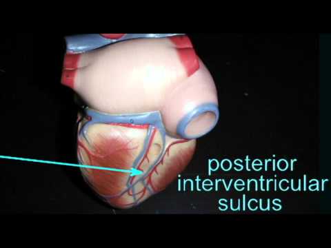
Posterior Interventricular Sulcus
Separates ventricles, outside posterior view
Interatrial Septum
Septum between atriums
Interventricular Septum
Septum separating ventricles
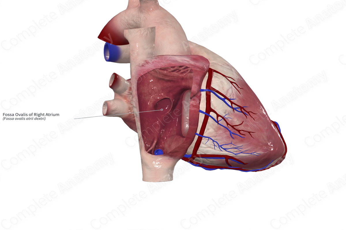
Fossa Ovalis
Location of fetal foramen ovale, tiny hole that closes up in the right atrium
Pectinate Muscle
Anterior atrial wall muscle
Trabeculae Carnae
Internal ventricle wall muscles

Papillary Muscles
Anchor chordae tendineae

Pulmonary Semilunar Valve
Separates right ventricle and pulmonary trunk

Aoritic Semilunar Valve
Separates left ventricle and entrance into the aorta
Coronary Circulation
Supply heart wall, branches of the ascending aorta
Posterior Interventricular Artery
Supplies posterior side of the ventricles
Anterior Interventricular Artery
Supplies anterior surface of ventricles and interventricular septum

Circumflex Artery
Supplies left atrium and ventricle
Myocardial Infarction (Heart Attack)
Damage to (or death of) myocardium that results from occluded coronary artery (lack of blood flow)

Great Cardiac Vein
Anterior left side of heart

Middle Cardiac Heart
Posterior middle heart

Small Cardiac Vein
Anterior right side of heart

Coronary Sinus
Big vein seen in posterior side of heart, opening in the right atrium
Intercalated Discs
Specialized cell to cell contacts that allow for muscle impulse passage
Conducting System
Specialized cells that initiate and conduct the muscle impulses for a heartbeat

Atrioventricular Bundle
Connects to the AV node and goes down the AV septum

Left and Right Bundles
Connects to the AV bundle and separates into left and right as it goes down the septum
Conducting System
Muscle impulse is generated at the SA node and spreads throughout the atria and to the AV node, AV node delays impulse, AV bundle conducts impulse into the AV septum, impulse splits into right and left bundles, impulse is delivered to the purkinje fibers and distributed to the ventricular myocardium
Cardiac Plexus
Sympathetic and parasympathetic components
Brainstem Innervation
Increase/decreases the heartbeat
Heart Innervation: Sympathetics
Spinal Cord (T1-T5), preganglionic axons enter the sympathetic trunk, go up to the thoracic and cervical portions and synapse, postganglionic axons travel directly to the heart through cardiac nerves
Heart Innervation: Parasympathetic
From the medulla oblongata via the vagus nerve, in the thoracic cavity, vagus nerve branches that supply to the heart
Atrial Systole
Atria contract, blood goes through the open AV valves into the ventricles, blood continues to enter the atria from their associated veins
Ventricular Systole
Atria relax and ventricles contract, blood pushes the AV valve cusps closed, semilunar valves are opened, blood enters the pulmonary trunk and aorta
Ventricular Diastole
Atria and ventricles are relaxed, blood passively flows through the AV valves from atria to ventricles
Arrhythmia
Any abnormality in the rate, regularity or sequence of the cardiac cycle
Ventricular Fibrillation
Uncoordinated contractions