Causes + tx of cardiac arrhythmias
 Call Kai
 Learn
Learn Practice Test
 Spaced Repetition
 Match
Match Flashcards
 Knowt Play
1/57

There's no tags or description
Looks like no tags are added yet.
Name | Mastery | Learn | Test | Matching | Spaced | Call with Kai | Chat |
|---|
No analytics yet
Send a link to your students to track their progress
58 Terms
How can arrhythmias be classified?
Brady/Tachy
Paraxysmal (episodic)/ sustained
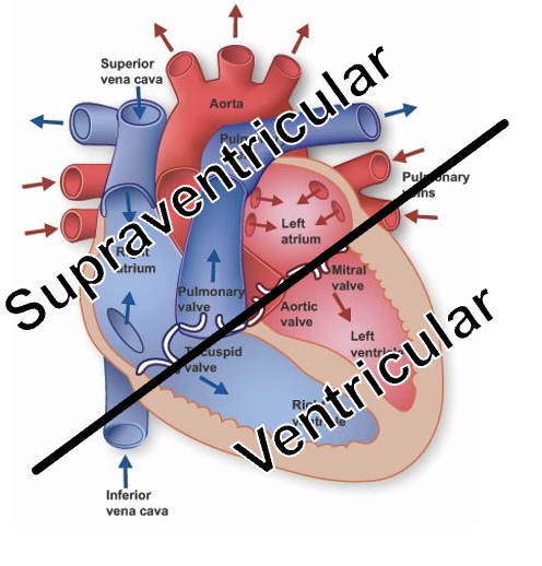
Tachy can be supraventricular/ Ventricular
How can arrhythmias be monitored?
Standard ECG (2-3 minutes)
Holter monitor (24h-7days)
Implantable device (years)
What is the general cardiac cause of weakness and syncope?
Compromised cardiac output
What can cause a compromised cardiac output leading to weakness and syncope?
Congenital heart disease
DCM
HCM
Cardiac tamponade
Neoplasias
Arryhthmias
tachy —> too fast not enough time for ventricles to fill for adequate SV
brady —> too slow not enough O2
What are the two types of action potentials in the heart? What is their main difference?
Pacemaker (nodal) cells action potentials
Primary due to influx of Ca
Cardiac myocytes
Primarily due to influx of Na
Recall the vaughan williams classification of antiarrhythmic drugs?
Class I- Na channel blocker (1° myocardial cells) —> slow upstroke of depolarisation
Class II- Beta blocker (1° nodal cells) —> slow depolarisation
Class III- K channel blocker (1° myocardial) —> lengthen AP duration
Class IV- Ca channel blocker (1° nodal cells) —> slow depolarisation
Misc: Digoxin —> slow conduction across AVN
'Never Buy Kennel Club Dogs'
What is this ecg showing?
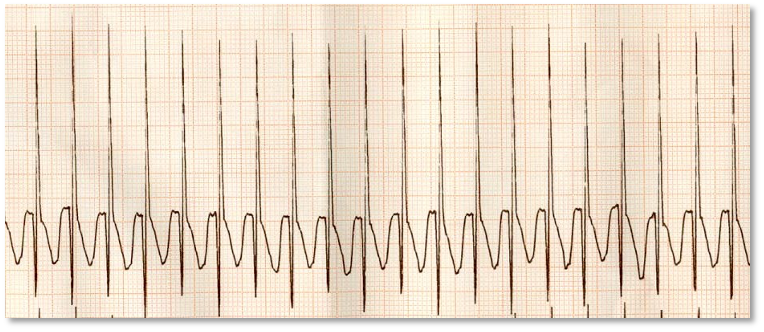
Supraventricular tachycardia
from above ventricles
Fast conduction because only above ventricle affected so narrow complexes
regular rhythm (except AFib)
Narrow QRS
What is arrhythmias are classed as SVTs?
sinus tachy
focal atrial tachy
multi-focal atrial tachy
orthodromic atrioventricular reciprocating tachy
AFib
atrial flutter
focal junctional tachy
junctional ectopic tachy
atrioventricular node reentry tachy
When could a SVT look wide not narrow?
If bundle branches are damaged?
What is the most common pathological arrhythmia in animals?
AFib
What is AFib associated with?
Most commonly atrial stretch from underlying heart disease (MMVD, DCM)
Can occur in abscence of heart disease if mass of atria is large in larger animals (lone AFib)
Explain what happens during AFib?
‘Fibrillating’ atria with chaotic depolarisation
No coordinated atrial depolarisation so absence of p wave
May see fibrillating baseline
Timing of depolarisation reaching AVN is irregular (ventricular rate irreg)
Irregular R-R interval
irregularly irregular
What is this ecg showing?

AFib
How is AFib treated?
Manage underlying condition
don’t try to convert to sinus rhythm (likely AFib with reoccur)
AIM control the ventricular response rate to AFib (decr HR) —> <140bpm, but ideally <125bpm
Drugs that slow conduction through AV node
Class 4s- Ca channel blocker —> Diltiazem
Digoxin
Better heart rate control with combination Digoxin + Diltiazem
What are beta blocker typically not use to tx AFib?
would worsen already poor systolic function
(NEVER use in uncontrolled CHF)
What effect does diltiazem have?
slow conduction across the AVN
Negative inotrope (clinically insignificant)
What effect does digoxin have?
Negative chronotrope (slows heart rate)
‘Vagomimetic’ —> enhances vagal tone and slows conduction across the AV node
Weak +ve inotrope
Blocks Na+/K+ ATPase → Ca2+ accumulation (inprove contract.)
What should you be careful of when using Digoxin?
Digoxin toxicity
Start with cautious dose and check plasma levels
GI signs

What is Orthodromic atrioventricular re-entrant tachycardia (OAVRT)?
Wolf-Parkinson White syndrome
Abnormal connection between atria and ventricles (band of muscle)
Conduction from the atria to the ventricles can occur across the pathway
ACCESSORY PATHWAY MEDIATED SVT
How does OAVRT present on ECG?
Short P-R in sinus rhythm
slurred QRS upstroke = delta wave (ventricular pre excitation)
Rapid narrow QRS complex tachycardia

What is the typical presentation of OAVRT?
Labrador retrievers + GR
Typically young (1-2 yr/o)
Syncope and exercise intolerance
Very fast HR
Can present in heart failure bc tachycardia induce cardiomyopathy
How can OAVRT be treated?
Radiocatheter ablation (burn accessory pathway)
How is SVT treated in an emergency situation?
Vagal manoeuvre
Ocular compression
Carotid massage
Gag reflex
IV esmolol (beta blocker)
IV diltiazem (IV verapamil if diltiazem unavailable)
Oral diltiazem if IV preparation not available
Oral sotalol can also be effective
In first opinion try oral options whilst evaluating- probably will work
What does a ventricular arrhythmia look like on an ecg?
Typically, wide QRS complexes
not conducted along the specialised conduction fibres therefore slow conduction
ventricular ectopics = VECs + VPCs
What is the difference between ventricular escape complex and ventricular premature complexes?
Look the same on ECG
Ventricular escape complexes occur after a long pause (‘escape’ death)
Ventricular premature complexes (VPCs) occur before you’d expect them (premature)

What does this ECG show?
ventricular premature complex (triplet + couplet)
What are the different classifications of monomorphic VTACH?
Non-sustained <30s
Sustained >30s
Incessant >12h
(wide QRS all look the same)

How does a VTACH present on ecg?
wide QRS
R-R interval constant (regular rhythm on ausc)
± P wave (not associated with the QRS or hidden within QRS)

What are the two most likely mechanisms of monomorphic VTACH?
1. Re-entry - a small circuit of electricity racing around and around an area of unhappy myocardium
2. Triggered activity – an unhappy island of ventricular myocardial cells discharging inappropriately at a fast rate
How does polymorphic VTACH differ from monomorphic?
More malignant cause – more than one region of unhappy ventricular myocardium
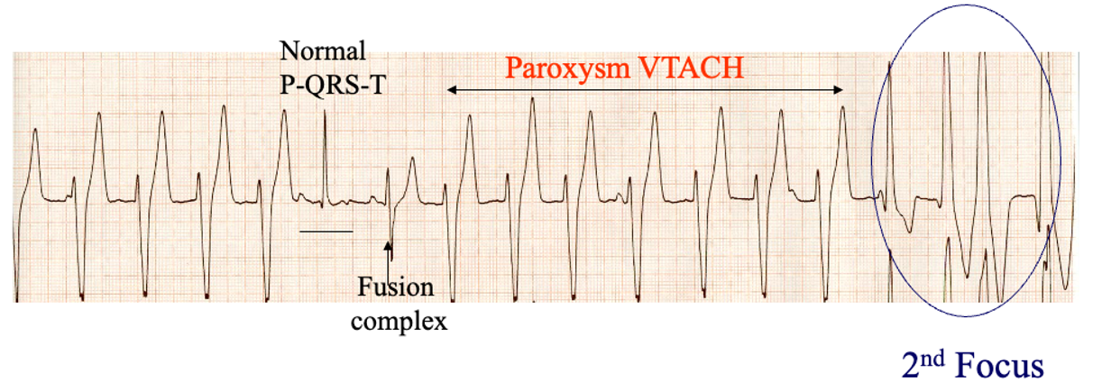
What signs of malignancy can be seen on ECG along side polymorphic ventricular tachy?
Faster rates (the faster the worse)
R on T phenomenon – two or more consecutive complexes so close together as to start their R wave on the preceding complex T wave
What can ventricular tachy trigger?
VFib
(tx of ventricular tachy aim to prevent VFib)

What does this ECG show?
VFib
What is the difference between accelerated idioventricular rhythm and ventricular tachy?
Same morphology on ECG
Only difference is rate
>180-200= VTACH
<180 = AIVR
AIVR typically not haemodynamically compromised
Typically don't require treatment
Most often extra cardiac cause (eg: severe GI disease, splenic lesion, pancreatitis)

What does this ECG show?
accelerated idioventricular rhythm
(wide QRS, HR<180, BP normal)
List some differentials of causes of ventricular arrhythmias
Underlying cardiac disease – CHF, cardiomyopathies, myocarditis, myocardial hypoxia, ischaemia etc.
Catecholamines – stress, pain
Acidosis
Hypokalaemia
Abdominal disease – GDV, splenic lesions (not just haemangiosarcoma), sepsis, pancreatitis etc.
Thoracic trauma (myocardial trauma)
Drug induced - eg: digoxin toxicity
How do you decide whether to treat ventricular arrhythmias?
Only treat if necessary so:
Treat if haemodynamically significant (eg: hypotensive, collapsed etc.)
Treat if very fast / close coupling intervals / ‘R on T’
Treat if multifocal
What do you do before treating ventricular arrhythmias?
Ensure no electrolyte or acid-base disturbances
How do you treat ventricular arrhythmias in an emergency case?
Lidocaine (Class IB)
Na+ channel blocker —> act on myocardial cells
bolus then cardiac resynchronization therapy
Esmolol (Class II- not used much)
short acting beta blocker
Amiodarone CRI (risk of anaphylaxis)
class 3 —> K channel blocker
Less common tx
Magnesium chloride
If completely refractory, electrical DC cardioversion (requires GA and a defibrillator)
What treatment do you use for a non emergency ventricular arrhythmia?
Sotalol - Class 3
Mexiletine - Class 1B
Amiodarone- Class 3 (similar to digoxin)
Risks of hepatotoxicity and thyroid dysfunction (long term)
Atenolol - Class 2
risk if underlying heart dz
List some of the causes of bradyarrythmias
Sinus bradycardia (normal)
Sinus arrest (no P wave)
Sinoventricular rhythm (hyperkalaemia – think urinary obstruction in cats)
AV Block
What tends to be the underlying cause of atrial standstill
Hyperkalaemia (K+)
Urinary obstruction
renal failure (oliguric / anuric patients)
Hypoadrenocorticism (Addison’s disease)
How does atrial standstill occur and how does it present on ecg?
Sinoatrial node is driving the rhythm as normal BUT there is no atrial depolarisation
Atria more sensitive to K+ than ventricles
Absent P waves, spikey T waves, mild prolongation of the QRS complex
(atrial myocardium are more sensitive to high K+ than ventricular myocardium)

What is this ECG showing?
atrial standstill
What is this showing?
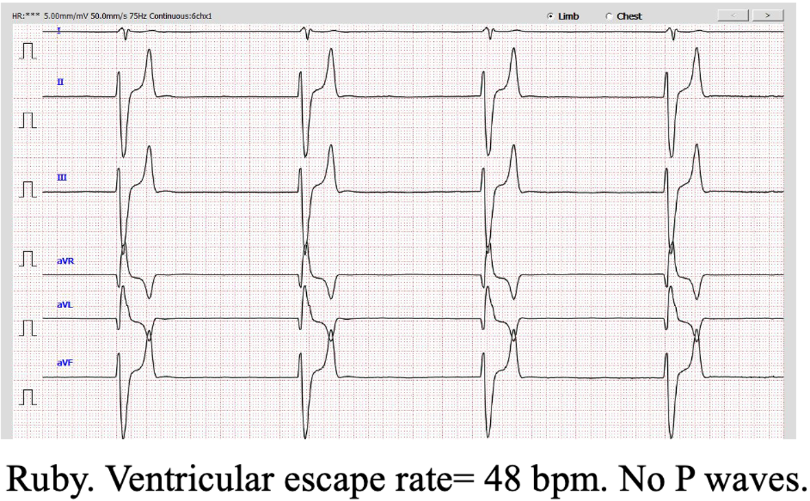
Atrial standstill with ventricular escape complexes
(no P wave, wide QRS + slow HR)
What are some of the causes of sinus arrest?
Sick sinus syndrome (’Brady – Tachy syndrome – think West Highland White Terriers)
Sinus node dysfunction
Drugs (eg: amiodarone, digoxin, sotalol)
Electrolyte changes
Atrial myocarditis
(a sinus pause lasting for more than 3 times the normal P-P interval)

What does this ECG show?
sinus arrest
What are the classes of AV block?
First degree
P: QRS normal but prolonged P-R interval
P for every QRS but elongated gap between P + QRS
Second degree
P:QRS normal but some non conducted P wave
Mobitz 1- no pattern and tends to physiological
Mobitz 2 - pattern and more pathological
Third degree
No association between P waves and QRS complexes, often fast sinus rate (freq p waves, wide QRS (ventricular escapes), HR often 20-40 in dogs

What does this ECG show?
Ist degree AV block
P:QRS = 1:1 (just like normal sinus rhythm)
Prolonged P-R interval (>130ms in a dog)
What are some of the causes and treatment of first degree AVB?
High vagal tone
Drug effect
Breed morphology related (brachy)
Tx- typically incidental with no treatment required
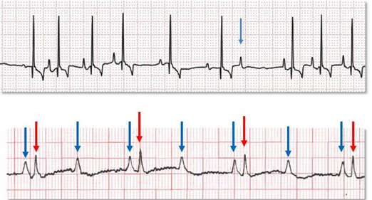
What do these ECGs show?
2nd degree AV block —> non-conducted P waves
top = Mobitz 1 (Wenkebach phenomenon)
bottom = Mobitz 2
How do the 2 types of second degree AV block present on ECG?
Mobitz 1
time for the signal to pass through the heart gets longer and longer with each beat until a QRS is completely dropped (P-QRS get progressively longer until QRS dropped)
Mobitz 2
not a prolongation of the P-R interval before the complete AV block/P-wave with missing QRS complex
often a pattern
2:1 = 2 P waves for every 1 QRS
4:3 = 4 P waves for every 3 QRS
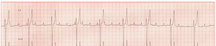
What does this ECG show?
third degree AVB
No association between P waves and QRS complexes (electricity cannot get through the AV node)
fast sinus rate —> frequent P wave
QRS wide (VECs)
HR abt 20-30bpm (dog) 100-130 bpm (cats)
How do you treat third degree AVB in dogs vs cats?
Dogs- emergency, refer for pacemaker ASAP
Cats- cats HR often 100-130 better at compensating so often incidental finding
How do you treat bradyarrhythmia?
Treat underlying disorder
Test response to anticholinergics (atropine) if vagally mediated
If HR increases suggests underlying disease causing high vagal tone
If life threatining refer for pacemaker if poss or trial a B agonist (terbutaline)
Oral meds
List some possible oral meds used in the treatment of bradyarryhtmias
B agonist- terbutaline
Anti-cholinergics – propantheline
Beta sympathomimetics – terbutaline
Xanthin derivatives – theophylline
Pimobendan – more blood per heartbeat
Explain what happens in atropine response test
Before= Sinus arrhythmia, sinus arrest, and ventricular escapes
30 minutes post atropine = If sinus tachycardia – proves bradycardia was vagally mediated
What are the two approaches to implant a pacemaker?
Transvenous- via right jugular pacing lead passed through the right atrium, across the tricuspid valve into the right ventricle
Epicardial- transabdominal pacing lead attached to epicardium (usually in cats bc size)
(gold standard tx for 3rd degree AVB or atrial standstill)