ch 10Skeletal Muscle Anatomy, Sarcomeres & Sliding Filament Theory
 Call Kai
 Learn
 Practice Test
 Spaced Repetition
 Match
 Flashcards
 Knowt Play
1/192

There's no tags or description
Looks like no tags are added yet.
193 Terms
What is the main function of muscle tissue?
Muscle tissue consists of specialized cells that contract to produce movement within the body and movement of the body itself.
What are the three types of muscle tissue?
Skeletal muscle, cardiac muscle, and smooth muscle.
What are the four common properties of muscle tissue?
Excitability (responding to stimuli), contractility (shortening), extensibility (stretching), and elasticity (returning to resting length).
What is excitability?
The ability of muscle tissue to receive and respond to a stimulus, such as a chemical signal from a nerve, by changing its membrane potential.
What are the main characteristics of skeletal muscle tissue?
Skeletal muscle attaches to and covers bones, contains long striated fibers, is under voluntary control, contracts rapidly, and can fatigue.
What structures make up a skeletal muscle organ?
Skeletal muscle fibers, connective tissues, blood vessels, and nerves. Each muscle fiber is one skeletal muscle cell.
What are the three connective tissue coverings of skeletal muscle?
Epimysium, perimysium, and endomysium. They support, protect, and organize muscle fibers and help prevent damage during strong contractions.
What is the epimysium?
A layer of dense irregular connective tissue surrounding the entire skeletal muscle. It separates the muscle from surrounding tissues and helps reduce friction.

What is the perimysium?
A layer of dense irregular connective tissue surrounding each fascicle, which is a bundle of skeletal muscle fibers.

What is the endomysium?
A flexible connective tissue layer surrounding individual muscle fibers. It contains capillaries, nerve fibers, and myosatellite cells involved in muscle repair.
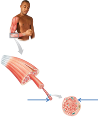
What is the structural organization of skeletal muscle from largest to smallest?
Whole muscle → fascicle → muscle fiber → myofibril → myofilaments.
What is the difference between a tendon and an aponeurosis?
A tendon is a cordlike bundle of collagen fibers (the three layers), while an aponeurosis is a broad, flat sheet. Both help attach skeletal muscles to bones and transmit contraction forces.

Why do skeletal muscles require extensive blood vessels and nerves?
Blood vessels deliver oxygen and nutrients and remove wastes. Nerves stimulate muscle fibers to contract.
How do motor nerves travel through skeletal muscle?
Axons enter through the epimysium, branch through the perimysium, and reach individual muscle fibers through the endomysium.
What is the neuromuscular junction (NMJ)?
The specialized site where a motor neuron's synaptic terminal communicates chemically with a motor end plate of the skeletal muscle fiber to initiate contraction.

What is the motor end plate?
A specialized region of the muscle fiber's sarcolemma at the neuromuscular junction that receives signals from a motor neuron.
What is the sarcolemma?
The plasma membrane of a skeletal muscle fiber. It surrounds the sarcoplasm and conducts electrical signals that initiate contraction.

What is the sarcoplasm?
The cytoplasm of a muscle fiber, containing organelles, myofibrils, glycogen reserves, and myoglobin.
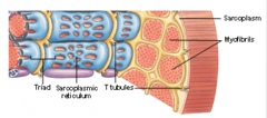
What is the resting membrane potential of skeletal muscle?
An electrical difference across the sarcolemma in which the inside is more negative than the outside. A sudden change in membrane potential can initiate contraction.
Why are skeletal muscle fibers multinucleated?
Multiple embryonic myoblasts fuse during development to form one large muscle fiber containing many nuclei just beneath the sarcolemma.

What are myosatellite cells?
Stem cells derived from myoblasts that remain in adult skeletal muscle and help repair or regenerate damaged muscle fibers.

What are glycosomes and myoglobin?
Glycosomes store glycogen that provides glucose for ATP production. Myoglobin is a red protein that stores oxygen inside muscle fibers.
Found in the sarcoplasm
What two intracellular tubular systems regulate skeletal muscle contraction?
T tubules and the sarcoplasmic reticulum (SR).
What are T tubules?
Deep inward extensions of the sarcolemma that carry action potentials into the interior of muscle fibers to stimulate contraction.
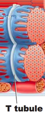
What is the sarcoplasmic reticulum (SR)?
A specialized membrane network surrounding myofibrils that stores and releases calcium ions to regulate muscle contraction.

What are terminal cisternae and a triad?
Terminal cisternae are enlarged calcium-storage chambers of the SR. A triad consists of one T tubule positioned between two terminal cisternae.
How does the sarcoplasmic reticulum control contraction and relaxation?
It releases calcium to initiate contraction and uses calcium pumps to return calcium to storage during relaxation.
What are myofibrils?
Long cylindrical structures inside muscle fibers composed of repeating sarcomeres. Their shortening produces skeletal muscle contraction.

What are myofilaments?
Protein filaments that make up myofibrils. Thin filaments contain actin, while thick filaments contain myosin.

What are the three major categories of myofilament proteins?
Contractile proteins (actin and myosin), regulatory proteins (troponin and tropomyosin), and structural/accessory proteins (such as titin and nebulin).
What is the structure of a myosin molecule?
Each myosin molecule contains two heavy and four light polypeptide chains, forming a rodlike tail, flexible hinge, and two globular heads.
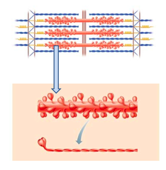
What is the function of myosin heads?
Myosin heads attach to actin, form cross-bridges, and pivot to generate force that pulls thin filaments during contraction.

What are G-actin and F-actin?
G-actin consists of globular subunits containing myosin-binding sites. Many G-actin subunits join to form long F-actin filaments (in thin filamenst)
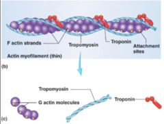
What is tropomyosin?
A regulatory protein that covers the myosin-binding sites on actin in relaxed muscle, preventing cross-bridge formation on thin filaments
rod like protein made up of 2 twisted chains

What is troponin and what are its three subunits?
Troponin is a regulatory protein complex in thin filaments. TnI binds actin and inhibits interaction, TnT binds tropomyosin, and TnC binds calcium.

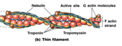
What is nebulin?
A structural protein that runs along thin filaments, holds the F-actin strand together, and may act as a ruler to determine thin filament length.
What is dystrophin?
A structural protein that links thin filaments to proteins of the sarcolemma, helping stabilize muscle fibers and prevent damage. Its absence causes Duchenne muscular dystrophy.

What is titin (connectin)?
An elastic structural protein extending from thick filaments toward Z discs. It maintains thin and thick filament alignment, restores resting sarcomere length, and resists overstretching.
What is desmin?
An intermediate filament protein that connects myofibrils at Z discs and links them to the sarcolemma, helping transmit forces produced by myofibrils and maintain muscle fiber strength.
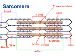
What are striations?
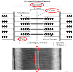
Alternating dark A bands and light I bands caused by the organized arrangement of thick and thin filaments in skeletal muscle.

What is a sarcomere?
The smallest functional contractile unit of skeletal muscle, extending from one Z disc to the next Z disc.
Interactions between the thick and thin filaments within sarcomeres are responsible for muscle contraction.

What is the function of Z discs?
Z discs form the boundaries of each sarcomere and anchor thin filaments and titin, helping maintain structural organization.
What is the A band?
The dark central region of a sarcomere containing the entire length of thick myosin filaments, including regions where thick and thin filaments overlap.
does NOT change in length during contractions

What is the I band?
The lighter region of a sarcomere containing only thin actin filaments. It is bisected by a Z disc.
DISAPPEARS at maximal muscle shortening

What is the H zone?
The central portion of the A band containing only thick myosin filaments, without overlapping thin actin filaments.
DISAPPEARS at maximal muscle shortening

How do the A band, I band, and H zone differ?
A band contains the full length of thick filaments; I band contains only thin filaments; H zone contains only thick filaments in the center of the A band.
Why are sarcomeres essential for skeletal muscle contraction?
They organize actin and myosin into repeating units, allowing coordinated filament sliding that shortens myofibrils and produces muscle tension.
What is the M line?
The M line is located in the center of the H zone and contains proteins such as myomesin that help stabilize and align thick myosin filaments.
What is the sliding filament theory?
The theory that muscle contraction occurs when thin actin filaments slide past thick myosin filaments, increasing overlap and shortening sarcomeres.
Do actin and myosin filaments shorten during contraction?
No. The filaments maintain their original lengths but slide past one another, shortening the sarcomere (Z lines closer together)
What is the role of myosin heads in the sliding filament theory?
Myosin heads attach to actin, form cross-bridges, and pivot to pull thin filaments toward the center of the sarcomere.
What happens to the A band, I band, and H zone during contraction?
The A band stays the same length, while the I band and H zone become shorter as actin-myosin overlap increases.
What happens to the Z discs and zone of overlap during contraction?
Z discs move closer together, and the overlap between actin and myosin increases.
Why does sarcomere shortening produce whole-muscle contraction?
Thousands of sarcomeres shorten together, causing myofibrils, muscle fibers, and ultimately the entire muscle to shorten.
What are the major events required for skeletal muscle contraction?
1. The fiber must be activated, that is, stimulated by a nerve ending so that a change in membrane potential occurs.
▪ 2. Next, it must generate an electrical current, called an action potential, in its sarcolemma.
▪ 3. The action potential is automatically propagated along the sarcolemma.
▪ 4. Then, intracellular calcium ion levels must rise briefly, providing the final trigger for contraction.
What are somatic motor neurons?
Nerve cells that stimulate skeletal muscle fibers to contract. Their axons branch to form neuromuscular junctions.
What is the synaptic cleft?
The narrow gap separating a motor neuron's synaptic terminal from the motor end plate of a skeletal muscle fiber.
What are synaptic vesicles?
Small membrane-bound sacs in the motor neuron's axon terminal that store acetylcholine until it is released.
What is acetylcholine (ACh)?
The neurotransmitter released by somatic motor neurons that binds receptors on the motor end plate and initiates muscle fiber excitation. (controls skeletal muscle contraction)
What causes acetylcholine release at the neuromuscular junction?
An action potential reaching the axon terminal triggers calcium entry into the terminal, causing synaptic vesicles to release acetylcholine.
What happens when acetylcholine binds to receptors on the motor end plate?
Chemically gated ion channels open, allowing mainly sodium to enter the muscle fiber and causing membrane depolarization.
What is depolarization of the sarcolemma?
A change in membrane potential that makes the inside of the muscle fiber less negative, potentially triggering an action potential.
What is the correct sequence of events at the neuromuscular junction?
Motor neuron action potential arrives → ACh is released → ACh binds receptors → sodium enters → motor end plate depolarizes → muscle action potential develops.
How does a muscle action potential reach the interior of a muscle fiber?
It travels along the sarcolemma and enters the T tubules, carrying the electrical signal deep into the fiber.
What is excitation-contraction coupling?
The process connecting an action potential in the sarcolemma to calcium release from the sarcoplasmic reticulum and the beginning of muscle contraction.

Where does excitation-contraction coupling occur in skeletal muscle?
At triads, where a T tubule lies between two terminal cisternae of the sarcoplasmic reticulum.
What happens when an action potential reaches the T tubules?
Voltage-sensitive proteins trigger calcium release channels in the SR, allowing calcium to enter the sarcoplasm.
Why is calcium necessary for skeletal muscle contraction?
Calcium binds troponin C, causing tropomyosin to move away from myosin-binding sites on actin so cross-bridges can form.
What happens when calcium binds to troponin C?
Troponin changes shape, shifting tropomyosin away from actin's active sites and allowing myosin heads to attach.
What prevents cross-bridge formation in relaxed skeletal muscle?
Tropomyosin blocks myosin-binding sites on actin when calcium levels are low.
How does calcium concentration change during excitation-contraction coupling?
Calcium concentration in the sarcoplasm rises rapidly when the SR releases stored calcium, allowing contraction to begin.
What is the sequence of excitation-contraction coupling?
Action potential travels through T tubules → SR releases calcium → calcium binds troponin → tropomyosin shifts → actin binding sites are exposed → cross-bridges form.
What are cross-bridges?
Temporary connections between myosin heads and actin filaments that allow myosin to generate tension and pull thin filaments.
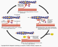
What is cross-bridge cycling?
The repeated process of myosin binding to actin, performing a power stroke, detaching, and reenergizing to generate muscle tension.
What is the function of ATPase in myosin heads?
Myosin ATPase breaks down ATP into ADP and phosphate, providing energy to position the myosin head for contraction.
What happens when ATP is hydrolyzed by myosin?
Energy is released, and the myosin head moves into an energized or cocked position with ADP and phosphate attached.
What does it mean when a myosin head is cocked?
The myosin head is energized and positioned to bind actin and generate a power stroke.
What is cross-bridge formation?
The attachment of an energized myosin head to an exposed binding site on an actin filament.
What is the power stroke?
The pivoting movement of an attached myosin head that pulls the thin actin filament toward the center of the sarcomere.
What happens to ADP and phosphate during the power stroke?
Phosphate release helps trigger the power stroke, and ADP is released as the myosin head completes its movement.
What causes a myosin head to detach from actin?
A new ATP molecule binds to the myosin head, causing it to release actin.
How does a detached myosin head become ready for another cycle?
ATP is hydrolyzed into ADP and phosphate, recocking and reenergizing the myosin head.
What two conditions are required for continued cross-bridge cycling?
Sufficient ATP must be available, and calcium must remain elevated to keep myosin-binding sites exposed.
What is the correct sequence of the cross-bridge cycle?
ATP hydrolysis energizes myosin → myosin binds actin → power stroke occurs → ATP binds and causes detachment → ATP hydrolysis recocks myosin.
Why is ATP necessary for both contraction and relaxation?
ATP powers myosin cycling, allows myosin to detach from actin, and supplies energy for calcium pumps that restore calcium to the SR.
What initiates skeletal muscle relaxation?
Nervous stimulation stops, acetylcholine is broken down, calcium release channels close, and calcium is pumped back into the SR.
What is acetylcholinesterase (AChE)?
An enzyme that breaks down acetylcholine in the neuromuscular junction, preventing continued stimulation of the muscle fiber.
What happens to calcium during relaxation?
ATP-dependent calcium pumps return calcium to the sarcoplasmic reticulum, decreasing calcium concentration in the sarcoplasm.
What happens to troponin and tropomyosin during relaxation?
Calcium dissociates from troponin, allowing tropomyosin to cover myosin-binding sites on actin and stop cross-bridge formation.
What is calsequestrin?
A calcium-binding protein inside the sarcoplasmic reticulum that allows large amounts of calcium to be stored.
What determines how much tension a muscle fiber produces?
The number of myosin heads performing power strokes, which depends partly on the overlap between actin and myosin filaments.
What is the length-tension relationship?
The relationship between the resting length of a sarcomere and the amount of active tension it can produce during contraction.
What is optimal sarcomere length?
The resting sarcomere length that provides sufficient actin-myosin overlap and room for filament sliding and frequency of stimulation, allowing maximum active tension.
What happens when a sarcomere is stretched too far?
Actin-myosin overlap decreases, fewer cross-bridges can form, and active tension decreases.
Why does excessive sarcomere shortening reduce tension?
There is less room for additional filament sliding, reducing the sarcomere's ability to generate effective tension.
What is a myogram?
A graphical recording of muscle contraction showing changes in muscle tension over time, usually measured using an isolated muscle attached to a strain gauge.
What is a muscle twitch and what are its three phases?
A single contraction-relaxation response to one stimulus. Its phases are the latent period, contraction period, and relaxation period.
What happens during the latent period of a muscle twitch?
A brief delay of approximately 1-2 milliseconds between stimulation and measurable tension while excitation-contraction coupling occurs.
is the time required for calcium to diffuse out of the SR

What happens during the contraction period of a muscle twitch?
Calcium binds troponin, cross-bridges form, and muscle tension increases to its peak.
What happens during the relaxation period of a muscle twitch?
Calcium returns to the SR, tropomyosin blocks actin, and muscle tension decreases toward resting levels. The slides describe this phase as approximately 25 milliseconds.