ana/phy exam 2
 Call Kai
 Learn
Learn Practice Test
 Spaced Repetition
 Match
Match Flashcards
 Knowt Play
1/102

There's no tags or description
Looks like no tags are added yet.
Name | Mastery | Learn | Test | Matching | Spaced | Call with Kai | Chat |
|---|
No analytics yet
Send a link to your students to track their progress
103 Terms
epiphyseal plate
thin layer of hyaline cartilage that allows for growth of a bone
compact bone
osteons
spongy bone
trabeculae
cancaliculi
small passageways in the bone
blood cell formation in…
red bone marrow - red, white, and platelets
hydroxyapatite
most salt is crystals of this
ossification
forming new bones w/ osteoblasts
bones start as what in the womb
cartilage
development of endochondral long bone
begins as hyaline cartilage model
primary ossification center: area in center of diaphysis where bone tissue first replaces cartilage
replacement with bone tissue proceeds towards ends of bone
osteoblasts from periosteum deposit compact bone around its primary center
secondary ossification centers: area of epiphyses where spongy bone forms later in development
epiphyseal plate: band of cartilage that persists between ossification centers
bones stop growing at what age
25
bone resoption
removal of bone by osteoclasts
bone deposition
formation of bone by osteoblasts
vit. D
calcium absorption; deficiency causes rickets
vit. A
osteoblast and osteoclast activity; deficiency retards bone develpment
vit. C
collagen synthesis; deficiency results in slender, fragile bones
thyroid hormone
causes replacement of cartilage with bone in epiphyseal plate, osteoblast activity
parathyroid hormone
stimulates osteoclasts, bone breakdown
sex hormones
promote bone formation; stimulate ossification of epiphyseal plates
simple fracture
has not come thru skin
compound
bone has come out of skin
steps in fracture repair
hematoma: blood clot forms
cartilaginous callus: fibrocartilage is produced, osteoblasts invade and produce spongy bone, phagocytes remove debris
bony callus: fibrocartilage is replaced by bone, forming the bony callus
remodeling: bone is restored close to original shape as osteoclasts remove excess bone
fontanels in baby
30
vertebrae sections

pubic symphesis

joint is also called
articulation
bursa
in between ligaments filled w/ synovial fluid
extra fibrous joints on a baby’s head
cranial sutures
What are the different ridges and nooks on bones for?
different muscles to be attached
Axial skeleton
skull and main area of body
appendicular skeleton
limbs
long bones + examples
a bone that is longer than it is wide; humerus and femur
short bones + examples
= in length and width (cube-shaped); wrist bones
flat bones + examples
plate-like w/ broad surfaces; in the skull
irregular bones + examples
don’t fit other categories; vertebrae
structure of long bone: epiphysis
expanded end
structure of long bone: diaphysis
bone shaft
structure of long bone: articular cartilage
covers epiphysis (protects from bones rubbing)
structure of long bone: periosteum
sheet of protective covering
structure of long bone: medullary cavity
hollow chamber in shaft and contains marrow
structure of long bone: endosteum
lines inner surfaces
structural unit of compact bone +flexible or solid?
osteons+ strong and solid
spongy bone structural unit and flexible or solid?
somewhat flexible
what are osteocytes?
mature bone cells
what do osteocytes start as?
osteoblasts
canaliculi
chambers connecting osteocytes
where are mature bone cells housed?
lacunae (little lake)
what is extracellular matrix made of?
collagen fibers and inorganic salts
Major functions of bones (6)
shape to body
support structures
protect structures
aid body movements
produce blood
store inorganic salts
blood cells formation called what + occurs where
hematopoiesis + in red bone marrow
bone matrix is 70% what
inorganic mineral salts
loss of bone mineralization
osteoporosis
parathyroid hormone
regulates calcium level in blood
what activates osteoblasts?
calcitonin
process of enochondral ossification
begin as hyaline cartilage models
chondrocytes enlarge and lacunae grow
matrix breaks down, chondrocytes die
function of joints (5)
functional connections between bones
bind parts of skeletal system together
make bone growth possible
permit parts of the skeleton to change shape during birth
enable body to move in response to skeletal muscle contractions
fibrous
held together by dense connective tissue containing many collagen fibers, found in bones in close contact
cartilaginous
held together by fibro/cartilage
synovial
have a complex structure
synarthrotic
immovable
amphiarthrotic
slightly moveable
diarthrotic
freely moveable
syndesmosis (type of fibrous)
bound by sheet of dense connective tissue or bundle, amphiarthrotic, between tibia and fibula
suture (type of fibrous)
thin layer of connective tissue, connects bones, synarthrotic, between flat bones of skull
gomphosis (type of fibrous)
cone-shaped bony process in a socket, synarthrotic, root of tooth
synchondrosis (type of cartilaginous)
bands of hyaline cartilage unite bones, synarthrotic, between manubrium and the first rib
symphysis (type of cartilaginous)
pad of fibrocartilage between bones, articular surfaces covered by hyaline cartilage, amphiarthrotic, pubic symphysis
s.j. articular cartilage
ends of bone, cushion and glide
s.j. joint capsule
tough, covers joint, reinforced w/ ligaments
s.j. ligaments
connect bone to bone
s.j. synovial membrane
inside the joint capsule, secretes synovial fluid “lubricant”
s.j. synovial cavity
fluid filled space between bones
s.j. meniscus (in some joints)
cartilage pads, shock absorbers; found in knees
s.j. bursa (in some joints)
help tendons glide over joints
ball and socket joint
round head in cup shaped cavity, widest range of motion; hip and shoulder
condylar joint
oval condyle fits into elliptical cavity, back+forth + sidetoside movements; between metacarpals and phalanges
plane joint
flat/slightly curved, wrist + ankle
hinge joint
convex surface fits into concave surface of other bone; elbow
pivot joint
cylindrical surface rotates within ring of other bone, rotation only; neck and forearm
saddle joint
both bones have concave and convex surfaces; meta/carpal of thumb
lateral flexion
bending to the side
dorsiflexion
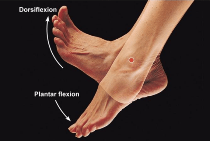
plantar flexion

circumduction

rotation

medial rotation

lateral rotation

supination

pronation

inversion

eversion

protraction
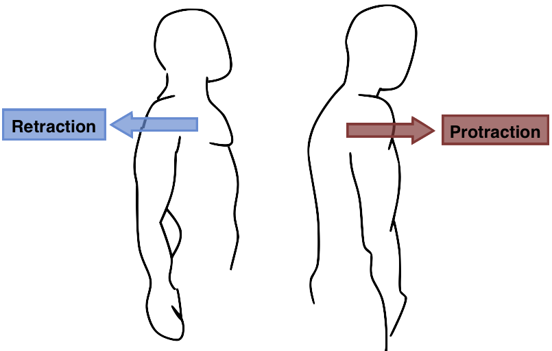
retraction

knee joint consists of what bones?
femur, tibia, patella
dislocation
displacement of articulating surfaces; shoulder, knee, fingers
sprain
tearing of connective tissue in joint, without bone dislocation
torn ligament
result from twist, overextension, lifting heavy, or fall
gout
type of inflammatory arthritis caused by uric acid crystalizing in joints
fascia
thin covering of connective tissue around a muscle
tendon
cord-like mass of connective tissue that connects muscle to bone
aponeurosis
sheet-like mass of connective tissue that connects a muscle to bone, skin, or another muscle