Plant Pathology Lab Practical
 Call Kai
 Learn
 Practice Test
 Spaced Repetition
 Match
 Flashcards
 Knowt Play
1/44

There's no tags or description
Looks like no tags are added yet.
45 Terms

Identify Symptoms (2) and Sign (1) of (indian hawthorn LEAF SPOT)
Symptoms: Necrosis and Chlorosis
Sign: Acervulus

Identify Symptom (2) and Sign (1) of (lirope lead w/ TIP NECROSIS)
Symptom: Necrosis and Chlorosis
Sign: Acervulus

Identify Symptom (3) and Sign (CUCUMBER MOSAIC VIRUS on american beautyberry)
Symptoms: Mosaic, wilt (leaf curl), malformations
Sign: None, it’s viral

Identify Symptom (1) and Sign (2)
Symptom: Necrosis/Rot
Sign: White and Black fungus, Septated hyphae (compound microscope)

Identify Symptom (1) and Sign
Symptom: Rot necrosis
Sign: N/A at this time

Identify Symptom (1) and Sign (2)
Symptom: Rot necrosis
Sign: White mycelium and green spores


Identify Symptom (1) and Sign (1)
Symptom: Rot necrosis
Sign: White mycelium (compound microscope image of hyphae with septations)


Identify fungal structure
ASCOMYCETE’s pseudothecium

Identify fungal structure
ASCOMYCETE’s acervulus

Identify fungal structure
ASCOMYCETE’s chasmothecium

Identify fungal structure
ASCOMYCETE’s pycnidium

Identify fungal structure
ASCOMYCETE’s apothecium

Identify fungal structure
ASCOMYCETE’s sporodochium

Identify fungal structure
ASCOMYCETE’s chasmothecium

Identify fungal structure
ASCOMYCETE’s external conidiophores

Identify fungal structure
ASCOMYCETE’s asci

Identify symptom and sign
Symptom: Gall
Sign: Black Mycelium (BASIDIOMYCETE)

Identify fungal structure
Teliospore (BASIDIOMYCETE)

Identify fungal structure
Cercosporoid sp. thin elongated conidia (ASCOMYCETE)

Identify symptom and sign
Symptom: Chlorosis
Sign: White and black mycelium

Identify fungal structure
Telia (BASIDIOMYCETE)

Identify fungal structure
Aecia (BASIDIOMYCETE)
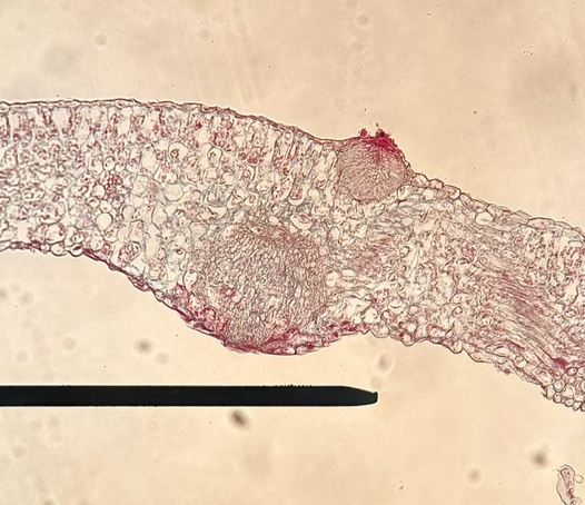
Identify fungal structure
Spermogonia (top)
Aecium (bottom)
(BASIDIOMYCETE)

Identify fungal structure
Uredinia (BASIDIOMYCETE)

Identify fungal structure
Urediniospore (BASIDIOMYCETE)

Identify symptom and sign
Symptom: N/A from this view
Sign: Uredinia (orange fungal pustules on bottom of leaf)

Identify fungal structure
Urediniospore (BASIDIOMYCETE)

Identify symptom and sign
Symptom: N/A from this view
Sign: aeciospores (fungal pustules on top of leaf) (BASIDIOMYCETE)

Identify fungal structure
Sporangium and sporangiophore (OOMYCETE)

Identify fungal structure
Oospores (OOMYCETE)

Identify fungal structure
Sporangium, sporangiophore, haustoria (OOMYCETE)

Identify fungal structure
Haustoria (OOMYCETE)

Identify fungal structure
Teliospores (BASIDIOSPORE)

Identify fungal structure
Sporodochia with conidium (ASCOMYCETE)

Anthracnose symptom and sign
condia in acervuli, often with setae and ooze (ASCOMYCETE)

Xanthomonas bacterial leaf spot symptom and sign
bright yellow colonies on plate (BACTERIA)

Alternaria leaf spot symptom and sign
unique conidia (ASCOMYCETE)

Pythium root rot symptom and sign
Coenocytic hyphae and round zoosporangia (OOMYCETE)

Root knot symptom and sign
Galls on roots; presence of nematode under microscope. Knots are swollen roots, knobs can be flicked off) (NEMATODE)
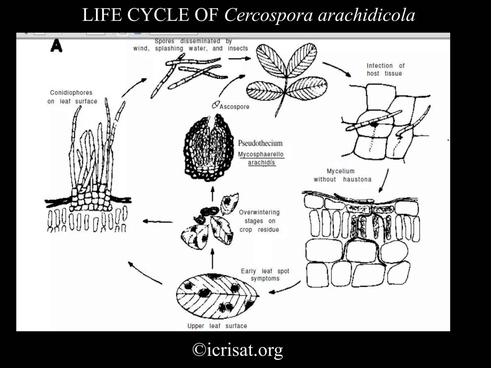
Early/Late Leaf Spot symptom and sign
conidia and sporodochia; pseudoperithecium (ASCOMYCETE)

Sclerotium rolfsii (white mold) symptom and sign
Septated hyphae and clamp connections; round sclerotia (BASIDIOMYCETE)

Puccinia symptom and sign
Presence of fungus indicated by orange-red spots; uredinia and urediniospores (BASIDIOMYCETE)

Lesion nematode symptom and sign
Lesions; migratory endoparasite (NEMATODE)

Neopestalotiopsis symptom and sign
Pestalotia: Opportunistic endophyte, unique spores with flagellum (ASCOMYCETE)

Tomato Spotted Wilt Virus (TSWV) symptom and sign
Mosaic chlorosis (VIRUS)