chp 3Anemia Evaluation: CBC Indices, Smear Findings & RPI
 Call Kai
 Learn
Learn Practice Test
 Spaced Repetition
 Match
Match Flashcards
 Knowt Play
1/26

There's no tags or description
Looks like no tags are added yet.
Name | Mastery | Learn | Test | Matching | Spaced | Call with Kai | Chat |
|---|
No analytics yet
Send a link to your students to track their progress
27 Terms
3 main steps to evaluate anemia & the labs/indices used for each:
Confirm anemia: Hemoglobin, Hematocrit, RBC count
Define cell pattern: MCV, MCHC, RDW, peripheral smear
(Mean Corpuscular Volume)
(Mean Corpuscular Hemoglobin Concentration)
(Red Cell Distribution Width)
Judge marrow response: Reticulocyte count, RPI (Reticulocyte Production Index)
3 decreased lab values indicating anemia AND the condition causing symptoms despite normal Hb:
Indicators: Decreased Hemoglobin, Hematocrit, or RBC count
Normal Hb but symptomatic: Abnormal oxygen affinity (hemoglobin doesn't release oxygen to tissues properly)
Standard units for Hb concentration AND impacts of
: 1. Age, 2. Altitude, 3. Hydration, 4. Heart issues
Units: g/dL (not mg/dL)
Age: Children typically have higher values
Altitude: Chronic hypoxia increases Hb levels
Hydration: Changes in plasma volume affect RBC/Hb concentration
Heart issues: Affects overall oxygen delivery to tissues
Acute vs. Chronic onset speed: Effect on compensation and danger level
Chronic (Slow): Compensatory mechanisms have time to develop; moderate anemia produces fewer symptoms.
Acute (Rapid): Sudden drop makes the exact same Hb level much more dangerous/deadly.
Compare the symptoms and key clinical signs of
Acute vs. Chronic anemia
like tolerance or suggest reduced volume
Chronic: Gradual symptoms (fatigue, headache, weakness, exertional dyspnea).
:Tolerance is reduced by baseline cardiopulmonary disease.
Acute: Early signs (tachycardia, dyspnea, hypotension).
:Orthostatic changes indicate reduced circulating volume.
Cardiovascular and Respiratory compensation for anemia:
Cardiovascular: Increased heart rate and cardiac output; blood flow is redistributed toward vital organs.
Respiratory: Ventilation increases with exertion; dyspnea appears when respiratory reserve is exceeded.
how do Compensation for hypoxia/chronic anemia:
1. Kidneys
2. Bone marrow
3. Hemoglobin
Kidneys: Renal erythropoietin rises
Bone Marrow: Output increases (if nutrients/stem cells are adequate)
Hemoglobin: Increased 2,3-DPG supports oxygen unloading
RBC Indices, RDW & Histograms (Practice Exam Integration)
What three things do RBC indices and RDW classify?
If an RBC volume histogram becomes much wider than normal, which specific index correlates directly with this change?
Classification: Size, hemoglobinization, and size variation.
Histogram Correlation (Exam Q24): RDW (Red Cell Distribution Width), because it is a mathematical expression of size variation (anisocytosis).
What 4 features are evaluated on a peripheral blood smear during anemia diagnosis?
What does the reticulocyte count measure, and what are the two common follow-up calculations?
Morphology
Inclusions
Polychromasia
Other lineages
Measures: Effective marrow output.
Follow-up: Corrected count and RPI (Reticulocyte Production Index).
On an automated analyzer, which 3 CBC values are directly MEASURED, and which 3 are CALCULATED?
Measured: Hemoglobin, RBC count, and MCV.
Calculated: Hematocrit, MCH, and MCHC.
If hematocrit is derived from the RBC count and cell volume, what is the estimated hematocrit if a patient's RBC count is 4.0 x 10^12/L and the MCV is 90 fL?
36%
Formula: (RBC x MCV) / 10
Calculation: (4.0 x 90) / 10 = 36
What are the three broad mechanisms that produce anemia?
What patient history finding immediately raises concern for gastrointestinal blood loss
Blood loss, inadequate production, and accelerated destruction (or abnormal synthesis).
Melena (or a history of ulcers).
Provide the formulas for calculating
ABD WHAT EACH MEANS
1. MCV,
2. MCH,
3. MCHC.
4.RDW
MCV: (Hct x 10) / RBC-Average cell volume
MCH: (Hgb x 10) / RBC.-Hemoglobin mass per cell
MCHC: (Hgb x 100) / Hct.- Hemoglobin concentration in packed RBCs
4.RDW: Variation in cell size
In impedance cell counting, what do the pulse number and pulse amplitude specifically measure?
Pulse number: Estimates the cell count.
Pulse amplitude: Reflects the cell volume (MCV).
Why can a normal MCV falsely conceal a mixed population of cells, and what index helps reveal this?
MCV is an Average cell volume
a mix of small (microcytic)
large (macrocytic) cells can average out to a normal volume.
An increased RDW reveals the mixed population by showing high size variation.
What smear findings distinguish megaloblastic macrocytosis from nonmegaloblastic macrocytosis?
Megaloblastic: Oval macrocytes and hypersegmented neutrophils.
Nonmegaloblastic: Round macrocytes.
What is the classic morphologic triad seen on a peripheral smear for megaloblastic anemia?
Oval macrocytes
Hypersegmented neutrophils
Howell-Jolly bodies

What do hypochromia and polychromasia indicate, and how do they appear on a smear?
LOOK AT IMAGE
Hypochromia: Reduced hemoglobinization; central pallor exceeds one-third of the cell.
Polychromasia: Young RBCs entering circulation (increased marrow output); cells appear blue/purple.
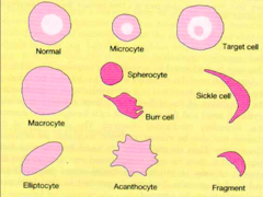
Define anisocytosis and poikilocytosis.
Anisocytosis: Variation in cell size (correlates with an increased RDW).
Poikilocytosis: Variation in cell shape.
Name the composition and the required stain for:
1. Howell-Jolly bodies,
2. Basophilic stippling.
Howell-Jolly bodies: DNA remnants; Wright stain.
Basophilic stippling: Aggregated RNA; Wright stain.
Name the composition and the required stain for
1. Pappenheimer bodies
2. Heinz bodies.
Pappenheimer bodies: Iron-containing granules; Wright stain (confirmed by Iron/Prussian blue stain).
Heinz bodies: Denatured hemoglobin; requires a Supravital stain.
Why is a raw reticulocyte percentage misleading in anemia, and what is the formula to correct it?
Why it's misleading: The reduced RBC total (denominator) makes the reticulocyte percentage look falsely high.
Formula: Observed Retic % x (Patient Hct / Reference Hct).
What is the formula for the Reticulocyte Production Index (RPI), and what RPI value indicates an inadequate marrow response?
Formula: Corrected retic % / Maturation correction factor.
Inadequate response: An RPI below 2.0.
What broad mechanisms are indicated by: 1. A low/inadequate RPI (
Low RPI: Nutrient deficiency, marrow failure, renal disease, ineffective erythropoiesis.
High RPI: Blood loss, hemolysis, or recovery after effective therapy.
A patient has MCV 68 fL, increased RDW, and RPI 0.8. Smear shows microcytosis, hypochromia, and anisocytosis. What is the leading hypothesis?
Iron deficiency (low RPI supports inadequate production; high RDW and microcytosis support iron restriction).
A patient has MCV 92 fL, RPI 4.1, increased indirect bilirubin, decreased haptoglobin, and spherocytes on smear. What is the primary diagnosis?
Hemolysis (likely immune or a membrane disorder due to the presence of spherocytes).
A patient has Hgb 7.2 g/dL, high RPI, low platelets, increased LDH, and numerous schistocytes. What urgent process does this pattern indicate?
Microangiopathic Hemolytic Anemia (MAHA) / fragmentation hemolysis.