ORAL ANATOMY - In Class Test Q's
 Call Kai
 Learn
Learn Practice Test
 Spaced Repetition
 Match
Match Flashcards
 Knowt Play
1/42

There's no tags or description
Looks like no tags are added yet.
Name | Mastery | Learn | Test | Matching | Spaced | Call with Kai | Chat |
|---|
No analytics yet
Send a link to your students to track their progress
43 Terms
List the bones of the skull which contain the paranasal sinuses
frontal, maxilla, sphenoid, ethmoid
b) List the functions of the paranasal sinuses. (2 marks)
equilibrium, lighten the skull, balance
c) Describe the changes in the paranasal sinuses with age. (2 marks)
Childhood: sinuses grow progressively, especially during puberty
adulthood: sinuses reach full size and function
older age: sinus walls may thin, and mucociliary function can decline, increasing susceptibility to infections
d) Individuals who suffer from hay fever can suffer from acute maxillary sinusitis.
Explain why this occurs, with reference to the position of the openings of the sinus
into the nasal cavity and the type of epithelium which lines the maxillary sinuses. (2
marks)
The maxillary sinus drains into the middle nasal meatus via the osmium (drainage whole). When inflammation or mucous blocks the opening this causes a blockage making drainage difficult.
Lined with pseudostratified ciliated columnar epithelium (respiratory epithelium) which relies on ciliary movement to clear mucous. If ciliary function is impaired due to inflammation, this leads to mucous build up and infection
e) What is meant by the term oroantral communication and how might it arise / occur
as part of dental treatment? (2 marks)
Oroantral communication refers to an opening or direct pathway from the oral cavity to the maxillary sinus. As the maxillary molar roots are in close proximity to the maxillary sinus floor, there may be a chance during extraction that the maxillary sinus is exposed.
1. Describe the distribution of minor salivary glands within the oral cavity.
soft palate
labial mucosa
buccal mucosa
ventral surface of the tongue
floor of the mouth
2. Choose one of the major salivary glands. Describe the anatomical location and
relationship of the gland and its associated duct to bones and muscles.
Parotid Salivary Gland:
anterior and inferior to the ear
superior boundary - zygomatic arch
inferior boundary - angle and lower border of the mandible
anterior boundary - master muscle
Parotid duct: (stenson’s duct) pierces the buccinator muscle, empties via parotid papilla positioned on buccal mucosa at maxillary molar region.
Submandibular gland:
located submandibular triangle of the neck
superficial to the mylohyoid muscle
Submandibular duct (Wharton’s duct) emerges from the deep part of the gland. travels anteriorly, passing over the mylohyoid muscle. opens into the oral cavity at the sublingual caruncle, near the base of the tongue
3. Explain why the side effect of some medications is a dry mouth
Xerostomia is a common side affect of some drugs due to their impact on salivary gland function. Anticholinergic drugs (antidepressants, antihistamines, antipsychotics) block acetylcholine is the primary neurotransmitter released by the parasympathetic nervous system to stimulate saliva.
radiation therapy and chemotherapy can damage salivary glands directly
decongestants and muscle relaxants may reduce saliva flow
This reduction can lead to: dysphasia (difficulty swallowing), increased risk of decay and oral infections, and altered taste sensation
a) Describe some of the common symptoms and signs of temporomandibular joint
dysfunction. (2 marks)
crepitus
tenderness
pain
trismus (limited jaw opening)
headaches/migraines
change in occlusion
b) Describe how you would go about the examination of the temporomandibular joint
and associated muscles for a patient as part of an extra-oral examination. (2 marks)
Find the head of the mandibular condyle with your fingers, prompt the patient to open jaw. depress mandible, elevate MD, lateral excursion, protrude, retract
feel for crepitus, deviation, tenderness
palpate the muscles of mastication (masseter, lateral/medial pterygoid, temporals) feel for tenderness or abnormalities.
c) Can you tell if a patient has TMJ dysfunction by examining their panoramic
radiograph? Explain your answer. (2 marks)
An OPG radiograph may support a TMJ diagnosis if it shows asymmetry, condylar erosion or flattening, however, it cannot confirm with the X-ray on its own. Clinical assessment and possibly advanced imaging (MRI) are needed for a definitive diagnosis
a) Draw a simple cross section diagram of the temporomandibular joint from a lateral
view. Label the diagram with the following features: (6 marks)
I. Articular eminence of the temporal bone
II. Articular disc
III. Articular fossa of the temporal bone
IV. Attachment of the muscle
V. Condyle of the mandible
VI. Upper and lower synovial cavities

1. Describe the anatomical location of the pterygomandibular fold and explain its
significance to dental treatment. 3 marks
Anatomical location: This fold is a mucosal ridge that stretches from the hamulus of the sphenoid bone to the posterior end of the mylohyoid line of the mandible. It overlies the pterygomandibular raphe, a tendinous band connecting the buccinator and superior pharyngeal constrictor muscles.
Serves as an anatomical landmark for the IAN Block injection (Inferior Alveolar Nerve)
2. Describe the anatomical location, appearance, colour and texture of the incisive
papilla. 1 mark
Found on the midline of the hard palate just posterior to the maxillary central incisors. Appearance is round/oval, raised, pink in health,
3. Describe the location, appearance of shape and contour, colour and texture of the
retromolar pad. 1 mark
Located posterior to the last mandibular molar. Triangular pad of tissue. Pink in health similar to adjacent tissue. Texture is soft and compressible
4. Draw a diagram of the dorsal surface of the tongue and label it with the following
for ½ mark each:
a) Anterior 2/3 and Posterior 1/3
b) Apex
c) Median sulcus
d) Distribution of the filiform papillae
e) Distribution of the fungiform papillae
f) Position and distribution of the follate papilla
g) Position and distribution of the circumvallate papilla
h) Terminal sulcus.
i) Lingual tonsils
j) Foramen caecum

This question relates to the sensory innervations of the oral cavity. Name the nerve
which supplies the following dental structures. (Don’t peek at the previous tables)
a) Pulps of mandibular teeth, periodontal ligaments and supporting bone.
b) Buccal gingiva and mucosa adjacent to the mandibular premolars.
c) Buccal gingival and mucosa adjacent to the mandibular molars.
d) Lingual gingival and mucosa on the mandible and floor of the mouth.
e) Skin and mucosa of the lower lip and chin adjacent to the mandibular anteriors.
f) Maxillary 2nd permanent molar and adjacent supporting structures.
g) Maxillary premolars and adjacent supporting structures.
h) Soft palate and uvula.
i) Palatal supporting tissue of the palate in the region posterior to the maxillary
canines and incisors.
j) Skin and mucosa of the upper lip.
a) Inferior alveolar nerve
b) Long buccal nerve
c) long buccal nerve
d) Lingual nerve
e) Mental nerve (branch of IAN)
f) Superior Posterior Alveolar Nerve
g) Superior Middle Alveolar Nerve
h) Lesser palatine
i) Greater palatine
j) Infraorbital nerve
a) Name the three periods of prenatal development and their timespan. Which of the
three periods of prenatal development is most critical and why? 2 marks
Germinal period (weeks 1-2): begins at conception and lasts until the zygote implants in the uterine wall
Embryonic period (weeks 3-8): Major organs and structures begin to form, this is most critical period because the foundations for all major body systems are established
Fetal period (week 9 to birth): Growth and maturation of tissues and organs, the foetus becomes more viable and functional over time
The embryonic period is most critical: This is when basic structures are occurring. Disruption during this phase can cause major structural defects (Teratogens)
b) Briefly describe what pharyngeal/branchial arches are in the human embryo.
Which pharnyngeal/branchial arch forms the muscles of facial expression? 2 marks
A series of mesodermal outpourings in the developing embryo that contribute to the formation of the head and neck structures. There are 6 arches but the fifth is rudimentary (very basic) and often not considered.
Muscles of facial expression are derived from the second pharangeal arch, which is associated with the facial nerve
(not in question but may be an alternative Ceinwen could ask): Muscles of mastication are derived form the first pharyngeal arch, which is associated the the trigeminal nerve (CN V)
c) Give two examples of teratogens and explain which period in gestation these
factors would have the most influence on facial or palatal malformation. 2 marks
Teratogens are substances or factors that can cause development malformations.
Alcohol: can lead to Fetal alcohol spectrum disorders. Includes facial abnormalities like a smooth philtrum, thin upper lip, and small palpebral fissures. Most harming during embryonic period (3-8 weeks) when facial structures are forming
Retinoic acid (vitamin A derivatives): found in acne medications, can cause cleft palate, craniofacial defects, ear malformations. most critical during weeks 4-7 when the palate and facial prominences are developing
a) At what age (on average), are the first signs of mineralization in the mandibular
first permanent molar? 1 mark
Around birth (late in utero approximately 30-36 weeks gestation)
b) At the time of birth there are no erupted teeth, but teeth will be in various stages of
development within the child’s jaws. How many teeth will be present in various
stages of development. 1 mark
Calcification of all primary teeth (20) will be in various stages, calcification of permanent first molar (4) be in initial stages. There will be 44 teeth present in various stages of development and calcification
c) Describe the stage of root formation when a permanent tooth erupts into the oral
cavity. 1 mark
When a permanent tooth erupts, the root is about two-thirds formed. Root development continues after eruption
d) Briefly describe the process of apexification and the approximate time this takes
after the tooth has erupted. 2 marks
Specification is the process by which the root apex (tip) completes formation and closes after the tooth erupts. It involves continued deposition of dentin and cementum
as the tooth matures, specification occurs gradually tapering this large opening down to a small hole. takes about 2-3 years after eruption.
e) On average, at what age is root formation and apexification of the permanent
central incisor complete. 1 mark
Around 9-10 years of age
1. Enamel can be described as permeable. Briefly explain the clinical significance of
the permeability of enamel and the microscopic features in enamel which allow this
permeability. (2 marks)
Enamel permeability allows fluoride ions and other remineralising agents to penetrate, helping prevent and reverse early caries. It also permits acid diffusion, contributing to subsurface demineralisation in caries development.
Microscopic features includeL Enamel rods which create microporosites (extremely tiny holes/void, less than 2 nanometers), enamel tufts, lamellae and rod sheaths which contribute to permeability pathways.
2. Describe the variations in thickness of enamel over the crown of the tooth. What is
the maximum thickness of enamel in millimetres? (2 marks)
Enamel is thickest at cusp tips and incise edges where functional stress is greatest. Becomes thinner towards the cervical margin of the crown.
Maximum thickness: Up to 2.5mm, typically on molar cusps
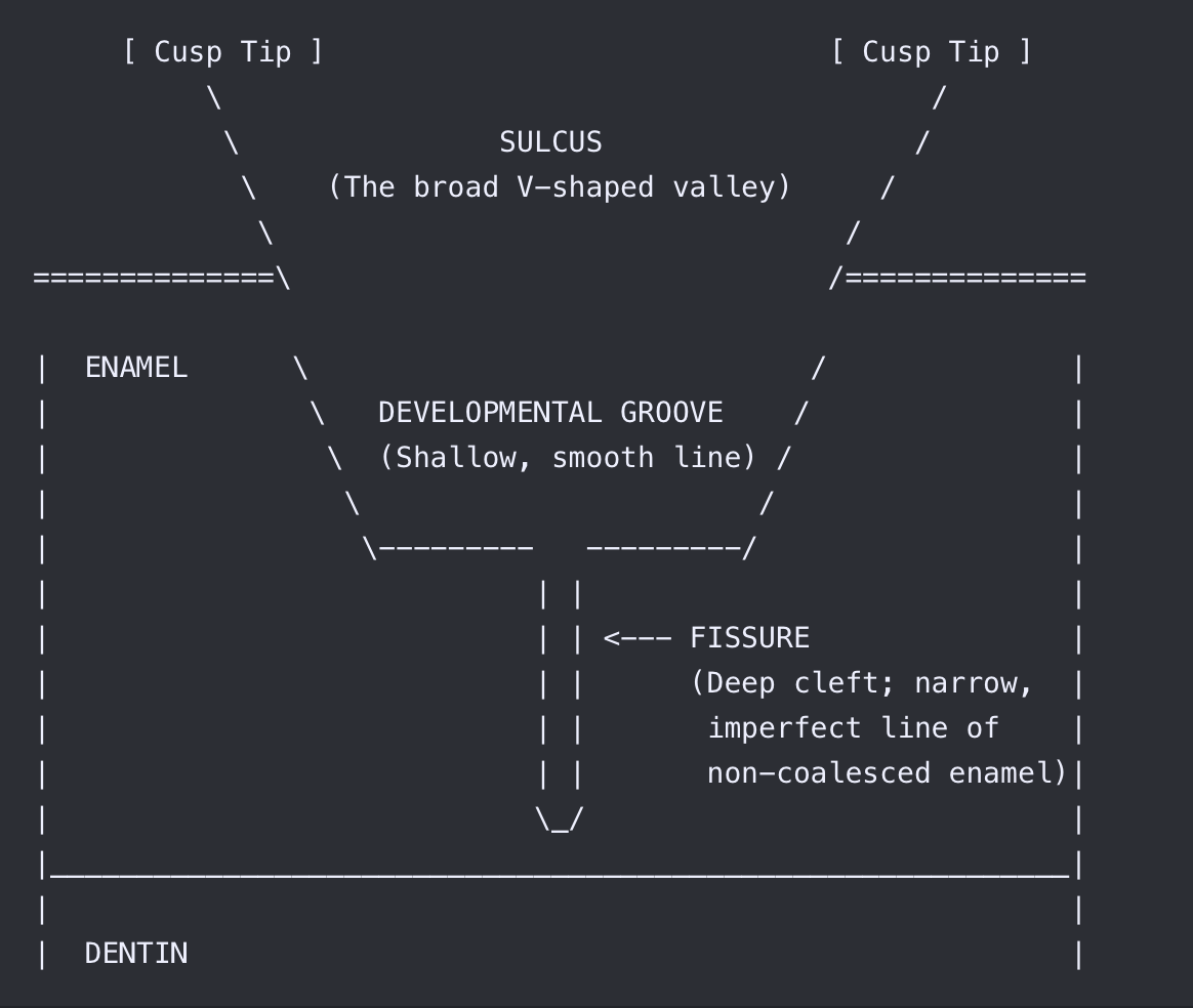
3. Describe the difference between developmental grooves and fissures by drawing
a labelling a diagram. Explain the difference between a developmental groove and
fissure and how they develop. Discuss the clinical significance of fissures. (3 marks)
Developmental grooves: shallow linear depressions marking the fusion of developmental lobes.
Fissures: Deep narrow clefts formed when lobes fail to fuse completely during development.
Grooves develop from normal lobe fusion, fissures not incomplete lobe fusion
4. Describe the appearance of perikymata and explain how these features develop.
Where on the surface of a tooth would these features be most visible? (3 marks)
Appearance: Fine, horizontal lines on enamel surface
Development: Result from the incremental deposition of enamel, corresponding to the Striae of Rezius.
Location: most visible on the facial surfaces of anterior teeth, especially near the cervical third

a) Explain the difference between the basal bone, alveolar process and alveolar
bone as discussed in the lectures. Draw a diagram to illustrate your answer (2
marks)
Basal bone: Osseous tissue that lies beneath the alveolar process, it provides the body of the mandible and maxilla.
Alveolar process: Extending from the basal bone, is the alveolar process which is a thickened ridge of bone which provides support which surrounds the sockets of the teeth.
The alveolar bone proper is the area which lines the tooth socket, where the PDL attaches the tooth to the bone
b) Compare the buccal and lingual cortical bone on the alveolar process of the
maxillary bone and the mandibular bone. How does this influence the delivery and
effectiveness of local anaesthesia? (4 marks)
In the maxilla bone, the buccal cortical bone is more porous and then compared to the lingual cortical bone which is more dense and thick. Because the buccal bone is thin, LA can diffuse easily through it. Making buccal infiltration most effective for anaesthetising the pulp.
When injecting on the palate, this is more to numb the surrounding tissue. (can be more painful/sensitive for the pt due to thickness)
When considering the mandible buccal and lingual cortical bone (posteriorly), it is very dense therefore infiltration technique is not effective to anaesthetise pulps (better of achieving IAN block). Moving anteriorly, infiltration can be used if needed as coritcal bone becomes thinner in the anterior/buccal/labial region.
c) Briefly describe how a tooth moves through bone when horizontal orthodontic
forces are placed on the tooth with reference to the periodontal ligament, cementum
and lamina dura. (4 marks)
When horizontal orthodontic forces are placed on the tooth, the tooth moves through bone by the remodelling of the PDL and surrounding alveolar bone.
On the pressure side, the PDL is compressed. This stimulates osteoclasts to resorb the adjacent bone including the bone forming the lamina dura, creating space for the tooth to move
On the tension side, the PDL is stretched. This stimulates osteoblasts to deposit new bone along the lamina dura, maintaining support as the tooth moves.
The cementum covering the root generally remains intact and provides attachment for the PDL fibres, which reorganise as the tooth changes position.
1. In some areas of the oral cavity the oral mucosa is described by the term
mucoperiosteum. Where in the oral cavity is this type of mucosa and why is it
described in this way? Your answer should describe the microscopic layers of oral mucosa. 2 marks
The mucoperiosteum can be found on the hard palate and attached gingiva. It is called this as it Is where mucosa is bound with the periosteum in the absence of the submucosal layer. It is comprised of a keratinised outer epithelium lining the loose connective tissue called lamina propria which is connected directly attached to the periosteum of the underlying bone beneath the mucosal membrane.
2. Drugs used to treat angina are often administered by placing the medication under the tongue on the floor of the mouth. Describe the type of oral mucosa and contents of the submucosa here and why this route of administration is effective. 3 marks
The epithelium is typical non-keratinised lining mucosa which is thin with underlying laminate propria and submucosa. The epithelium is composed of a stratum basal (basal layer of cuboidal cells), stratum spinous (second layer or oral flattened cells). The submucosa here is thick and loose, containing salivary glands, blood vessels, nerves and adipose tissue. This means that administering medication in this area under the tongue is effective as it can effectively and quickly reach the blood stream due to the thin epithelium and through the high vascularity of the underlying connective tissue.
3. Discuss the function of the junctional epithelium and how it cahnges during tooth
eruption and with disease. 3 marks
Function: The junctional epithelium attaches the gingiva to the tooth and forms a protective seal. It is permeable allowing crevicular fluid and immune cells such as neutrophils to pass into the sulcus to help defend against bacteria
During eruption: The JE initially forms from the reduced enamel epithelium, which fuses with the oral epithelium as the tooth erupts
During disease: Inflammation increases the flow of crevicular fluid and immune cells. With periodontitis the junctional epithelium can detach and migrate apically resulting in attachment loss and periodontal pocket formation.
4. Draw a diagram to demonstrate the gingival sulcus, sulcular epithelium and
junctional epithelium. 2 marks

1. How much saliva do we produce in a day and how does the secretion of saliva
vary during the day and night? 2 marks
500-1500ml per day on average. Stimulation is low during night/sleep in comparison to during the day
2. What factors might influence the volume of saliva we produce? 2 marks
Salivary gland secretion is regulated by the sympathetic and parasympathetic nervous system, with cell bodies located within specific ganglia of the head and neck
Afferent information from the sight, smell and thoughts of food as well as the action of mastication are sent to the brain and in turn parasympathetic stimulation occurs. Salivary secretion depends on taste and masticatory stimulation with chemoreceptors and mechanoreceptors.
Other factors include sleep, postural position, level of hydration, anticholinergic drugs (antidepressants, antihistamines, antipsychotics), disease present, xerostomia, radio-chemo therapy, age etc.
3. Explain the terms stimulated and unstimulated saliva and the difference in
composition and pH between stimulated and unstimulated saliva. 2 marks
Unstimulated saliva is the constant flow that is produced at rest throughout the day, while stimulated saliva is activated via masticatory, gustatory, olfactory stimulus, and/or physical palpation. The composition of unstimulated is 99% = water and 1% = 1% range of different ions, specialized glycoproteins and immunoglobulins with a pH of 6.5. When stimulated the pH rises to 7.61 and bicarbonate levels are more than 60 times higher than the low level in unstimulated saliva. The state of unstimulated saliva is more viscous, while stimulated saliva is more watery.
a) What is meant by the term odontogenic infection? What other terms are used
instead of odontogenic infection? (2 marks)
An odontogenic infection is an infection involving the teeth and its associated tissues, including the alveolar process, oral mucosa, paranasal sinuses, adjacent structures to the oral cavity. These infections can also be named as dental infections, dento-alveolar infections and dento-facial infections
b) The pus from an infected periapical region of a tooth will often drain via a tract
onto the outside of the alveolar process adjacent to the tooth root. What is the tract
called? What is the name for the opening of the tract onto the mucosa? What is a
common name for the opening of the track? Explain why the pus drains in this
direction. (2 marks)
c) A patient has a swollen face as result of an infected tooth. The swelling is located
beneath the eye and over the cheek. The skin overlying the swelling is slightly red
and feels warm to touch. What is the name for this diffuse swelling? With your
knowledge of the spread of infection, which teeth or groups of teeth are possibly
responsible? (2 marks)
d) A patient complains of toothache in a maxillary molar, but you suspect the cause
of their pain is not their tooth but infected sinuses. What are the typical signs andsymptoms of acute sinusitis and what symptoms might they complain of regarding
their teeth? (2 marks)