CHAPTER 25: MANAGEMENT OF FACIAL FRACTURES
 Call Kai
 Learn
Learn Practice Test
 Spaced Repetition
 Match
Match Flashcards
 Knowt Play
1/70

Earn XP
Description and Tags
ppt / book
Name | Mastery | Learn | Test | Matching | Spaced | Call with Kai | Chat |
|---|
No analytics yet
Send a link to your students to track their progress
71 Terms
evaluation of mandible
bimanual palpation by placing pressure over the mandible posterior and anterior to the fracture area
evaluated by extraorally palpating all areas of the inferior and lateral borders and the temporomandibular joint
evaluation of maxilla
to assess mobility:
patient’s head should be stabilized by using pressure over the forehead with one hand.
With the thumb and forefinger of the other hand, the maxilla is grasped; firm pressure should be used to elicit maxillary mobility

general signs and symptoms
bleeding from the mouth
limitation in mouth opening
bony discontinuity, lacerations
blood in external auditory canal
swelling, pain, crepitus, tenderness
ecchymosis, fractured, subluxed, luxated teeth
neurosensory deficits of the inferior alveolar nerve
changes in facial contour or mandibular arch form
periorbital ecchymosis
bruising around the eye.
especially when associated with subconjunctival hemorrhage.
may indicate:
orbital rim fracture
zygomatic complex fracture
battle sign
bruising behind the ear.
suggests a basilar skull fracture
floor-of-mouth ecchymosis
usually suggests an anterior mandibular fracture
anisocoria
refers to unequal pupil sizes
in a lethargic patient → may suggest:
intracranial bleeding
subdural hematoma
epidural hematoma
intraparenchymal bleeding
other intracranial injury
irregular/asymmetric pupil
especially if not round
may suggest globe (eyeball) perforation.
abnormal eye movements
may indicate:
central neurologic injury involving CN III, IV, or VI
mechanical restriction of eye muscles due to orbital fractures
radiographic examination
cephalometric
submentovertex
jug handle view
water’s view
reverse-Towne
cephalometric
lateral projection of the sagittal or median plane
obtained to evaluate the relationship of the oral and facial structures

submentovertex
shows base of the skull
jug handle view
shows zygomatic arches (a modification of the submentovertex view)

water’s view
shows maxillary sinus, frontal and ethmoidal sinuses

reverse-Towne
shows the posterior view of both condylar heads and necks (similar to postero-anterior view)

mandibular bone fractures
comprised 10–25% of all facial fractures
classified and named as to:
anatomic region
type of fracture or fracture pattern
terms associated with TMJ fractures
direction of muscle pull and angle of fracture
mandibular bone fractures as to anatomic region
condylar neck → most commonly fractured
angle of the MN → second most common
coronoid process → least common

condylar fracture
fracture extending from the sigmoid notch to the posterior border of the ramus
ideal anatomic reduction is not always necessary.
minimal to moderate displacement may still result in good function and occlusion if proper occlusion is maintained during healing
angle
more easily treated through an extraoral approach
fracture between distal aspect of second molar and the posterior attachment of the masseter muscle extending through the inferior border of the mandible
ramus
more easily treated through an extraoral approach.
fracture extending horizontally through the anterior-posterior borders of the ramus
or extending vertically from the sigmoid notch to the inferior border of the mandible distal to the second molar
coronoid process
fracture involving the coronoid process
symphysis
usually accessible through an intraoral approach
fracture involving the area between the lateral incisors extending vertically through the inferior border of the mandible subcon-axillary
parasymphysis
fracture between the mental foramen and the mesial aspect of the canine extending through the inferior border of the mandible
body
may use a combination approach: intraoral incision + small trocar/cannula through the skin.
fracture between the mental foramen and the distal aspect of the second molar extending through the inferior border of the mandible
alveolar
fracture confined to the tooth-bearing segment of bone
anteromedially
A fracture of the condylar neck (subcondylar fracture), will displace the head in what direction
deviation of the mandible on opening = towards the side of the fracture / ipsilateral
mandibular bone fractures as to type of fracture or fracture pattern
simple
compound
greenstick
comminuted
complex
simple fracture
a closed fracture
complete transection of the bone
minimal fragmentation at the fracture site
fracture does not communicate with the external environment

compound fracture
an open fracture
any jaw fracture in a tooth-bearing segment is considered this fracture
fracture communicates with external environment either thru skin or the PDL of a tooth
communication may occur thru:
mucosal tears
sinus lining
overlying skin laceration
gingival sulcus/periodontal ligament

greenstick fracture
bone remains somewhat flexible.
usually has minimal mobility when palpated
common in children due to high organic content
a bone bends and cracks instead of breaking completely

comminuted fracture
ex: gunshot wounds, penetrating objects
bone is broken into multiple segments/fragments
multiple segments of bone as a result of greater force

complex
fracture involving damage to adjacent structures
mandibular bone fractures as to direction of muscle pull and angle of fracture
favorable fracture
unfavorable fracture
favorable fracture
does not separate the fragments
often managed by closed reduction
the fracture line and muscle pull resist displacement.
when muscle contracts, the fractured segments come closer

unfavorable fracture
open reduction may be needed
fracture line and muscle pull displaces the fracture
make a flap through either skin or mucosa to see the bone
separate the fragments and increase the gap of the fracture
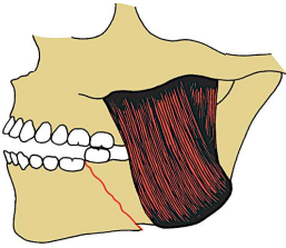
line of fracture
determines whether muscles will displace fractured segments from their original position
mandibular bone fractures as to terms associated with TMJ fractures
displaced
dislocated
extracapsular
intracapsular
displaced TMJ fracture
movement of condylar (proximal) segment relative to remainder of the mandible
although the condyle is malpositioned, it is still within the confines of the glenoid fossa and has not ruptured the capsule of the TMJ
dislocated TMJ fracture
the condylar (proximal) segment is no longer within the confines of the glenoid fossa
extracapsular TMJ fracture
fracture not involving capsule of the TMJ
intracapsular TMJ fracture
fracture occurring within the TMJ capsule
Le Fort I midface fractures
aka: Guerin fracture, transmaxillary fracture
usually caused by a horizontal force to the maxilla.
key features:
floating jaw
fracture passes through:
alveolar ridge
lateral nose
inferior wall of the maxillary sinus
separates the maxilla from:
pterygoid plates
nasal structures
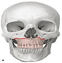
Le Fort II midface fractures
pyramidal fracture, with the teeth at the pyramid base, and nasofrontal suture at its apex, force is applied more superiorly.
key features:
CSF rhinorrhea
battle’s sign (ecchymosis in mastoid region)
fracture arch passes through:
posterior alveolar ridge
lateral walls of maxillary sinuses
inferior orbital rim and nasal bones
separates maxilla + attached nasal complex from:
orbital and zygomatic structures

Le Fort III midface fractures
aka: craniofacial disjunction fracture
horizontal force is applied at an even higher level
separation of midface from the cranial base → craniofacial disjunction
key features:
dish face or pan face
raccoon eyes (bruising of the eyelids)
CSF rhinorrhea and otorrhoea → highest rate of CSF leak
transverse fracture line passes through:
nasofrontal, maxillo-frontal, orbital walls
zygomatic arch or zygomaticofrontal sutures
separates NOE complex + zygomas + maxilla from:
the cranial base

zygomatic complex fracture
the most common type of midfacial fracture
fracture of nasal bones, orbital rim or floor areas
usually caused by a blow to the lateral cheek
key features:
bruising, swelling, blow-out fracture
results when:
an object such as fist or baseball strikes the lateral aspect of the cheek
isolated fracture of nasal bones, orbital rim or orbital floor areas
tripod fracture, fracture sites are:
zygomaticofrontal (ZF) suture
zygomaticomaxillary (ZM) buttress
zygomatic arch
main treatment goals, restore:
ocular function
nasal function
masticatory function
facial esthetics
usually treated with open reduction.

enophthalmos (sunken eye)
caused by blunt trauma to the eye leads to blow-out facture of the orbital floor
timing of treatment
as soon as possible
delayed treatment can cause:
increased risk of infection and malunion
more difficult or incomplete anatomic reduction
increased edema, especially during the first 2–3 days
treatment may be delayed when:
the patient has more serious injuries requiring immediate treatment.
severe neurologic injury or other conditions increase surgical/anesthetic risk.
a short delay is needed for severe edema to decrease.
surgical principles for tx of facial fractures
reduction of fracture → restoration of the bony segments to their proper anatomic location
open reduction (surgical)
closed reduction (non-surgical)
fixation of fracture → immobilization of bony segments at the fracture site
internal fixation (inside skin)
external fixation (outside skin)
restore the patient's preoperative occlusion.
prevent or eliminate infection at the fracture site.
bony repair should precede soft-tissue repair.
order of fracture repair:
traditionally: mandible → midface
layered closure:
close the wound from inside outward:
oral mucosa → muscle → subcutaneous tissue → skin
surgical principles for tx of mandibular fractures
reduce the fracture properly or place segments into proper relationship with each other
for fractures of tooth-bearing bones, teeth must be placed into the preinjury occlusal relationship
establishing a proper occlusal relationship by wiring teeth together:
intermaxillary fixation (IMF)
maxillomandibular fixation (MMF)
IMF or MMF techniques
ivy loops
continuous loop wiring
arch bar fixation → most common
arch bar fixation
most common
prefabricated arch bars are shaped and attached to teeth and secured by circumdental wiring teeth or acid-etch bonding

ivy loops
individual wire passed around specific teeth to create loops

continuous loop wiring
One wire loops around several teeth in a continuous chain

open reduction and internal fixation (ORIF)
best tx for mandibular fractures

surgical principles for tx of midface fractures
basis: bony structures within the face provide primary support or buttress in vertical and anteroposterior directions
these buttresses are reduced then fixated by means of:
direct wiring
bone plates
suspension wiring techniques
buttresses responsible for anteroposterior support
frontal bar
zygomatic arch & zygoma
maxillary and mandibular segments
mnemonic: fucking zebra zit my mug
buttresses responsible for vertical support
zygomatic
nasomaxillary
pterygomaxillary
mnemonic: pinched nipple zummy
complications of fracture during healing
malunion
non-union
delayed union
malunion
fracture that has healed in nonanatomic alignment
non-union
failure of a fracture to heal within the expected time frame
delayed union
slower than expected fracture healing, but eventual union occurs
naso-orbito-ethmoid (NOE) injuries
can cause:
spreading of nasal bones
displacement of medial canthal ligaments
increased intercanthal distance
produces:
traumatic telecanthus (normal 30–35mm)
widening of the medial intercanthal distance
treatment goals, restore:
normal nasolacrimal function
normal ocular function
properly reposition nasal bones
properly reposition medial canthal ligaments
achieve good facial esthetics
usually requires open reduction.
coronal flap is commonly used because it:
provides wide exposure of the upper face and nasoethmoidal complex.
closed reduction
treatment using MMF alone, without directly exposing the fracture surgically
if adequate reduction is achieved, MMF may provide sufficient stabilization during initial healing.
open reduction
direct surgical exposure and reduction of the fracture through an incision
when is it needed?
unfavorable fractures
continued displacement of fractured segments.
may be preferred when rigid fixation can reduce or eliminate the need for prolonged MMF.
wire osteosynthesis
traditional method of bone fixation.
uses direct intraosseous wiring.
usually requires MMF for about 4–6 weeks until adequate healing occurs
rigid internal fixation
uses:
bone plates
bone screws
provides rigid stabilization during healing.
usually allows little or no MMF
maxillomandibular fixation (MMF)
duration:
adults: maximum 2–3 weeks
children: 10–14 days
after MMF → aggressive functional rehabilitation.
prolonged MMF can cause:
fibrosis, bony ankylosis
severe limitation of mouth opening
fractures that do not necessarily affect occlusion:
isolated zygoma
zygomatic arch
NOE (naso-orbito-ethmoid) complex
suspension wiring
stabilizes fractured bones by attaching them to a more stable bone superiorly.
purpose:
prevent downward movement of the maxilla caused by the mandible during opening
limitations:
wires provide less rigidity.
they may not maintain proper facial contours.
they may not resist muscular forces throughout healing.
rigid plating has largely eliminated the need for suspension wiring
miniature / micro bone plates
usually made of titanium alloy, used with screws.
thickness: approximately 0.6–1.5 mm.
advantages:
more rigid stabilization
helps reconstruct facial pillars
useful for comminuted fractures
better restoration of normal bony contours
allows stabilization of multiple small bone fragments
titanium plates
biocompatible.
usually do not need removal unless they become:
palpable, infected
interfere with later reconstructive surgery
resorbable plates
made from polymers such as:
polyglycolic acid
polylactic acid
may be useful in:
children, skull trauma
situations where growth or repeated CT imaging is a concern
not routinely used because of:
mechanical limitations
need for tapping, cost
bone grafting
also improves stabilization
plates and screws also allow immediate bone grafting.
useful for replacing:
missing bone
severely comminuted bone segments