1 + 2 - Renal System
 Call Kai
 Learn
Learn Practice Test
 Spaced Repetition
 Match
Match Flashcards
 Knowt Play
1/16

There's no tags or description
Looks like no tags are added yet.
Name | Mastery | Learn | Test | Matching | Spaced | Call with Kai | Chat |
|---|
No analytics yet
Send a link to your students to track their progress
17 Terms
What are the 2 major compartments in which body fluids are housed in
Extracellular Fluid (ECF) - Volume : 15L , 20% of Body weight
Interstitial fluid (IF) - Volume : 12L - 80% of ECF
Plasma - Volume : 3L - 20% of ECF
Na+ and Cl- key ions in ECF
Intracellular Fluid (ICF) - Volume : 25L , 40% of body weight
K+ and phosphates in ICF
What are the gross features of the kidneys
Two functional regions:
Cortex - outmermost layer
Medulla - inner part
Renal pyramids house tubular structures that process filtrate and collect urine (nephrons)
Hillman - point of insertion of vessels, nerves and the uterus into which kidney excretions (urine) drain
What are the nephrons briefly
Structural and functional units of the kidney
Rich network of tubules and vasculature across cortex and medulla through which filtrate is formed
Two functional parts:
renal corpuscle - glomerus & capsule
Rena tubules - Proximal convoluted tubule (PCT), nephron loop, distal convoluted tubule (DCT)
Describe the path of blood flow through the kidney
RENAL ARTERY : oxygen rich blood, solutes + waste
Branches toward renal cortex → CORTICAL RADIATE ARTERY → AFFERENT ARTERIOLE
GLOMERULUS : fluid leaves capillary bed, moves into the tubules
EFFERENT ARTERIOLES brands into the capillary network, supplying the nephron tubules
Reclaimed water, nutrients returned to systemic circulation via the RENAL VEIN
What are the cortical and juxtamedullary nephrons
CORTICAL NEPHRON
short nephron loop
Glomerulus further from the cortex medulla junction
Efferent arteriole supplies peritubular capillaries
JUXTAMEDULLARY NEPHRON
long nephron loop
Glomerulus closer to the cortex-medulla junction
Efferent arteriole supplies vasa recta
How do the capillary beds reclaim filtrate
EFFERENT ARTERIOLE branches into the capillary networks supplying the nephron tubules.
PERITUBULAR CAPILLARIES:
low-pressure, porous capillaries adapted for absorption of water and solutes
Wrapped around cortical renal tubes
VASA RECTA:
long, thin-walled vessels, run parallel to long nephron loops of juxtamedullary nephrons
Contribute to formation of concentrated urine
What are the specialised cells involved in regulation of filtration rate and blood pressure
MACULA DENSA: proximal portion of the DCT lies adjacent to arterioles, modified epithelial cells:
Chemoreceptors monitor Na+ content of fluid in DCT
Sensory cilia detect fluid flow rate
secrete Paracrine agents (ATP, adenosine) when these increase.
GRANULAR CELLS: modified smooth muscle cells lining afferent arteriole
vasoconstriction of afferent arteriole in response to increase in arteriole BP, via Paracrine signalling from macula densa Paracrine.
Secretes renin - enzyme involved in systemic blood pressure regulation
EXTAGLOMERULAR MESANGIAL CELLS:
pass Paracrine signals between macular densa and granular cells
What are the 3 functional regions of the renal tubules & collection ducts
Proximal convoluted tubule -
majority of reabsorption ; some acid-base balance
Apical surfaced lined by microvili - increases surface area for water & solute reabsorption, and secretions
Nephron Loop -
descending (thin) & ascending libs alter filtrate osmolarity by reabsorbing water and NaCl
Distal convoluted tubule -
regulated reabsorption of ions, passive diffusion of water follows
Secretion of H+ contributes to acid- base balance
What are the 2 cell types in the collecting ducts
PRINCIPAL CELLS -
more numerous
Blunt, fewer microvili
Receptors for antidiuretic hormone (ADH) and aldosterone for water and Na+ reabsorption
INTERCALATED CELLS -
more microvili
Used for acid-base balance
What is the first major renal process
Glomerular Filtration -
Glomerular Filtration Rate (GFR): volume of plasma filtered (filtrate formed) per minute by both kidneys.
Of the plasma volume entering the afferent arteriole:
20% filtered by glomerulus and enters renal tubules
80% continues back out efferent arteriole into capillaries
>99% of filtrate reabsorbed.
Glomerular filtration is passive, nonselective process.
What pressures affect glomerular filtration
OUTWARD PRESSURES (promote filtrate formation)
Bowman’s Space Glomerular Hydrostatic Pressure (GBHP)***
the blood pressure inside the glomerular capillaries. Main driving force pushing water and solutes into bowman’s capsule.
Bowman’s Space Colloid Osmotic Pressure
normally no plasma proteins filter through the membrane, so this osmotic pull is zero.
INWARD PRESSURES (inhibit filtrate formation)
Blood Colloid Osmotic Pressure (BCOP)***
osmotic pressure created by non-filterable plasma proteins remaining in the capillary blood. Pulls water back into the circulatory system
Capsular Hydrostatic Pressure (CHP)***
back-pressure exerted by fluid already inside bowman’s capsule and the renal tubule, which resists incoming filtrate.
Increase blood volume at afferent arteriole → increase HP → increase filtrate
NFP = (GBHP-CHP) - BCOP
How is renal blood flow regulated
SYMPATHETIC innervation of afferent arteriole smooth muscle
at rest, minimal sns input → arterioles dilated, increased glomerular flow
Under stress, increase sns input → vasoconstriction + reduction in flow and GFR; blood shunted away from kidneys
MYOGENIC STRETCH of afferent arteriole smooth muscle
volume/pressure of fluid entering arterioles alters arteriole diameter
Vascular smooth muscle contracts when stretched, relaxes when not stretched
High blood pressure stretches arteriole walls triggering vasoconstriction → decrease flow of blood entering glomerulus, maintain appropriate GFR.
TUBULOGLOMERULAR FEEDBACK
macula densa cells monitor filtrate flow rate and NaCl in DCT
High GFR reduces time for solute reabsorption at PCT, osmolarity stays high by DCT
Vasoactive Paracrine (ATP, adenosine) secreted → vasoconstriction of afferent arterioles to reduce blood flow and GFR.
Describe what Tubular reabsorption is
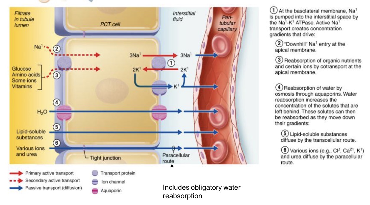
Substances moved by passive/active transport mechanisms along, sometimes against, their concentration gradient
From TUBULES → CAPILLARIES
Movement of water from tubules→nephron capillary beds influenced by osmolarity and concentration gradients
osmosis by aquaporins and obligatory water reabsorption with Na+
Sodium is actively transported against/with concentration gradients
glucose, amino acids, vitamins, ions, co-transported out of lumen alongside various Na+ transporters
Describe what secretion is
From CAPILLARIES/INTERSTITIAL SPACE → TUBULES
What is ADH and what does it do
Antidiuretic Hormone (AD) regulates renal water and solutes handling to maintain blood osmolarity, volume and pressure
ADH - synthesized in hypothalamus, released from posterior pituitary
increased osmolarity detected by hypothalamic osmoreceptors → secretion of ADH
Fall in blood volume/pressure → loss of baroreceptor stimulation → secretion of ADH
Effects:
ADH binds to receptors on principle cells of collecting ducts
Aquaporins inserted, increased water reabsorption
Reduced volume and more concentrated urine
What does aldosterone do
Aldosterone regulates renal water and solutes handling to maintain blood osmolarity, volume and pressure
Initiated by RENIN SECRETION FROM GRANULAR CELLS of afferent arterioles (3 ways this can occur) :
Decrease in renal perfusion detected by granular cells of afferent arterioles
Drop in blood volume/pressure → reduced GFR (slow movement of glomerulus) → more time for NaCl reabsorption at PCT, nephron loop, DCT → macula densa cells signal granular cells
Increased sympathetic activity directly stimulates granular cells
Effect:
renin release → RAAS cascade → Na+ reabsorption (+water follows)
Decrease Na+ and water loss in urine

How does the osmolarity gradient adjust urine composition
At nephron loop…
DESCENDING LIMB :
water moves out by osmosis into the increasingly hyperosmotic medulla; filtrate becomes more concentrated
ASCENDING LIMB :
NaCl pumped out into the medullary interstitium (limb impermeable to water); filtrate becomes more dilute
At the collection duct…
UREA
some moves from the collecting duct into the deep medullary institium, contributing to the osmotic gradient
