Lymph node and spleen histology
 Call Kai
 Learn
 Practice Test
 Spaced Repetition
 Match
 Flashcards
 Knowt Play
1/27

There's no tags or description
Looks like no tags are added yet.
28 Terms

Pictured is a lymph node, what are the black arrows pointing at
Capsule

Pictured is a lymph node, what are the white arrows pointing at
Trabecula
Recall which type of CT proper provides structural scaffolding to the lymphocytes within the lymph node and spleen
Reticular CT network

Outlined in white of this lymph node is
Nodular cortex

Outlined in pink of this lymph node is
Paracortex
Which lymphocyte predominates in the nodular cortex
B lymphocytes
Which lymphocyte predominates in the diffuse paracortex
T lymphocytes

Pictured is a nodular cortex, what is pink?
Mantle

This is a nodular cortex, what is in blue?
Germinal center
Germinal center
Has proliferating B lymphocytes that are clonally dividing in response to antigenic stimulation.

In this lymph tissue, what is yellow?
Medullary sinus

In this lymph tissue, what is purple?
Medullary cord
Medullary cords
More densely populated with lymphocytes, plasma cells, and macrophages
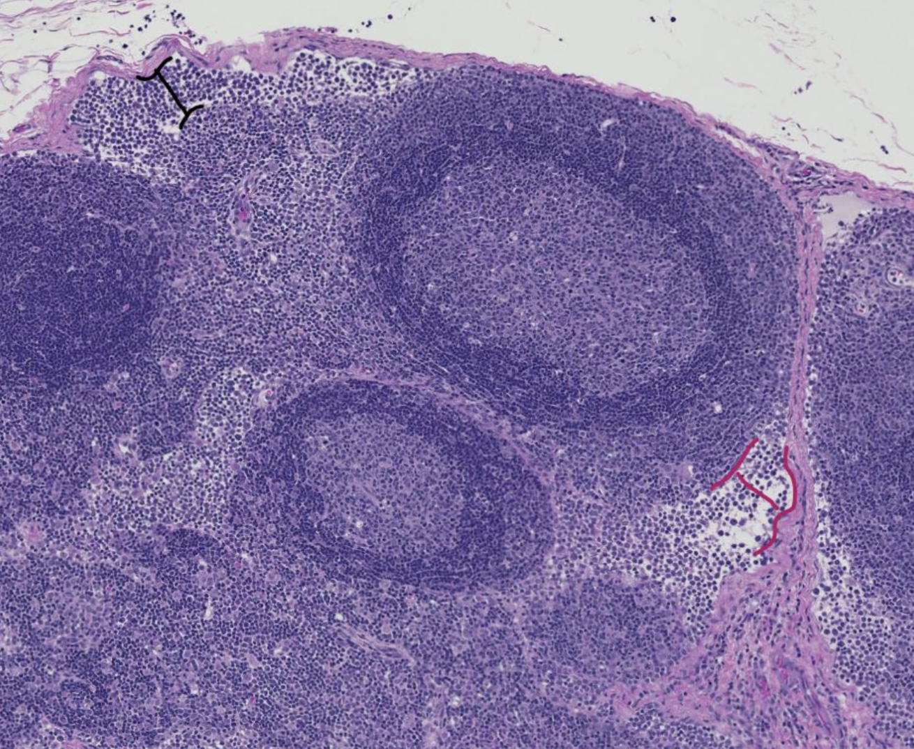
In this lymph tissue, what is bracketed in black?
Subcapsular sinus

In this lymph tissue, what is bracketed in magenta?
Cortical sinus
Basic flow of lymph through a lymph node
lymph enters node from afferent lymphatics
lymph flows through subcapsular sinus
lymph flows into cortical sinuses
lymph flows to medullary sinuses
lymph flows out of the node via the efferent lymphatics at the hilus
List two ways that lymphocytes are delivered to a lymph node.
Through the blood, via high endothelial venules (HEVs)
Through the lymph, via afferent lymphatic vessels

Pictured is spleen tissue, what is in green?
Trabecula with smooth muscle

Pictured is spleen tissue, what is in purple?
Capsule (spleen)
Do capsule or trabeculae contain smooth muscle in the spleen?
They both can, it varies with the species

Pictured is spleen, what is in blue?
Red pulp

Pictured is spleen, what is pink?
White pulp
T lymphocytes are predominant in this spleen tissue
periarterial lymphatic sheath (PALS) (diffuse lymphoid tissue)
B lymphocytes are predominant in this spleen tissue
splenic follicles (nodular lymphoid tissues)

This is red pulp, what is orange?
Splenic cord

This is red pulp, what is pink?
Splenic sinus
Determine which direction red blood cells (RBCs) are passing between these structures during the filtering process that removes senescent RBCs. (cords→sinuses or sinuses→cords)
Cords → sinuses
The spleen receives the blood that is delivered to the organ, then “subdivides the labor” between the two pulps. Which pulp functions like a lymph node and processes foreign antigens? Which pulp functions to “capture and recycle” old red blood cells?
White pulp
Red pulp