exam 4, not entirely
 Call Kai
 Learn
 Practice Test
 Spaced Repetition
 Match
 Flashcards
 Knowt Play
1/131

There's no tags or description
Looks like no tags are added yet.
132 Terms
What is acute pancreatitis?
inflammation of the pancreas caused by digestive enzymes being activated but not being released. dysfunction of the ACINAR cells. this leads to an inflammatory response that causes Increased permeability: causing hemorrhage, edema, and necrosis
What are the three functions of the pancreas?
ENDOCRINE (secrete insulin + glucagon to stabilize glucose balance), EXOCRINE (produce digestive enzymes which helps break down food), and DIGESTIVE (protease, lipase, amylase)
acute pancreatitis s/s
sudden onset of LUQ Abdominal and back pain, distention, guarding, N/V, fever, Anorexia, Ascites, Jaundice

Cullen's sign
ecchymosis in smbilical area, seen with pancreatitis

turner's s
flank grayish blue (turn around to see your flanks) pancreatitis

What labs can be drawn to help diagnose acute pancreatitis?
lipase, amylase, calcium ALT/AST, WBC, glucose, potassium, cholesterol + triglycerides
lipase in acute pancreatic
Breaks down fat.
amylase in acute pancreatitis
#1 indicator: breaks down carbs.
LOOK FOR ELEVATION 3 TIMES THE NORMAL LIMIT
calcium in acute pancreatitis
low with fat necrosis
ALT/AST in acute pancreatitis
elevated with gallstones
alkaline phosphate in acute pancreatitis
elevated with biliary disease
WBC in acute pancreatitis
increased due to inflammation and infection
glucose in acute pancreatitis
elevated with islet cell damage
potassium in acute pancreatitis
low with severe vomiting, high with acute renal failure
What scans can be done to diagnose acute pancreatitis?
CT scan + x-ray + ultrasound
why do we use a CT scan in acute pancreatitis?
can assess for severe cases of pancreatitis
-pseudocyst
-ascites
why do we use an X-ray(KUB) in acute pancreatitis?
can visualize calcifications. can indicate chronic pancreatitis.

why do we use a ultrasound in acute pancreatitis?
can detect gallstones
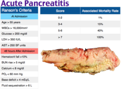
ranson criteria
Used to assess severity + mortality of pancreatitis
0-2=minimal mortality
3-5= 10-20% mortality
6+= 50% mortality
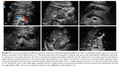
ERCP
Diagnoses pancreatitis: Endoscopy, looks at bile and pancreatic duct to see if it is emptying. If it is not, the catheter goes into the duct to determine blockage and place a stent if needed
causes of acute pancreatitis
#1= Biliary tract obstructed, #2= ETOH, Use of corticosteroids, thiazide diuretics, and sulfonamides, Trauma, hyperlipidemia, ERCP

acute pancreatitis treatment
ERCP,
IV hydration (crystalloids or colloids),
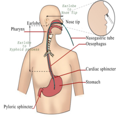
Nutrition (Usually NPO, NG tube + TPN if severe),
Pain management,
Antibiotics if severe,
Surgery(removal of gallbladder or pancreas),
analgesics (PCA),
decompression of intra-abd pressure (paracentesis)

complications of acute pancreatitis
Increased abdominal pressure
Respiratory distress or failure,
Sepsis,
(DIC),
Renal Failure,
Acute Necrotizing Pancreatitis
DKA (Diabetic keto acidosis)
hyperglycemia, no insulin going into the cell (cellular starvation), burns fat for energy, ketone production, metabolic acidosis, K+ exchange, kussmaul respirations. causes a osmotic shift, increased UO, K+ loss, dehydration

DKA s/s
D-ehydration
K-etones in urine/blood, Kussmauls and K+
A-cidosis, Acetone breath, Anorexia d/t nausea, visual distubances
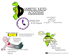
DKA treatment
insulin drip, fluid replacement(LR+ NS or plasmalyte), potassium, bicarb only to bring PH to 7.1 (metabolic acidosis)

HHS (Hyperglycemic Hyperosmolar State)
commonintype2becauseintype2wehavefew
-increase in glucose (hyperglycemia) + thick and dehydrated blood (osmotic shift) + increased urine output + potassium loss
-no keynotes
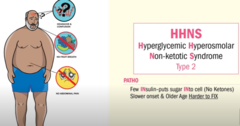
HHS treatment
"HSS"
-Hydration= FLUID REPLACEMENT (0.45% or 0.9% NS) + potassium
-Stabilize the sugar with INSULIN (continuous infusion)
-Stabilize the BP with PRESSERS

DKA + HSS nursing considerations
education on managing blood glucose, diet, exercise, medication, assess their access to care
-Hourly VS
-Monitor I&O's
-Monitor neuro

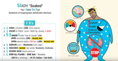
SIADH (syndrome of inappropriate antidiuretic hormone)
overproduction of the antidiuretic hormone ADH, leading to bloating, water retention, and electrolyte imbalance. VASOCONSTRICTION. action on the smooth muscles of the arterioles

what causes SIADH?
CNS disorders, head injury, anoxic encephalopathy infection, hemorrhage, surgery, stroke, cancer producing ADH, failure of normal regulatory feedback loops, too much ADH

How is SIADH diagnosed?
hyponatremia in the blood, elevated urine sodium
-may decrease serum BUN, Cr, Albumin due to hemodynamics
-hypertonic urine compared to plasma

treatment for mild to moderate SIADH
(Na level: 125-135)
fluid restriction, monitor serum sodium
treatment for severe SIADH
(Na level
causes of burns
thermal injury (flame, scald, and contact)
chemical, electrical, inhaltion injury

superficial partial-thickness burn (2nd degree)
damage to the epidermis and portion of the dermis
Moist pink, molted
Pain, blisters, blanches

how do superficial partial-thickness burns appear? (2nd degree)
reddened, blanches with pressure, minimal edema, possible blisters. Takes 7-10 days to heal

how long does it take for a superficial burn to heal?
normally heals on it's own within 3-7 days

Deep partial thickness burn (2nd degree)
epidermis + dermis + portion of deeper dermis
Blistered, moltted red base, weeping surface edema
GRaft
few days to demarcate
2-3wks

how does a deep partial thickness burn appear?
blistered, mottled, red base, weeping surface, edema, Can be grafted

how long does it take for a deep partial-thickness burn to heal?
Has the potential to heal on its own in 2-4 weeks

full thickness burn
all 3 layers of the skin a destroyed
thick, leathery, eschar, painless, will not blanch

how does a full thickness burn appear?
dry, white, leathery, or charred, edema, NO PAIN

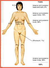
rule of 9
way to determine the extent of burns. 25-30% is considered a major burn.

pathophysiology of burns
burn->
inflammation ->
Histsmine release→
increase capillary permeability and vasoconstriction->
increase protein leakage.
Increased BP/BLOOD FLOW
Capillary leak
Edema
Burrns less than 25% total body surface area produces a local response. There is swelling, constriction, and hypovolemic.

Resuscitative phase of a burn
-From onset to completion of fluid resuscitation. 72 hours
nursing care of the Resuscitative phase
(NOTE:electrical burns should have an ECG)
Please Avoid Naughty Officers; Little Freaks Really Crave Attention, Honestly.
Prevent Injury to rescuer/ stop injury
ABC’s
NG
O2
large bore IV’s (iv meds only)
foley
Remove restrictive clothes
Cover Wound
Assessment
HTX
what is our goal during the Resuscitative phase of a burn?
Respiratory + prevent shock
systolic BP over 100
urine output 30-50 ml/hr
K+ & Na levels to near normal
Iv access
Hypothermia
myoglobinuria (in electrical)
when do we insert a NG tube in a burn patient?
if the burns are of 25%

what s/s do we expect burn patients to have?
“Dirty Hippos Hate Drinking Icy Lemonade; Hungry Tigers Misbehave.”
dehyration
hyperkalemia
hyponaterimia
decreased blood volume
(increased Hct)
low UO
hypotension
tachycarida
metabolic acidosis
parkland formula
2-4 ml x kg x %burn
Acute/intermediate phase of burn care
-From beginning of diuresis to near completion of wound closure, 48-72 hours
what is happening during the Acute/intermediate phase of burn
Fluid re-enters the vascular space
hemodilution(decreased Hct)
increased U/O
K+ and Na lowers with diuresis,
metabolic acidosis
Rehabilitation phase of burns
major wound closure to return of individuals optimal early as possible
-priority: wound healing, psychosocial support, self-image, lifestyle, restoring function
-pt may need reconstructive surgery to improve function and appearance
how should home care look during the rehabilitation phase?
Mental health
skin
wound care
exercise
nutrition
pain
thermoregulation
sexual issues
no tanning
wound care for burns
Cleaning (hydrotherapy)
topical agents
debridement
dressings, dressing changes,
skin grafting

debridement of burns
Natural, mechanical, chemical (Santyl), and surgical

burn grafts
Autograft- permanent w/ own skin

homo (allo)
temporary from living or recently deceased humans.

hetero graft
temporary from an animal

Biobrane graft
synthetic dressing

how would someone with acute pancreatitis describe their pain?
steady and dull
what causes acute pancreatitis
"GET SMASHED"
Gallstones
Ethanol (alcohol)
Trauma
Steroid use
Mumps
Autoimmune
S
Hyperlipedemia + Hypercalcemia
ERCP
Drugs (sulfonamides + thiazide diuretics)

what effect does acute pancreatitis have on the lungs?
-infection and increased abd pressure
-ARDS, arterial hypoxia, pleural effusion, pneumonia, atelectasis

treatment for ARDS and pleural effusion
intubation (ARDS), deep breathing, supplemental oxygen, antibiotics
other complications of acute pancreatitis
sepsis, septic or hypovolemic shock, DIC, Acute renal failure

acute pancreatitis and acute necrotizing pancreatitis
HIGH MORTALITY RATE
enzymes spill over into the pancreas infection, pseudocyst

DKA nursing considerations
- insulin lowers K+ so monitor and check (K+ has to be >3.3 to give insulin)
-when giving fluids check renal function
-LR has electrolytes so it's important to monitor electrolytes
HHS s/s
"HHHNS"
Higher fluid loss: extreme dehydration (tachycardia, ↓ Skin turgor, dry mucous membranes, flushed + dry skin)
Hypotension + shallow respirations
Head change: neuro changes (confusion or altered LOC)
No abdominal pain, No ketones, No acid-base imbalance No kussmaul
Slower onset + Stable potassium

nursing considerations for HHS
vitals signs q1h,
monitor I/O,
monitor neuro status
give tx: IV fluids, obtaining labs, monitor blood glucose
nursing considerations for SIADH
-monitor I&O's (fluid restriction)
-high sodium diet
-assess Nero and signs of HF

what does an increase in capillary permeability cause after the burn
causes fluid to leak out which leads to 3rd spacing which leads to SHOCK

Lund and Browder chart
Correlates body surface area with age-related proportions

burns less than 25%
produces a local response
burns more than 25%
MAJOR BURNS
-may produce a systemic response
Abdominal compartment syndrome
the abdomen has become a fixed compartment with increased pressure resulting in ischemia and organ dysfunction
-pressure greater than 20 could indicate dead gut syndrome

SIADH s/s in the CNS
-weakness
-lethargy
-confusion
-headaches
-seizures
SIADH s/s in the GI
-N/V
-anorexia
-decreased BoSo (bowel sounds)
SIADH s/s in the heart
edema + HTN

SIADH pulmonary s/s
increased RR, dyspnea, crackles, diminished or absent lung sounds, pulmonary edema, pulmonary effusion, pink + frothy sputum
how do we determine fluid replacement?
2-4 mL x kgs %TBSA
how long do nurse preform fluid and electrolyte replacement for?
administer 1/2 in the first 8 hours and the other half in 16 hours
paralytic illeus (from burns)
burn trauma causes decreased peristalsis
Curling's ulcer (from burns)
gastric bleeding caused by massive stress from burns
Abdominal compartment syndrome (from burns)
patient's with large TBSA burns
patient's with circumferential burns at the trunk

what happens if a full thickness burn does not heal?
grafting

what are some of the most problematic areas to get burned?
head + neck, hands + feet, perineum
what does the severity of burns depend on?
-age (children + elderly)
-depth of burn
-% of TBSA burned
-medical history (diabetes)
-location of burn
-inhalation injury
-presence of other injuries
will cholesterol and triglycerides be elevated or decreased in acute pancreatitis?
elevated
-indicates etiology
When is the rehabilitation phase started?
from major wound closure to return of the persons optimal level of physical and psychosocial adjustmetn
how much potassium do we add to the IV influsion during DKA
20-30 mEQ
what does the nurse expect to see on the monitor when the potassium is high?
Peak T waves + ST elevation
what does the nurse expect to see on the monitor when the potassium is low?
Flat T waves + ST depression + U wave
hemodynamics of burns
high afterload, HR
Decrased BP, CO, contractility
LOW PRELOAD
Ranson criteria ON ADMISSION for pancreatitis
each scored as one point
-Older than 55
-WBC >16,000
-blood glucose >200
-serum LDH >350
-AST > 250

Ranson Criteria developing during the first 48 hours for pancreatitis
-hct fall of more than 10%
-BUN level increase by more than 8%
-serum calcium less than 8
-PaO2 less than 60
-base deficit higher than 4
-estimated fluid sequestration higher than 6

how do we monitor perfusion in acute pancreatitis?
IAP (intra-abdominal pressure)
what kind of precautions do we put patients on who have SIADH?
seizure precautions + fall risk due to their low sodium levels
what are the causes of both DKA & HHS
"SSSS"
-Sepsis (infection)
-Steroids
-Surgeries
-Skiping insulin

s/s of both DKA & HHS
flushed + dry skin, dry mucus membranes, decreased skin tumor, hypotension, electrolyte abnormalities, increased BUN + CR