3.6 Static neuroimaging techniques

0.0(0)
Studied by 0 people
call kaiCall Kai
learnLearn
examPractice Test
spaced repetitionSpaced Repetition
heart puzzleMatch
flashcardsFlashcards
GameKnowt Play
Card Sorting

1/12

flashcard set

Earn XP

Description and Tags

Static neuroimaging techniques

Last updated 7:17 AM on 9/25/26
Name
Mastery
Learn
Test
Matching
Spaced
Call with Kai
Chat

No analytics yet

Send a link to your students to track their progress

13 Terms

1
New cards

Static neuroimaging techniques

  • X-RAYS

  • PNEUMOENCEPHALOGRAPHY

  • COMPUTED TOMOGRAPHY (CT SCAN)

  • MAGNETIC RESONANCE IMAGING (MRI)

  • MAGNETIC RESONANCE SPECTROSCOPY (MRS)

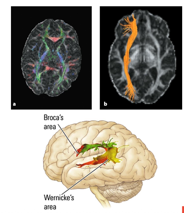
  • DIFFUSION TENSOR IMAGING (DTI)


2
New cards

X-RAYS

  • First method for imaging the brain

  • Produced static 2-D image of the brain

Involves → Conventional radiography


<ul><li><p><strong><mark data-color="purple" style="background-color: purple; color: inherit;">First method for imaging the brain</mark></strong></p></li><li><p><strong><mark data-color="purple" style="background-color: purple; color: inherit;">Produced static 2-D image of the brain</mark></strong></p></li></ul><p><strong>Involves → Conventional radiography</strong></p><p></p>
3
New cards

Conventional radiography

  • Passing X-rays through the skull onto an X-ray sensitive film

  • Differenttypes of tissue absorb the rays to different degrees, producing an image that shows the tissue location

  • Still used for examining the skull for fractures & looking for gross brain abnormalities


4
New cards

PNEUMOENCEPHALOGRAPHY

  • Small amount of cerebrospinal fluid (CSF) is removed from the subarachnoid space & replaced by air

→ X-rays are not absorbed by air

  • X-rays are taken as the air moves up the spinal cord & into ventricular system

  • Ventricles stand out in image due to air in them

  • Is painful & invasive

  • Not currently used much


<ul><li><p><strong><mark data-color="purple" style="background-color: purple; color: inherit;">Small amount </mark></strong>of cerebrospinal fluid (<strong><mark data-color="purple" style="background-color: purple; color: inherit;">CSF</mark></strong>) <strong><mark data-color="purple" style="background-color: purple; color: inherit;">is removed from the subarachnoid space &amp; replaced by air</mark></strong></p></li></ul><p class="p2">→ <strong><em>X-rays are not absorbed by air</em></strong></p><ul><li><p class="p1"><strong><mark data-color="purple" style="background-color: purple; color: inherit;">X-rays are taken as the air moves up the spinal cord &amp; into ventricular system</mark></strong></p></li><li><p class="p1"><strong><mark data-color="purple" style="background-color: purple; color: inherit;">Ventricles stand out in image due to air in them</mark></strong></p></li><li><p class="p1"><strong><mark data-color="purple" style="background-color: purple; color: inherit;">Is painful &amp; invasive</mark></strong></p></li><li><p class="p1"><strong><mark data-color="purple" style="background-color: purple; color: inherit;">Not currently used much</mark></strong></p></li></ul><p></p>
5
New cards

Arteriography

  • Asubstance that absorbs X-rays is injected into the bloodstream

  • X-ray produces an excellent image of the blood vessels

  • Can be dangerous & painful procedure


6
New cards

COMPUTED TOMOGRAPHY (CT SCAN)

  • Passes narrow X-ray beams through the brain at different angles to create many images; then combines the images to create a 3-D image of the brain

– Bone shows white; fluid & lesions shows darker

  • CT scan cannot discriminate between gray & white matter

  • Ventricles & major fissures can be seen → Damaged areas have fewer neurons & more fluid, so show dark


<ul><li><p><strong><mark data-color="purple" style="background-color: purple; color: inherit;">Passes narrow X-ray beams through the brain at different angles to create many images; </mark></strong>then combines the images to create a 3-D image of the brain</p></li></ul><p class="p2"><strong><mark data-color="green" style="background-color: green; color: inherit;">– Bone shows white;</mark></strong> <strong><mark data-color="blue" style="background-color: blue; color: inherit;">fluid &amp; lesions shows darker</mark></strong></p><ul><li><p class="p1"><strong><mark data-color="purple" style="background-color: purple; color: inherit;">CT scan cannot discriminate between gray &amp; white matter</mark></strong></p></li><li><p class="p1"><strong><mark data-color="purple" style="background-color: purple; color: inherit;">Ventricles &amp; major fissures can be seen </mark></strong>→ Damaged areas have fewer neurons &amp; more fluid, so show dark</p></li></ul><p></p>
7
New cards

MAGNETIC RESONANCE IMAGING (MRI)

  • Uses a large magnet & a specific radiofrequency pulse to generate a brain signal that produces an image

  • Image resolution derived from strength of magnetic field, measured in teslas

  • Magnets: 1.5 tesla in strength (1.5T)

  • High image resolution


<ul><li><p><strong><mark data-color="purple" style="background-color: purple; color: inherit;">Uses a large magnet &amp; a specific radiofrequency pulse to generate a brain signal that produces an image</mark></strong></p></li><li><p><strong><mark data-color="purple" style="background-color: purple; color: inherit;">Image resolution derived from strength of magnetic field</mark></strong>, measured in <strong><em><mark data-color="purple" style="background-color: purple; color: inherit;">teslas</mark></em></strong></p></li><li><p>Magnets: 1.5 tesla in strength (1.5T)</p></li><li><p><strong><mark data-color="purple" style="background-color: purple; color: inherit;">High image resolution</mark></strong></p></li></ul><p></p>
8
New cards

MAGNETIC RESONANCE IMAGING (MRI) →  T1-weighted

  • the CSF is dark, grey mater is darker than white mater


9
New cards

MAGNETIC RESONANCE IMAGING (MRI) → T2-weighted

  • the CF is brighter, grey mater is lighter than white mater


10
New cards

MAGNETIC RESONANCE IMAGING (MRI) → T2-flair

  • the CSF is dark, grey mater is lighter than white mater


11
New cards

MAGNETIC RESONANCE IMAGING (MRI) → observed lesion due to CSF

  • due to relaxation time of hydrogen atoms (CSF) → they manipulate the MRI sequence to attenuate any signal coming from the CSF & therefore, any lesion that might be abutting in the CSF can be seen.


<ul><li><p class="p1"><strong><mark data-color="purple" style="background-color: purple; color: inherit;">due to relaxation time of hydrogen atoms (CSF) → they manipulate the MRI sequence to attenuate any signal coming from the CSF &amp; therefore, any lesion that might be abutting in the CSF can be seen.</mark></strong></p></li></ul><p></p>
12
New cards

MAGNETIC RESONANCE SPECTROSCOPY (MRS)

  • 20% of brain tissue that an MRI is unable to image; does it by varying the frequency of the radio waves

  • Provides biochemical information

  • Detects N-acetylaspartate (neuron & glia) & creatin (neurons, not glia), etc…

  • Can detect Alzheimer’s, loss of myelin in MS, brain abnormalities…


<ul><li><p> 20% of brain tissue that an MRI is unable to image; <strong><mark data-color="purple" style="background-color: purple; color: inherit;">does it by varying the frequency of the radio waves</mark></strong></p></li></ul><ul><li><p class="p1"><strong><mark data-color="purple" style="background-color: purple; color: inherit;">Provides biochemical information</mark></strong></p></li><li><p class="p1"><strong><mark data-color="purple" style="background-color: purple; color: inherit;">Detects N-acetylaspartate</mark></strong> (neuron &amp; glia) &amp; <strong><mark data-color="purple" style="background-color: purple; color: inherit;">creatin</mark></strong> (neurons, not glia), etc…</p></li></ul><ul><li><p class="p1"><em>Can detect Alzheimer’s, loss of myelin in MS, brain abnormalities</em>…</p></li></ul><p></p>
13
New cards

DIFFUSION TENSOR IMAGING (DTI)

  • MRI method that images fiber pathways by detecting directional movements of water molecules in ventricles

  • Movement in nerve fibers tends to follow the longitudinal axis, a property called anisotropy

  • Can detect degeneration of axons & distortion of and damage to fibers

  • Can be combined with MRI, fMRI, & ERP


<ul><li><p>MRI method that <strong><mark data-color="purple" style="background-color: purple; color: inherit;">images fiber pathways by detecting directional movements of water molecules in ventricles</mark></strong></p></li><li><p class="p1"><strong>Movement</strong> in<strong><mark data-color="purple" style="background-color: purple; color: inherit;"> nerve fibers tends to follow the longitudinal axis, a property called anisotropy</mark></strong></p></li><li><p class="p1">Can <strong><mark data-color="purple" style="background-color: purple; color: inherit;">detect degeneration of axons &amp; distortion of and damage to fibers</mark></strong></p></li><li><p class="p1">Can be <strong>combined</strong> with <strong><mark data-color="purple" style="background-color: purple; color: inherit;">MRI, fMRI, &amp; ERP</mark></strong></p></li></ul><p></p>