approach to the atypical colic
 Call Kai
 Learn
 Practice Test
 Spaced Repetition
 Match
 Flashcards
 Knowt Play
1/34

There's no tags or description
Looks like no tags are added yet.
35 Terms
what is colic
describes a symptom of abd pain
what can cause colic
problem in git usually
impaction
displacement
torsion
strangulation
obstruction
there are non git causes too
list some non git causes of colic
renal disease
urogenital tract disorders
liver disease
peritonitis
thoracic disease
basic colic exam
major body system assessment
rectal
abdo us
peritoneal fluid anaysis
ng tube
15yo gelding
History
24 hour history of depression, anorexia and reduced faecal output
Physical exam
QAR, mild colic signs, membranes pale pink, HR84, RR24, T36.2oC
Rectal
Large, firm viscus palpable in the left ventral abdomen
Top differential??
pelvic flexure impaction?
causes of increased hr
hypovolaemia
reduced circ vol
sirs
inflam cytokine,s vasodilation ect
pain
stress response
dysrhythmias
electrolyte abnormalities, myocarditis etc
in this case what is more likely to be causing an incrs hr
Hypovolaemia
Clinical examination: poor pulse qual, crt 3s, slow jug refill, cold extremities
do PCV/TP/Lactate
SIRS? → need to do:
WBC
Acute phase proteins
What haem and biochem results would you expect with uncomplicated impaction?
-PCV high due to dehydration
-lactate to be normal to high
-SAA to be normal to high
-normal pulse quality
haem and biochem

abnormalities?
Manual PCV 21%
low→ blood loss or anaemia
Lactate 6.2mmol/l (normal <2mmol/l)
high→ poor tissue perfusion
SAA 84mg/l (normal <20mg/l)
systemic inflam
Fibrinogen 3.4mg/l (normal <4mg/l)
-impaction colic would not cause anaemia with hypoperfusion this severe
Abdominal ultrasound
Large quantity of echogenic free fluid
Peritoneal tap
Frank blood – PCV 50%
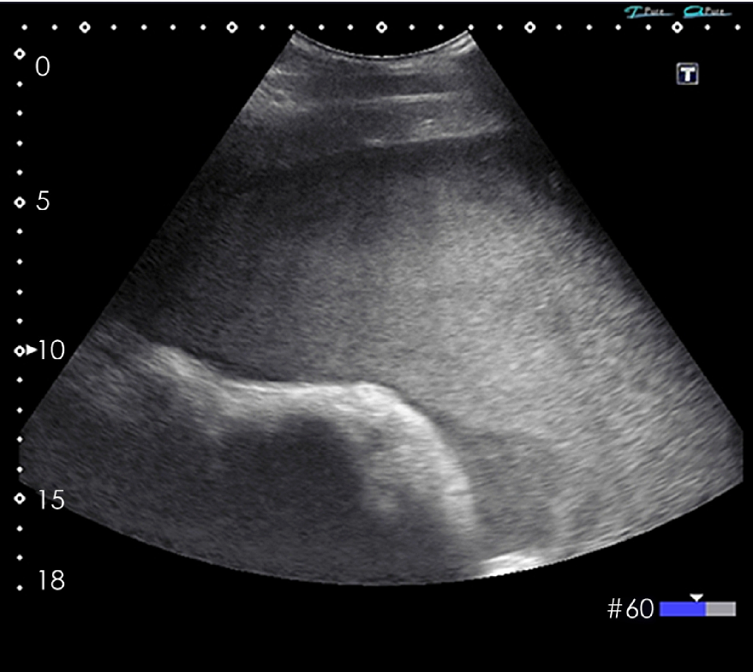
definitive diagnosis?
haemoabdomen
case 2
8yo gelding MWH
History
Reduced mentation and mild colic signs for 48 hours.
Physical exam
QAR, mild colic signs, injected membranes, HR44, RR36, T39.2oC
Rectal
No abnormalities detected
problem list? top ddx
reduced mentation
colic signs
injected mems
tachypnoea
high temp
sirs induced sepsis
causes of incrs rr
pain
stress response
primary lung disease
infectious inflammatory
pleural space
reduced tidal vol
how can we assess which is the likely cause of incrs rr in case 2
which diagnostics are needed
Pain
Response to analgesia
Primary lung disease
Tracheal wash/BAL
Radiography
Ultrasonography
Pleural space disease
Auscultation
Radiography
Ultrasonography
60 minutes after the administration of flunixin meglumine, reparatory rate and demeanor remain unchanged
radiography
Ambulatory machines unable to obtain good thoracic radiographs
Endoscopy unlikely immediately available
ausc
Increased respiratory noise dorsally, muffled ventrally, crackles audible over trachea
does this case need referral
yes want to image

thoughts and definitive diagnosis
endoscopy: excess mucus
us: pleural effusion
diagnosis: pleuropneumonia
case 3 13yo sport horse
History
Rolling, flank watching and kicking at abdomen for the last 3 hours
Physical exam
Agitated, constant kicking at abdomen. HR44, RR16, T37.9oC, gut sounds reduced but present in all quadrants
Rectal
No abnormalities detected
Colic signs persist following analgesia
problem list
colic signs
reduced gut sounds
colic persistent after analgesia
how can we assess an intestinal lesion
peritoneal fluid analysis
abd us→ flash scan
what is a flash scan

Fast Localised Abdominal Sonography of Horses
Haematology and Biochemistry
unremarkable
Peritoneal fluid
Lactate 0.7mmol/l
Nucleated cell count 1.2x10⁹ cells/l
Abdominal ultrasonography
NAD
Gastroscopy
No lesions identified
Posturing to urinate
Chewing at flanks
Half exteriorising penis
what do next?
inspect sheath → sedate to do this

case 4- ella, 5yo thoroughbred mare
History
Horse reported to be dull, off feed and showing mild but consistent colic signs
Physical exam
Horse is restless and flank watching, with some patchy sweating. HR is 60, RR20, T38.7oC. Gut sounds present but reduced.
Rectal
No abnormalities detected
Nasogastric intubation
16 liters of green fluid recovered form stomach
Top differential??
grass sickness
impaction→ but no abnormalities on rectal
strangulation
what are the limitations of palp per rectum
Cannot evaluate the entire abdomen per rectum

what other ways can we identify strangulated si
ultrasound
peritoneal fluid analysis
what is seen on us with strangulted si
disteneded loops
thickened loops
two distinct populations → normal part and abnormal part
what is seen on peritoneal flud analysis of strangulated si
devitalised tissue → lactate produced
if peritoneal lactate >2x peripherial → indicative
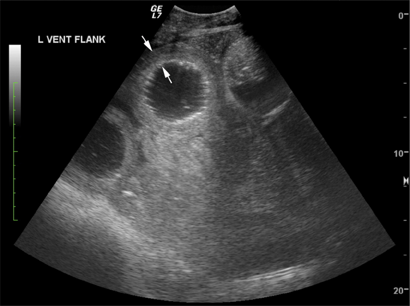
Peritoneal fluid:
Total protein 50g/l
TNCC 7x10⁹ cells/l
Lactate 3.9 mmol/l
(peripheral 3 mmol/l)
does not indicate strangulation
Ultrasonography=
Moderate distension of small-intestinal loops (2–4 cm diameter)
Wall thickness normal ( < 3 mm )
Motility sluggish
No free peritoneal fluid or distinct populations of loops
Peritoneal lactate < 2× systemic → not strongly supportive of a strangulating lesion.
Elevated protein + mild inflammation → leakage or inflammation, not necrosis.
Consistent with proximal enteritis / non-strangulating obstruction, rather than ischaemic strangulation.
Supports a diagnosis of proximal enteritis (DPJ) or functional ileus, rather than mechanical obstruction.
how would you manage this case 4
supportive care
analgesia
ivft
gastric decompression
prokinetics
sirs management
what to do or what could be wrong if your clinical findings disagree
Clinical examination findings:
Repeat them!
Clinical pathology results:
Machine error does happen
Repeat runs if necessary
Diagnostic imaging:
Get more pictures, spend more time
Consider other modalities
Diagnostic procedures:
Find other ways to investigate suspicious findings
E.g. markers of strangulation
How to treat proximal enteritis (DPJ) or functional ileus?
gastric decompression
ivft
anti inflam/ analgesia
antimicrobials
supportive care
what referral indications for proximal enteritis
Persistent or increasing pain despite decompression
Rising peritoneal lactate (> 2× systemic)
Worsening cardiovascular parameters or refractory ileus
what supportive care for proximal enteritis
-Anti-endotoxin therapy (flunixin, polymyxin B if severe).
-Muzzle or withhold feed until reflux resolves.
-Monitor for laminitis (systemic inflammatory risk).
-Serial lactate, PCV/TP, heart rate monitoring.
what abx for proximal enteritis
-Often indicated because DPJ may have bacterial or toxin component (e.g. Clostridial species).
-Broad-spectrum IV (penicillin + gentamicin or similar).
what analgesia for proximal enteritis
-Flunixin meglumine 1.1 mg/kg IV (anti-endotoxic + analgesic).
-Opioid (butorphanol) if further analgesia needed.
-Lidocaine CRI (1.3 mg/kg/h after loading dose 1.3 mg/kg IV) – promotes motility, reduces endotoxaemia.
what ivft for proximal enteritis
-Isotonic crystalloids (e.g. Hartmann’s) to replace losses from reflux and dehydration.
-Estimate: 20–40 L bolus initially, then maintenance + ongoing reflux replacement.
what gastric decompression for proximal enteritis
-Leave nasogastric tube in situ for intermittent drainage (every 1–2 h) to prevent gastric rupture.
what is pleuropneumonia often associated with
with shipping fever which is long-distance transport
causing stress and elevated head carriage which inhibits mucociliary clearance