Muscles of the Pelvic Limb OIA + Images
 Call Kai
 Learn
Learn Practice Test
 Spaced Repetition
 Match
Match Flashcards
 Knowt Play
1/76

There's no tags or description
Looks like no tags are added yet.
Name | Mastery | Learn | Test | Matching | Spaced | Call with Kai | Chat |
|---|
No analytics yet
Send a link to your students to track their progress
77 Terms
Tensor fasciae latae m.
Origin: tuber coxae
Insertion: fascia lata
Action: tense the fascia lata (flex the coxal joint and extend the genual joint)

Superficial gluteal m.
Origin: sacrotuberous ligament
Insertion: third trochanter of the femur
Action: abduct the limb (may have a minor extensor action on the coxal joint)

Middle gluteal m.
Origin: gluteal surface of the ilium
Insertion: greater trochanter of the femur
Action: extend the coxal joint; abduct the limb; rotate pelvic limb medially

Piriformis m.
Origin: sacrum and first caudal vertebra
Insertion: greater trochanter
Action: extend the coxal joint

Deep gluteal m.
Origin: body of ilium and ischiatic spine
Insertion: greater trochanter (deep to the middle gluteal m.)
Action: abduct the limb and extend the coxal joint

Articularis coxae m.
Origin: ilium
Insertion: neck of femur
Action: adjusts/protects coxal joint capsule

External obturator m.
Origin: ventral aspect of os coxae around obturator foramen
Insertion: trochanteric fossa
Action: adduct and/or rotate the limb laterally at the coxal joint

Internal obturator m.
Origin: dorsal aspect of os coxae around obturator foramen
Insertion: trochanteric fossa
Action: rotate the limb laterally at the coxal joint
Highlighted tendon belongs to this muscle

Gemelli mm.
Origin: lateral aspect of ischium
Insertion: trochanteric fossa
Action: rotate the limb laterally at the coxal joint

Quadratus femoris m.
Origin: ventral aspect of ischium
Insertion: caudal femur near the trochanteric fossa
Action: extend coxal joint; adduct and/or rotate the limb laterally at the coxal joint.

Biceps femoris m.
Origin: ischiatic tuberosity and sacrotuberous ligament
Insertion: patella, tibia, and tuber calcanei
Action: extend the coxal joint; variable action on the genual joint; extend the tarsocrural joint

Semitendinosus m.
Origin: ischiatic tuberosity
Insertion: tibia and tuber calcanei
Action: extend the coxal joint; variable action on the genual joint; extend the tarsocrural joint

Semimembranosus m.
Origin: on or near the ischiatic tuberosity
Insertion: distal femur and proximal tibia
Action: extend coxal joint; variable action on the genual joint

Sartorius m. (has cranial (purple) and caudal (red) bellies in the dog only)
Origin: crest of ilium
Insertion: medial aspect of the genual region
Action: flex the coxal joint, extend (cr.) or flex (cd.) the genual joint
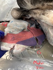
Gracilis m.
Origin: pelvic symphysis
Insertion: medial aspect of the genual region and tuber calcanei
Action: adduct the limb, extend the coxal joint; flex the genual joint; extend the tarsocrural joint

Pectineus m.
Origin: iliopubic eminence (prepubic tendon and cranial border of pubis)
Insertion: distal body of femur
Action: adduct the limb
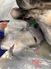
Adductor m.
Origin: ventral surface of os coxae
Insertion: caudal surface of femur
Action: adduct the limb and extend the coxal joint

Quadriceps femoris m.
4 heads:
Green = rectus femoris
Red = vastus lateralis
Blue = vastus intermedius
Yellow = vastus medialis
Origin: rectus femoris - ilium; other 3 heads - proximal femur Insertion: tibial tuberosity
Action: flex the coxal joint (rectus femoris); extend the genual joint (all heads)

Iliopsoas m. (iliacus m. (Red) + psoas major m. (Green))
Origin: ilium (iliacus) and lumbar vertebrae (psoas major).
Insertion: lesser trochanter
Action: flex the coxal joint

Cranial tibial m.
Origin: lateral aspect of proximal tibia
Insertion: plantar base of metatarsal bones 1 and 2
Action: flex the tarsocrural joint and rotate paw laterally

Long digital extensor m.
Origin: extensor fossa of femur
Insertion: extensor processes of the distal phalanges
Action: extend the genual joint; flex the tarsocrural joint; extend the digital joints

Lateral digital extensor m.
Origin: on and near the proximal aspect of the fibula
Insertion: lateral digits
Action: flex the tarsocrural joint; extend the digital joints

Fibularis (peroneus) longus m. (absent in eq).
Origin: on or near lateral collateral ligament of the genual joint
Insertion: 4th tarsal bone and plantar base of metatarsal bones
Action: flex the tarsocrural joint and rotate the paw medially

Fibularis (peroneus) brevis m.
Origin: lateral surface of distal tibia & fibula
Insertion: metatarsal V base
Action: Tarsal flexion

Gastrocnemius m. (medial and lateral heads)
*Medial head not pictured
Origin: distal caudal femur
Insertion: tuber calcanei of the calcaneus
Action: flex the genual joint; extend the tarsocrural joint

Superficial digital flexor m.
Origin: lateral supracondylar tuberosity of femur
Insertion: tuber calcanei and bases of middle phalanges
Action: flex the genual joint; extend the tarsocrural joint; flex the metatarsophalangeal and proximal interphalangeal joints
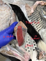
Deep digital flexor m.
Red = Lateral head
Green = Medial head
Origin: proximal caudal aspect of the tibia and fibula
Insertion: flexor tubercle of distal phalanges
Action: extend the tarsocrural joint; flex the digital joints

Popliteus m.
Origin: popliteal fossa of the femur
Insertion: proximal caudal aspect of tibia
Action: flex the genual joint; rotate leg medially

Caudal crural abductor
Origin: sacrotuberal ligament
Insertion: crural fascia
Action: lim abduction + stifle flexion

Femoral Triangle
Space through which femoral vessels run
Lies between caudal belly of the sartorius m. and the pectineus m.

Lateral collateral ligament of stifle

Tendon of long digital extensor

Lateral menisci

Popliteal lymph node

Common calcanean tendon
Muscle ligaments that contribute:
Gastrocnemius m.
Biceps Femoris m.
Gracilis m.
Semitendinosus m.
Superficial digital flexor m.
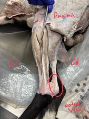
Calcaneal bursa

Patellar ligament

Crural extensor retinaculum

Tarsal extensor retinaculum (distal to crural extensor retinaculum)

Sacrotuberous ligament

Ligament of the head of the femur

Transverse acetabular ligament

Acetabular lip

Medial femoropatellar ligament (genual jt)

Lateral femoropatellar ligament (genual jt)

Transverse ligament (genual jt)

Meniscofemoral ligament (genual jt)
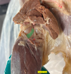
Cranial cruciate ligament (genual jt)

Caudal cruciate ligament (genual jt)
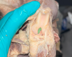
Cranial meniscotibial ligament

Tensor fasciae latae m.
Origin: tuber coxae
Insertion: fascia lata
Action: tense the fascia lata (flex the coxal joint and extend the genual joint)
Superficial gluteal m.
Origin: sacrotuberous ligament
Insertion: third trochanter of the femur
Action: abduct the limb (may have a minor extensor action on the coxal joint)
Middle gluteal m.
Origin: gluteal surface of the ilium
Insertion: greater trochanter of the femur
Action: extend the coxal joint; abduct the limb; rotate pelvic limb medially
Piriformis m.
Origin: sacrum and first caudal vertebra
Insertion: greater trochanter
Action: extend the coxal joint
Deep gluteal m.
Origin: body of ilium and ischiatic spine
Insertion: greater trochanter (deep to the middle gluteal m.)
Action: abduct the limb and extend the coxal joint
Articularis coxae m.
Origin: ilium
Insertion: neck of femur
Action: adjusts/protects coxal joint capsule
External obturator m.
Origin: ventral aspect of os coxae around obturator foramen
Insertion: trochanteric fossa
Action: adduct and/or rotate the limb laterally at the coxal joint
Internal obturator m.
Origin: dorsal aspect of os coxae around obturator foramen
Insertion: trochanteric fossa
Action: rotate the limb laterally at the coxal joint
Gemelli mm.
Origin: lateral aspect of ischium
Insertion: trochanteric fossa
Action: rotate the limb laterally at the coxal joint
Quadratus femoris m.
Origin: ventral aspect of ischium
Insertion: caudal femur near the trochanteric fossa
Action: extend coxal joint; adduct and/or rotate the limb laterally at the coxal joint.
Biceps femoris m.
Origin: ischiatic tuberosity and sacrotuberous ligament
Insertion: patella, tibia, and tuber calcanei
Action: extend the coxal joint; variable action on the genual joint; extend the tarsocrural joint
Semitendinosus m.
Origin: ischiatic tuberosity
Insertion: tibia and tuber calcanei
Action: extend the coxal joint; variable action on the genual joint; extend the tarsocrural joint
Semimembranosus m.
Origin: on or near the ischiatic tuberosity
Insertion: distal femur and proximal tibia
Action: extend coxal joint; variable action on the genual joint
Sartorius m. (has cranial and caudal bellies in the dog only)
Origin: crest of ilium
Insertion: medial aspect of the genual region
Action: flex the coxal joint, extend (cr.) or flex (cd.) the genual joint
Gracilis m.
Origin: pelvic symphysis
Insertion: medial aspect of the genual region
Action: adduct the limb, extend the coxal joint; flex the genual joint; extend the tarsocrural joint
Pectineus m.
Origin: iliopubic eminence (prepubic tendon and cranial border of pubis)
Insertion: distal body of femur
Action: adduct the limb
Adductor m.
Origin: ventral surface of os coxae
Insertion: caudal surface of femur
Action: adduct the limb and extend the coxal joint
Quadriceps femoris m. (4 heads: rectus femoris, vastus lateralis, vastus intermedius, and vastus medialis)
Origin: rectus femoris - ilium; other 3 heads - proximal femur
Insertion: tibial tuberosity
Action: flex the coxal joint (rectus femoris); extend the genual joint (all heads)
Iliopsoas m. (iliacus m. + psoas major m.)
Origin: ilium (iliacus) and lumbar vertebrae (psoas major).
Insertion: lesser trochanter
Action: flex the coxal joint
Cranial tibial m.
Origin: lateral aspect of proximal tibia
Insertion: plantar base of metatarsal bones 1 and 2
Action: flex the tarsocrural joint and rotate paw laterally
Long digital extensor m.
Origin: extensor fossa of femur
Insertion: extensor processes of the distal phalanges
Action: extend the genual joint; flex the tarsocrural joint; extend the digital joints
Lateral digital extensor m.
Origin: on and near the proximal aspect of the fibula
Insertion: lateral digits
Action: flex the tarsocrural joint; extend the digital joints
Peroneus longus m. (absent in eq).
Origin: on or near lateral collateral ligament of the genual joint
Insertion: 4th tarsal bone and plantar base of metatarsal bones
Action: flex the tarsocrural joint and rotate the paw medially
Gastrocnemius m. (medial and lateral heads)
Origin: distal caudal femur
Insertion: tuber calcanei of the calcaneus
Action: flex the genual joint; extend the tarsocrural joint
Superficial digital flexor m.
Origin: lateral supracondylar tuberosity of femur
Insertion: tuber calcanei and bases of middle phalanges
Action: flex the genual joint; extend the tarsocrural joint; flex the metatarsophalangeal and proximal interphalangeal joints
Deep digital flexor m.
Origin: proximal caudal aspect of the tibia and fibula
Insertion: flexor tubercle of distal phalanges
Action: extend the tarsocrural joint; flex the digital joints
Popliteus m.
Origin: popliteal fossa of the femur
Insertion: proximal caudal aspect of tibia
Action: flex the genual joint; rotate leg medially