Disorders of Muskuloskeltal System

0.0(0)
Studied by 0 people
call kaiCall Kai
learnLearn
examPractice Test
spaced repetitionSpaced Repetition
heart puzzleMatch
flashcardsFlashcards
GameKnowt Play
Card Sorting

1/46

encourage image

There's no tags or description

Looks like no tags are added yet.

47 Terms

1
New cards

Arthritis

Broad general term that means inflammation of 1 or more joints

Umbrella category that include such as:

  • Osteoarthritis (arthrosis)

  • Rheumatoid arthritis

  • Infectious arthritis

  • Gout


<p>Broad general term that means inflammation of 1 or more joints</p><p>Umbrella category that include such as: </p><ul><li><p>Osteoarthritis (arthrosis) </p></li><li><p>Rheumatoid arthritis </p></li><li><p>Infectious arthritis</p></li><li><p>Gout </p></li></ul><p></p>
2
New cards

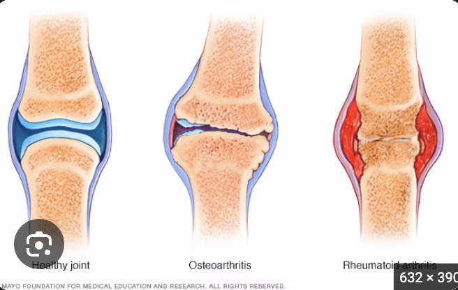
Osteoarthritis

Etiology

  • degenerative wear and tear joint diseases

  • protective cartilage that cushions the ends of your bones gradually wears away

Primary

  • May be result of increased weight bearing or lifting

Secondary

  • Trauma or repetitive use

Pathophys

  • Articular cartilage is damaged

  • Surface of cartilage becomes rough and worn

  • Tissue damage causes release of enzymes, accelerating disintegration of cartilage

  • Subchondral bone may be damaged

  • Cysts, osteophytes or new bones spur

  • osteophytes and cartilage break off

    • bone spurs are smooth, bony projections that develop along the edges of bones, most often where joints meet or on the bones of the spine

  • Joint space become narrower

  • Secondary inflammation on the surrounding

  • Loss of normal range of motion

  • Pain with weight bearing use

More common oon larger joints!

<p>Etiology</p><ul><li><p>degenerative wear and tear joint diseases</p></li><li><p>protective cartilage that cushions the ends of your bones gradually wears away</p></li></ul><p>Primary</p><ul><li><p>May be result of increased weight bearing or lifting</p></li></ul><p>Secondary</p><ul><li><p>Trauma or repetitive use</p></li></ul><p>Pathophys</p><ul><li><p>Articular cartilage is damaged</p></li><li><p>Surface of cartilage becomes rough and worn</p></li><li><p>Tissue damage causes release of enzymes, accelerating disintegration of cartilage</p></li><li><p>Subchondral bone may be damaged</p></li><li><p>Cysts, osteophytes or new bones spur</p></li><li><p>osteophytes and cartilage break off</p><ul><li><p>bone spurs are smooth, bony projections that develop along the edges of bones, most often where joints meet or on the bones of the spine</p></li></ul></li><li><p>Joint space become narrower</p></li><li><p>Secondary inflammation on the surrounding</p></li><li><p>Loss of normal range of motion</p></li><li><p>Pain with weight bearing use</p></li></ul><p>More common oon larger joints!</p>
3
New cards

Rheumatoid Arthritis

Autoimmune and chronic systemic inflammatory diseases

immune system mistakenly attacks the lining of the joints (the synovium), causing painful swelling, inflammation, and potential bone and cartilage destruction

Etiology

  • Unknown, family predispostion, infections

Pathophys

  1. Synovitis: market inflammation and cell proliferation

  2. Pannus Formation: granulation tissue spread

  • new, temporary connective tissue and microscopic blood vessels that form on the surface of a wound during the healing process

  1. Cartilage erosion: creates unstable joint

  2. Fibrosis: calcifies and obturate joint space

  3. Ankylosis: joint fixation and deformation development

Other changes around the joint:

  • atrophy of muscles

  • bone alignment shift

  • muscle spasms due to inflammation/pain

  • contracture deformation

    • Normally stretchy, flexible tissues are replaced or shortened by rigid, inelastic fiber-like tissue; restricts normal joint movement, reduces range of motion, and can cause permanent bending or twisting of the affected body par

Systemic Effect

  • marked fatigue, depression, malaise, anorexia, low grade fever, iron deficiency anemia

Still Disease (systemic form in adults)

  • lymphadenopathy, hepatomegaly, fever, rash

MOre common on smaller joints!!

<p>Autoimmune and chronic systemic inflammatory diseases</p><p>immune system mistakenly attacks the lining of the joints (the synovium), causing painful swelling, inflammation, and potential bone and cartilage destruction</p><p>Etiology</p><ul><li><p>Unknown, family predispostion, infections</p></li></ul><p>Pathophys</p><ol><li><p>Synovitis: market inflammation and cell proliferation</p></li><li><p>Pannus Formation: granulation tissue spread</p></li></ol><ul><li><p>new, temporary connective tissue and microscopic blood vessels that form on the surface of a wound during the healing process</p></li></ul><ol start="3"><li><p>Cartilage erosion: creates unstable joint</p></li><li><p>Fibrosis: calcifies and obturate joint space</p></li><li><p>Ankylosis: joint fixation and deformation development</p></li></ol><p>Other changes around the joint:</p><ul><li><p>atrophy of muscles</p></li><li><p>bone alignment shift</p></li><li><p>muscle spasms due to inflammation/pain</p></li><li><p>contracture deformation</p><ul><li><p>Normally stretchy, flexible tissues are replaced or shortened by rigid, inelastic fiber-like tissue; restricts normal joint movement, reduces range of motion, and can cause permanent bending or twisting of the affected body par</p></li></ul></li></ul><p>Systemic Effect</p><ul><li><p>marked fatigue, depression, malaise, anorexia, low grade fever, iron deficiency anemia</p></li></ul><p>Still Disease (systemic form in adults)</p><ul><li><p>lymphadenopathy, hepatomegaly, fever, rash</p></li></ul><p>MOre common on smaller joints!!</p>
4
New cards

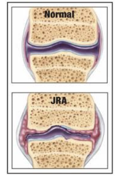
Juvenile Idiopathic arthritis JIA

AKA juvenile rheumatoid arthritis

joint inflammation of unknown cause that begins before the age of 16 and lasts for more than 6 weeks

develops before 16yrs

Several different types, onset more acute

May cause polyarticular inflammation

  • multiple joints inflamed

Some form involved fewer joints but cause uveitis (inflammation of the iris, ciliary body and choroid of the eye)
<p>AKA juvenile rheumatoid arthritis </p><p>joint inflammation of unknown cause that begins before the age of 16 and lasts for more than 6 weeks</p><p>develops before 16yrs</p><p>Several different types, onset more acute</p><p>May cause polyarticular inflammation </p><ul><li><p>multiple joints inflamed </p></li></ul><p>Some form involved fewer joints but cause uveitis (inflammation of the iris, ciliary body and choroid of the eye)</p>
5
New cards

Infectious Arthritis

AKA septic arthritis

joint infection in the joint fluid and surrounding tissues

develop in single joint

Joint red, swelling, decrease in movement, painful

Causes

  • Intro of bacteria into joint (Eg. trauma, non sterile injection, surgery)

  • Secondary infection due to bacteremia


<p>AKA septic arthritis </p><p>joint infection in the joint fluid and surrounding tissues</p><p>develop in single joint </p><p>Joint red, swelling, decrease in movement, painful </p><p>Causes</p><ul><li><p>Intro of bacteria into joint (Eg. trauma, non sterile injection, surgery) </p></li><li><p>Secondary infection due to bacteremia </p></li></ul><p></p>
6
New cards

Gout

Inflammatory arthritis caused by a buildup of uric acid crystals in the joints

Formation tophus—> large, hard nodule of urate crystals

Signs and Symptoms

  • include sudden intense joint pain

  • severe swelling

  • red or warm skin around affected area


<p>Inflammatory arthritis caused by a buildup of uric acid crystals in the joints</p><p>Formation tophus—&gt; large, hard nodule of urate crystals </p><p>Signs and Symptoms</p><ul><li><p>include sudden intense joint pain</p></li><li><p>severe swelling</p></li><li><p>red or warm skin around affected area </p></li></ul><p></p>
7
New cards

Musculoskeletal Diagnostic test

Bone disorders

  • radiograph (xray), bone scans (Dexa)

Muscular Disorders

  • Electromyography (EMG), biopsy

Joint disorders

  • radiography (xray), arthroscopy, magnetic resonace image (MRI), synovial fluid analyses


<p>Bone disorders</p><ul><li><p>radiograph (xray), bone scans (Dexa)</p></li></ul><p>Muscular Disorders</p><ul><li><p>Electromyography (EMG), biopsy </p></li></ul><p>Joint disorders</p><ul><li><p>radiography (xray), arthroscopy, magnetic resonace image (MRI), synovial fluid analyses </p></li></ul><p></p>
8
New cards

Fractures

Break in the integrity of a bone

Trauma, neoplasms, increased stress

Charted using the # sign

<p>Break in the integrity of a bone</p><p>Trauma, neoplasms, increased stress</p><p>Charted using the # sign </p>
9
New cards

Complete

Classification of fracture

____ fracture

  • bone broken forming separate pieces


10
New cards

Incomplete

Classification of fracture

____ fracture

  • bone only partially broken


11
New cards

Open (compound)

Classification of fracture

____ fracture

  • skin is broken


<p>Classification of fracture</p><p>____ fracture</p><ul><li><p>skin is broken </p></li></ul><p></p>
12
New cards

Closed

Classification of fracture

____ fracture

  • skin is not broken


<p>Classification of fracture</p><p>____ fracture</p><ul><li><p>skin is not broken </p></li></ul><p></p>
13
New cards

Simple

Classification of fracture

____ fracture

  • single break, maintaining alignment and postion


<p>Classification of fracture</p><p>____ fracture</p><ul><li><p>single break, maintaining alignment and postion </p></li></ul><p></p>
14
New cards

Comminuted

Classification of fracture

____ fracture

  • multiple fractures and bone fragments


<p>Classification of fracture</p><p>____ fracture</p><ul><li><p>multiple fractures and bone fragments </p></li></ul><p></p>
15
New cards

Compression

Classification of fracture

____ fracture

  • bone crushed or collapses into small pieces


<p>Classification of fracture</p><p>____ fracture</p><ul><li><p>bone crushed or collapses into small pieces </p></li></ul><p></p>
16
New cards

Impacted

Classification of fracture other types

____ fracture

  • one end forced into adjacent bone

  • broken ends of a bone are driven or compressed directly into each other by a strong axial or vertical force


<p>Classification of fracture other types</p><p>____ fracture</p><ul><li><p>one end forced into adjacent bone </p></li><li><p>broken ends of a bone are driven or compressed directly into each other by a strong axial or vertical force</p></li></ul><p></p>
17
New cards

Pathological

Classification of fracture other types

____ fracture

  • results from weakness, occurs with little stress


18
New cards

Stress

Classification of fracture other types

____ fracture

  • fatigue fracture

  • small, hairline crack in a bone caused by repetitive force, overuse, or a sudden increase in physical activity faster than the bone can rebuild itself


<p>Classification of fracture other types</p><p>____ fracture</p><ul><li><p>fatigue fracture </p></li><li><p>small, hairline crack in a bone caused by repetitive force, overuse, or a sudden increase in physical activity faster than the bone can rebuild itself</p></li></ul><p></p>
19
New cards

Depressed

Classification of fracture other types

____ fracture

  • skull fractured and forced into brain


<p>Classification of fracture other types</p><p>____ fracture</p><ul><li><p>skull fractured and forced into brain </p></li></ul><p></p>
20
New cards

Pathophysiology

_____ of bone fracture

  • fractures initiate inflammatory response and hemostasis

    • bleeding

    • edema causes stretching of periosteum (if intact) a swelling of soft tissue = PAIN ++

    • release of Bradykinin (vasodilator and pain mediator) and other chem mediators also contribute to pain

    • Clot forms at fracture site

    • Systemic signs of inflammation may occur


<p>_____ of bone fracture</p><ul><li><p>fractures initiate inflammatory response and hemostasis </p><ul><li><p>bleeding</p></li><li><p>edema causes stretching of periosteum (if intact) a swelling of soft tissue = PAIN ++</p></li><li><p>release of Bradykinin (vasodilator and pain mediator) and other chem mediators also contribute to pain </p></li><li><p>Clot forms at fracture site</p></li><li><p>Systemic signs of inflammation may occur </p></li></ul></li></ul><p></p>
21
New cards

Healing

____ of Bone Fracture

  1. Hematoma- fibrin network is formed

  2. Phagocytic cells remove debris

  3. Fibroblasts lay down new collagen fibers

  4. Chondroblasts form new cartilage

  5. Formation of procallus (fibrous collar)

  6. Osteoblasts generate new bone

  7. Procallus replaced by bony callus

  8. Remodeling of bone with return to use


<p>____ of Bone Fracture </p><ol><li><p>Hematoma- fibrin network is formed </p></li><li><p>Phagocytic cells remove debris </p></li><li><p>Fibroblasts lay down new collagen fibers</p></li><li><p>Chondroblasts form new cartilage </p></li><li><p>Formation of procallus (fibrous collar) </p></li><li><p>Osteoblasts generate new bone </p></li><li><p>Procallus replaced by bony callus </p></li><li><p>Remodeling of bone with return to use </p></li></ol><p></p>
22
New cards

Bone Healing

Factors affecting _____

  • amount of local damage

  • proximity of bone ends

  • Presence of foreign material or infection

  • Blood supply to fracture site

  • System factors—— age, nutrition, anemia, etc.

Complications

  • muscle spasms

  • infections

  • ischemia

  • fat emboli

    • Fat in the medullary cavity can escape and cause embolus

  • nerve damage

  • failure to heal or deformity during healing

  • residual effects such as osteoarthritis


23
New cards

Compartment Syndrome

  • Edema within 1 area of compartment of the limb that is between layers of dense CT (fascia)

  • Ischemia and infarctions (death of tissue due to low oxygen + blood supply) of tissue may occur due to compression of arterial blood supply

  • Dead tissue may become gangrenous requiring amputation

    • body tissue dies due to a loss of blood supply or a severe bacterial infection

  • A TIGHT CAST can cause ____

overgrowing of CT limit blood to certain areas of the body, can lead to hypoxia → necrosis → amputation

<ul><li><p>Edema within 1 area of compartment of the limb that is between layers of dense CT (fascia)</p></li><li><p>Ischemia and infarctions (death of tissue due to low oxygen + blood supply) of tissue may occur due to compression of arterial blood supply</p></li><li><p>Dead tissue may become gangrenous requiring amputation</p><ul><li><p>body tissue dies due to a loss of blood supply or a severe bacterial infection</p></li></ul></li><li><p>A TIGHT CAST can cause ____</p></li></ul><p>overgrowing of CT limit blood to certain areas of the body, can lead to hypoxia → necrosis → amputation </p>
24
New cards

Dislocation

  • Seperation of 2 bones at joint with loss of contact between articulating surfaces

  • Usually accompanied by significant soft tissue damage to ligamanets and tendons

  • Distortion of joint usually evident

  • May recur repeatedly, requiring surgery

  • occurs when extreme force, a fall, or a sports injury pushes the bones in a joint out of their normal position


<ul><li><p>Seperation of 2 bones at joint with loss of contact between articulating surfaces </p></li><li><p>Usually accompanied by significant soft tissue damage to ligamanets and tendons</p></li><li><p>Distortion of joint usually evident </p></li><li><p>May recur repeatedly, requiring surgery </p></li><li><p>occurs when extreme force, a fall, or a sports injury pushes the bones in a joint out of their normal position</p></li></ul><p></p>
25
New cards

Sprain

tear in a ligment

<p>tear in a ligment </p>
26
New cards

Strain

tear in tendon

<p>tear in tendon </p>
27
New cards

Avulsion

Ligaments or tendons completely separated from bony attachments

28
New cards

Immobilization

often used to prevent tissue damage and promote healing

  • Not moving


29
New cards

Osteoporosis

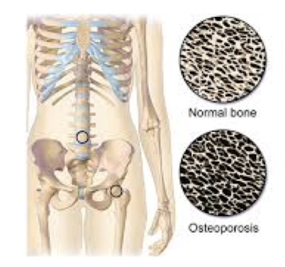
  • Most common disease that affect the skeleton

  • Characterized by low bone mineral density BMD, Microarchitectural deterioration of bone tissue with a consequent increase in bone fragility and susceptibility to fracture

  • Skeletal disorder characterized by compromised bone strength, predisposing individauls to increased risk of fracture

Two types- 1 and 2

Race/Ethnicity

  • can occur in persons of all races and ethnicities

  • In general whites (esp. northern European descent) and Asians are at increased risk

  • Non-hispanic white women and Asian women are at higher risk

Pathophysiology

  • metabolic bone disease that on cell level, result from osteoclastic bone resorption NOT compensated by osteoblastic bone formation. Causes bones to become weak and fragile, increasing risk of fractures

Diagnosis

  • utilizes bone mineral density as defintion (T score <2.5) surrogate marker

  • BMD falls 2.5 standard deviations below the mean (T score-2.5)

<ul><li><p>Most common disease that affect the skeleton</p></li><li><p>Characterized by low bone mineral density BMD, Microarchitectural deterioration of bone tissue with a consequent increase in bone fragility and susceptibility to fracture</p></li><li><p>Skeletal disorder characterized by compromised bone strength, predisposing individauls to increased risk of fracture </p></li></ul><p>Two types- 1 and 2</p><p>Race/Ethnicity</p><ul><li><p>can occur in persons of all races and ethnicities</p></li><li><p>In general whites (esp. northern European descent) and Asians are at increased risk</p></li><li><p>Non-hispanic white women and Asian women are at higher risk</p></li></ul><p>Pathophysiology</p><ul><li><p>metabolic bone disease that on cell level, result from osteoclastic bone resorption NOT compensated by osteoblastic bone formation. Causes bones to become weak and fragile, increasing risk of fractures</p></li></ul><p>Diagnosis</p><ul><li><p>utilizes bone mineral density as defintion (T score &lt;2.5) surrogate marker</p></li><li><p>BMD falls 2.5 standard deviations below the mean (T score-2.5)</p></li></ul><p></p>
30
New cards

Type 1

_____ Osteoporosis

  • also known as postmenopausal osteoporosis

  • develops mainly in women due to decrease in estrogen production

  • Can also be from testosterone deficiency

  • Ages 50-70 yrs


<p>_____ Osteoporosis </p><ul><li><p>also known as postmenopausal osteoporosis </p></li><li><p>develops mainly in women due to decrease in estrogen production </p></li><li><p>Can also be from testosterone deficiency</p></li><li><p>Ages 50-70 yrs </p></li></ul><p></p>
31
New cards

Type 2

____ Osteoporosis

  • also known as senile osteoporosis

  • Typically develops after age 70 in older men and women

  • It is associated with decreased bone formation and decrease in vitamin D production


32
New cards

Secondary osteoporosis

  • When the loss of bone is caused by certain lifestyle behaviors, diseases or meds

    • Cushings Disease

    • Hyperthyroidism

      • T3 and T4 increase osteoclast activity

      • too much energy in thyroid hormone production

    • Prolonged treatment with corticosteroids

    • excess alcohol and smoking


33
New cards

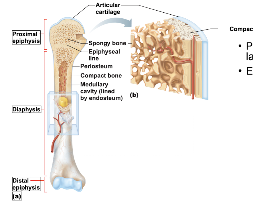
Periosteum

Outer fibrous layer of bone

  • Provides blood supply, nerve innervation, and mechanical protection.

  • Serves as an attachment site for tendons and ligaments.

  • Drives appositional bone growth (increasing thickness) and fracture healing


<p>Outer fibrous layer of bone</p><ul><li><p><span>Provides blood supply, nerve innervation, and mechanical protection.</span></p></li><li><p><span>Serves as an attachment site for tendons and ligaments.</span></p></li><li><p><span>Drives appositional bone growth (increasing thickness) and fracture healing</span></p></li></ul><p></p>
34
New cards

Endosteum

Inner layer of bone

thin, delicate, incomplete cellular layer containing flattened osteoprogenitor cells, osteoblasts, and osteoclasts.
<p>Inner layer of bone </p><p>thin, delicate, incomplete cellular layer containing flattened osteoprogenitor cells, osteoblasts, and osteoclasts.</p>
35
New cards

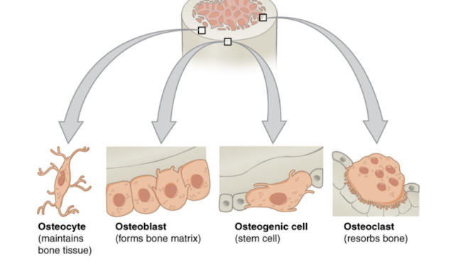
Osteoblasts

Bone building cells

36
New cards

Osteoclasts

Bone destroying cells

37
New cards

Osteogenic cells

stem cells of bone

  • undifferentiated biological stem cell found within bone tissue that can divide, multiply, and differentiate into specialized bone-forming cell


38
New cards

Nutrient Foramina

Nerve fibers, nutrient blood vessels, and lymphatic vessesls enter the bone via ____

39
New cards

Hormonal

What controls continual remodeling of bone?

  • ____ mechs that maintain calcium homeostasis in blood

  • Mechanical and gravitational forces


<p>What controls continual remodeling of bone?</p><ul><li><p>____ mechs that maintain calcium homeostasis in blood </p></li><li><p>Mechanical and gravitational forces </p></li></ul><p></p>
40
New cards

Mech

___ of calcium levels in serum

<p>___ of calcium levels in serum </p>
41
New cards

T score

Standard deviation- variation from the average

The score that you get from your bone density (BMD or DXA) test is measured as a standard deviation from that of a 30yr old adult

One standard deviation is equal to 10-12% difference in bone mass

If you are exactly qual to the peak bone mass of average 30yr, you do NOT deviate at all from the average so your t score would be 0 standard deviations (SD)

If your bones are stronger (T-score +1 or +2 SD) indicating that your bones have a mass 10-20% above that of the average 30 yr

If bones are less dense (t score -2 or -3) indicating that your bone mass is 20-30% below that of average 30yr

42
New cards

Osteopenia

Less severe form of osteoporosis

  • BMD T score between -1 and -2.5


43
New cards

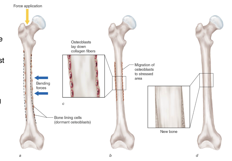
Resistance exercise

Standard adaptations to ______

Bone Modeling

  1. Application of a longitudinal weight bearing forces causes the bone to bend (as depicted by the dotted line) creating stimulus for new bone formation at the regions experiencing the greatest deformation

  2. Osteoblasts lay down additional collagen fibers

  3. Previously dormant osteoblasts migrate to the area experiencing the strain

  4. The collagen fibers become mineralized and bone diameter effectively increases

<p>Standard adaptations to ______</p><p>Bone Modeling</p><ol><li><p>Application of a longitudinal weight bearing forces causes the bone to bend (as depicted by the dotted line) creating stimulus for new bone formation at the regions experiencing the greatest deformation </p></li><li><p>Osteoblasts lay down additional collagen fibers </p></li><li><p>Previously dormant osteoblasts migrate to the area experiencing the strain </p></li><li><p>The collagen fibers become mineralized and bone diameter effectively increases</p></li></ol><p></p>
44
New cards

Connective Tissue

____ Adaptations

  • trabecular bone responds more rapidly to stimuli than cortical bone

  • Exercise is recommended for patients with osteoporosis or those at risk

    • both aerobic weight bearing program and resistive exercise program


<p>____ Adaptations</p><ul><li><p>trabecular bone responds more rapidly to stimuli than cortical bone </p></li><li><p>Exercise is recommended for patients with osteoporosis or those at risk </p><ul><li><p>both aerobic weight bearing program and resistive exercise program </p></li></ul></li></ul><p></p>
45
New cards

Rickets and osteomalacia

Result from deficit of Vit. D and Phosphate

Causes

  • dietary deficits

  • malabsorption

  • intake of phenobarbital

    • long-acting barbiturate medication used primarily as an anticonvulsant to prevent and control seizures, as well as a central nervous system depressant for sedation, anxiety, and alcohol or benzodiazepine withdrawa

  • lack of sun exposure

In kids

  • leads to weak bones and skeletal deformation

In adults

  • May lead to soft bones, resulting in compression fractures


<p>Result from deficit of Vit. D and Phosphate </p><p>Causes</p><ul><li><p>dietary deficits</p></li><li><p>malabsorption </p></li><li><p>intake of phenobarbital</p><ul><li><p>long-acting <strong>barbiturate medication</strong> used primarily as an anticonvulsant to prevent and control seizures, as well as a central nervous system depressant for sedation, anxiety, and alcohol or benzodiazepine withdrawa</p></li></ul></li><li><p>lack of sun exposure </p></li></ul><p>In kids</p><ul><li><p>leads to weak bones and skeletal deformation </p></li></ul><p>In adults</p><ul><li><p>May lead to soft bones, resulting in compression fractures </p></li></ul><p></p>
46
New cards

Bone Tumors

Common site of secondary tumors

Majority of primary tumors termed sacromas are malignant

Osteosarcoma most common

<p>Common site of secondary tumors </p><p>Majority of primary tumors termed sacromas are malignant </p><p>Osteosarcoma most common </p>
47
New cards

Osteosarcoma

Most common primary neoplasm of bone

  • occurs in shaft of ling bones of leg

  • bone pain at rest is warning sign