Anatomy 2300 Unit 4: Organization of the Nervous System
 Call Kai
 Learn
 Practice Test
 Spaced Repetition
 Match
 Flashcards
 Knowt Play
1/27

There's no tags or description
Looks like no tags are added yet.
28 Terms
Central Nervous System (CNS)
The CNS is made up of the brain and spinal cord, which lie along the midline of the body within the cranial cavity and vertebral canal.
It integrates information received from the PNS and coordinates and influences the activity of all parts of the body.

Peripheral Nervous System (PNS)
The PNS is all of the nervous tissue outside the brain and spinal cord, made up of 31 pairs of spinal nerves, 12 pairs of cranial nerves and their branches, plexuses, and ganglia.
It provides information to the CNS about the internal and external environment, and it is divided into the somatic and autonomic nervous systems.

Somatic Nervous System
The somatic nervous system is the division of the PNS that carries sensory information from the skin, muscles, and joints to the CNS.
It also carries motor commands from the CNS to skeletal muscle, and it is generally under voluntary control.

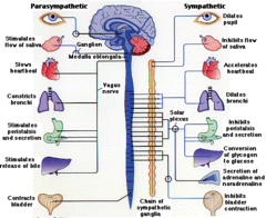
Autonomic Nervous System
The autonomic nervous system is the division of the PNS that controls involuntary functions.
It sends motor signals to smooth muscle, cardiac muscle, and glands, and it also carries sensory information from the internal organs. It is further divided into sympathetic and parasympathetic parts.
Plexus
(part of PNS)
A plexus is a network of intertwining spinal nerves in the PNS, where the anterior (ventral) rami of neighboring nerves merge and reorganize before branching out.
Plexuses are found in the neck, shoulder, lower back, and pelvis, and they distribute fibers to the limbs and body regions.

Neuron
Specialized cell transmitting electrical signals in the nervous system.
A neuron is an excitable cell of the nervous system that receives, processes, and transmits electrical signals.
It consists of one cell body (soma) and processes that extend from it, namely one or more dendrites and a single axon.

Neuroglial Cell
Neuroglial cells (glia) are the supportive, non-excitable cells of the nervous system that protect, nourish, and support neurons.
In the CNS they include oligodendrocytes, astrocytes, ependymal cells, and microglia.

Cell Body (Soma) (collections of it form?)
The cell body, also called the soma, is the central part of a neuron that contains the nucleus and most of the cell's organelles.
The dendrites and axon extend from it, and it is the neuron's metabolic center. Collections of cell bodies form gray matter nuclei in the CNS and ganglia in the PNS.

Dendrite
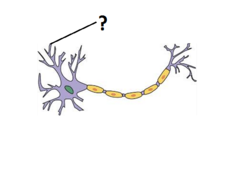
Dendrites are the branching processes that extend from the neuron's cell body, and a neuron has one or more of them.
Their function is to receive incoming signals from other cells and carry them toward the cell body.
Axon
The axon is the single long process that extends from the neuron's cell body.
It transmits signals away from the cell body toward other neurons, muscles, or glands. Many axons are wrapped in myelin, and collections of axons form tracts in the CNS and nerves in the PNS.

Myelin Sheath (what color does it give?)
The myelin sheath is a fatty, insulating layer wrapped around the axon in segments, with small gaps between them.
It speeds up signal transmission along the axon. It is formed by oligodendrocytes in the CNS (and Schwann cells in the PNS), and it gives white matter its color.

Axon Terminals (they release?)
Axon terminals are the small branched endings at the far end of an axon, away from the cell body. They release neurotransmitters at synapses to communicate with the next neuron, muscle fiber, or gland cell.

Nucleus (which system? Collections form what?)
In the nervous system, a nucleus is a collection of neuronal cell bodies located within the CNS (not to be confused with the nucleus inside a cell).
Nuclei are masses of gray matter found deep within the brain's white matter or within the spinal cord and brain stem.

Tract (which system? Collections form what?)
A tract is a collection of axons within the CNS that carries similar information between regions of the brain and spinal cord.
Because it is made of myelinated axons, a tract is part of the white matter. It is the CNS counterpart of a PNS nerve.

Fasciculus (which system? Collections form what?)
A fasciculus is another term for a collection of axons within the CNS, and it is often used for a bundle of axons or a named tract.
Like a tract, it forms part of the white matter and is the CNS equivalent of a PNS nerve.

Ganglion / Ganglia (which system?)
A ganglion (plural: ganglia) is a collection of neuronal cell bodies located in the PNS, outside the brain and spinal cord.
It is the PNS counterpart of a CNS nucleus. An example is the dorsal root ganglion, where sensory neuron cell bodies are found.

Nerve (which system?)
A nerve is a collection (bundle) of axons in the PNS, bound together by connective tissue and traveling outside the brain and spinal cord.
Nerves may be sensory, motor, or mixed, and they include the 12 pairs of cranial nerves and 31 pairs of spinal nerves.

White Matter Composition
White matter is the lighter nervous tissue of the CNS, composed primarily of myelinated axons.
The fatty myelin gives it a white appearance. It lies external to the gray matter in the spinal cord and deep to the cortex in the brain, and it forms the tracts that connect different CNS regions.

Gray Matter Composition
Gray matter is the darker nervous tissue of the CNS, composed of neuronal cell bodies, dendrites, unmyelinated axons, and neuroglia.
Because it lacks heavy myelination, it appears gray. It forms the inner core of the spinal cord, the cortex of the cerebrum and cerebellum, and nuclei scattered deep within the brain.

CNS Tissue Patterning (brain and spinal cord)
Basic CNS Pattern: A central cavity is surrounded by gray matter, which is surrounded by white matter.
Spinal cord: The central canal is surrounded by an inner core of gray matter, with white matter on the outside.
Brain: Follows the same general pattern but has additional gray matter:
- Nuclei: Clusters of gray matter located within white matter.
- Cortex: Outer layer of gray matter covering the cerebrum and cerebellum.

Dorsal Horn
The dorsal (posterior) horn is the posterior projection of the butterfly-shaped gray matter in the spinal cord.
It receives incoming sensory information from the dorsal root and is made up of interneuron cell bodies and unmyelinated axons.

Dorsal Root
The dorsal root is the sensory (afferent) root that attaches to the posterior side of the spinal cord. It carries sensory axons into the dorsal horn, and it contains the dorsal root ganglion along its path.

Dorsal Root Ganglion
The dorsal root ganglion is a swelling on the dorsal root, lying just outside the spinal cord in the PNS. It contains the cell bodies of sensory neurons, whose axons run in the spinal nerve and the dorsal root.

Ventral Horn
The ventral (anterior) horn is the anterior projection of the spinal cord's gray matter. It contains the cell bodies of somatic motor neurons, whose axons leave the cord through the ventral root to reach skeletal muscle.

Ventral Root
The ventral root is the motor (efferent) root that exits the anterior side of the spinal cord. It carries motor axons away from the ventral horn and joins the dorsal root to form the spinal nerve.

Spinal Nerve
A spinal nerve is a mixed nerve of the PNS, containing both sensory and motor axons, formed where the dorsal and ventral roots merge just lateral to the spinal cord. It exits through the intervertebral foramen and then splits into its rami. There are 31 pairs.

Dorsal Ramus
The dorsal (posterior) ramus is the smaller branch of a spinal nerve, extending toward the back. It is a mixed branch that supplies the skin and deep muscles of the back. (A)

Ventral Ramus
The ventral (anterior) ramus is the larger branch of a spinal nerve, extending toward the front and sides of the body. It is a mixed branch that supplies the anterior and lateral trunk and the limbs, and it forms the plexuses. (B)
