04research methods in biopsychology
 Call Kai
 Learn
 Practice Test
 Spaced Repetition
 Match
 Flashcards
 Knowt Play
1/29

There's no tags or description
Looks like no tags are added yet.
30 Terms
[A] What are the three primary functional categories of methods used to study brain functions?
Visualize brain structure or activity in brain areas
Measure brain activity and investigate behavior/cognition
Stimulate brain areas and investigate behavior/cognition
[A-ExQ] What is Computed Tomography (CT / CAT), what is its imaging principle, and what structures does it show best?
Terms: CT (Computed Tomography) and CAT (Computerized Axial Tomography) refer to the exact same technique.
Imaging Principle: Uses X-rays (absorption of X-rays) to create 3D representations of the brain.
Safety & Cost: Involves radiation exposure; shorter examination time (5–10 min) and lower cost than MRI.
Resolution: High detail for bony structures; lower detail for soft tissue compared to MRI.
Preferred areas: lung, abdomen, bone etc.
Weren’t used for brain function
[A] How does Structural MRI differ from CT in terms of imaging principle, safety, and soft tissue detail?
Imaging Principle: Uses strong magnetic fields and radio waves (no X-rays / no radiation exposure), to create images of inside the body
Examination Time & Cost: Longer examination time (15–45 min) and higher cost than CT.
Resolution: Provides higher detail in soft tissue (e.g., brain, spinal cord, junctions=preferred areas) compared to CT, lower detail in bony structures
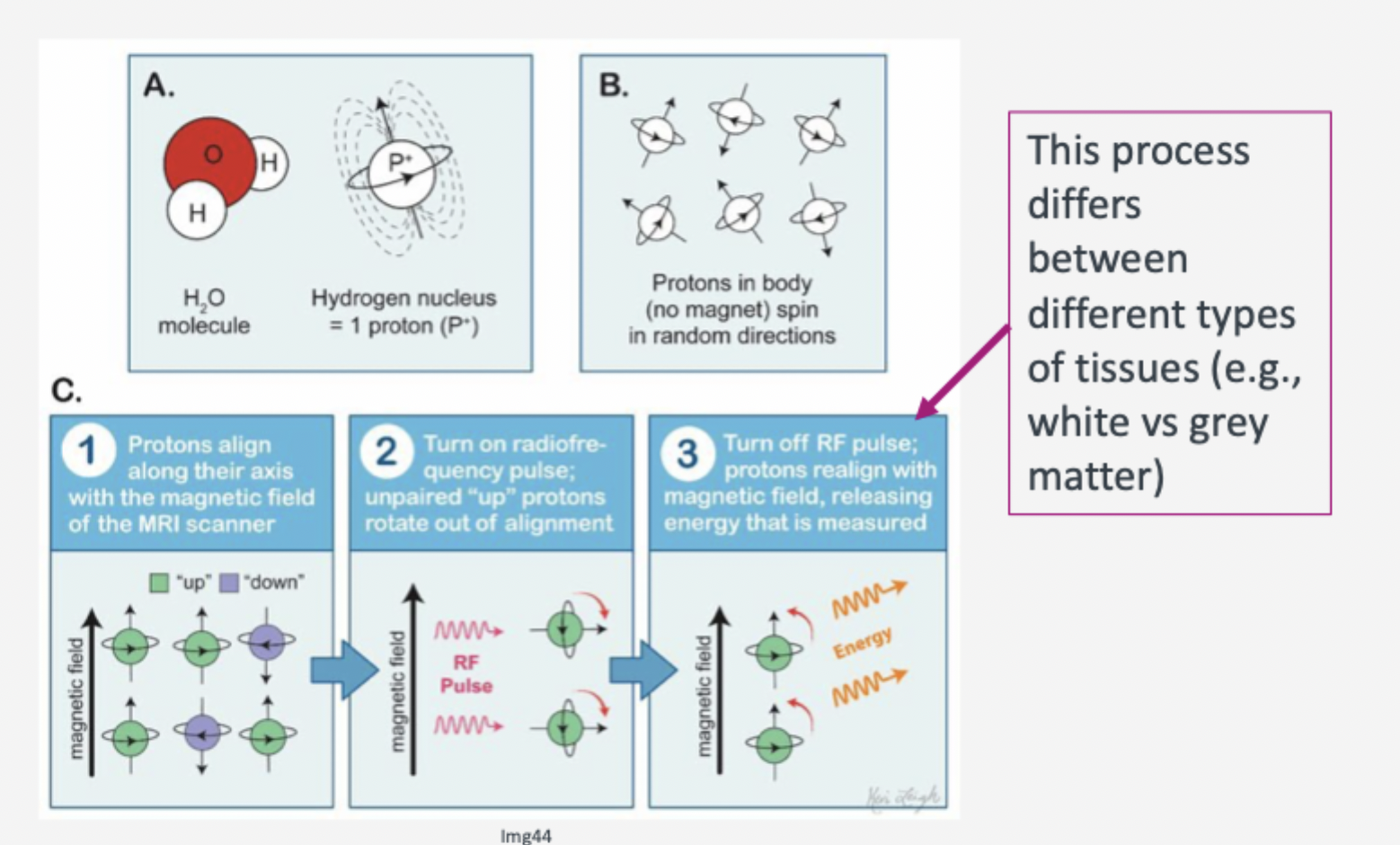
Why does MRI work?
Most of human body made up of water molecules, which consist of hydrogen and oxygen atoms
each hydrogen atom contains a proton which acts like tiny magnet and is very sensitive to magnetic fields
[B] What are the 5 primary EEG frequency bands (oscillations) and which functional/mental states are they associated with?
Delta (δ): Sleep.
Theta (θ): Drowsiness, learning and memory processes.
Alpha (α): Relaxed awake state (e.g., eyes closed).
Beta (β): Mental, physical, and psychological activity.
Gamma (γ): Representation and analysis of perceptual objects

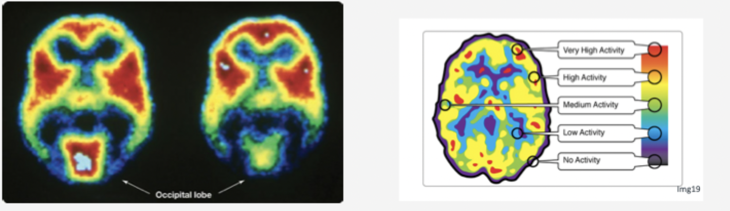
[A] How do radio-ligands in PET imaging visualize neurotransmitter receptors, and what does the color scale indicate?
Mechanism: Radio-ligands are radioactive molecules injected into the body that specifically bind to target structures (neurotransmitters, receptors, transporters, or amyloid plaques).
Example: Radio-ligand ¹¹C-WAY-100635 used for imaging serotonin receptors.
Color Scale:
Red / Warm colors: Highest radioactivity → The redder the color, the higher the receptor density / binding.
Blue / Cool colors: Low or no activity / binding.

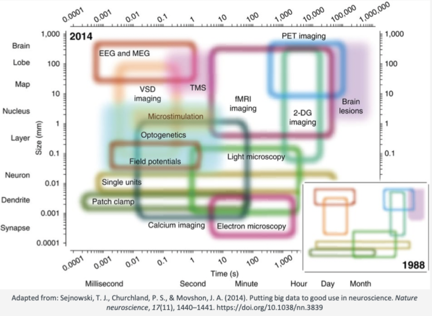
[B] Compare EEG, fMRI, PET, and TMS in terms of their spatial and temporal resolution based on the Sejnowski neuroscience methods matrix
EEG / MEG:
Temporal Resolution: Extremely HIGH (milliseconds range). Best for tracking fast real-time activity and ERPs.
Spatial Resolution: LOW (brain lobe level, 10–100 mm).
fMRI:
Temporal Resolution: LOW / SLOW (seconds range, delayed by BOLD hemodynamic response).
Spatial Resolution: HIGH (millimeter range, 1–3 mm). Best for precise anatomical localization of active regions.
PET Imaging:
Temporal Resolution: VERY LOW (minutes to hours).
Spatial Resolution: MODERATE to HIGH (5–10 mm). Best for mapping neurotransmitter receptor density.
TMS:
Temporal Resolution: HIGH (milliseconds pulse application).
Spatial Resolution: MODERATE (cortical target region). Provides direct causal testing.
[A] What is Transcranial Magnetic Stimulation (TMS), how does it work, and what are its 3 main advantages?
Definition: An electromagnetic coil is positioned next to the skull to temporarily turn off or on a specific area of the human cortex.
Main Purpose: Allows researchers to assess direct consequences of brain (in)activity on cognition and behavior (establishes causality).
3 Key Advantages:
Non-invasive method.
Serves as a direct measure of causal relations between brain (in)activity and behavior/cognition.
Can be used prior to surgery (e.g., tumor removal) to determine how much tissue can/should be safely removed

[A-keep in mind] What does Functional MRI (fMRI) measure, and how does the BOLD signal work?
BOLD Signal: Stands for Blood Oxygenation Level Dependent signal.
Mechanism:
Active brain areas experience increased energy demand and higher oxygen consumption.
Proportion of deoxyhemoglobin ( in blood) initially increases.
Vascular response increases local blood supply, causing oxygen concentration to increase
Hemodynamic Response Function: A mathematical model describing the temporal changes of the BOLD signal in a brain region following neural activity
Different magnetic susceptibility of oxygenated and deoxygenated blood → MR signal
(How magnetic changes depends on arounding tissue?- richtig verstanden?)

[A]What is Positron-Emission Tomography (PET), and how are radio-ligands used?
Mechanism: Involves injecting radioactive chemicals (e.g., 18F-DOPA or radioactive 2-deoxyglucose).
Chemical: 18F-DOPA = radioactive analog of L-DOPA → absorbed by cells that actively produce dopamine or serotonin
Radio-ligands: Ions or molecules that bind to other molecules, allowing researchers to identify the distribution of specific neurotransmitters, receptors, transporters, or amyloid plaques.
Example: 18F-DOPA is absorbed by cells actively producing dopamine or serotonin (used in studying Parkinson's disease)

Positron-emission tomography (PET) what is it similar to and by what is it taken up?
Chemical: Radioactive fluorodeoxyglucose (FDG) = similar to glucose → rapidly taken up by active (energy-consuming) cells
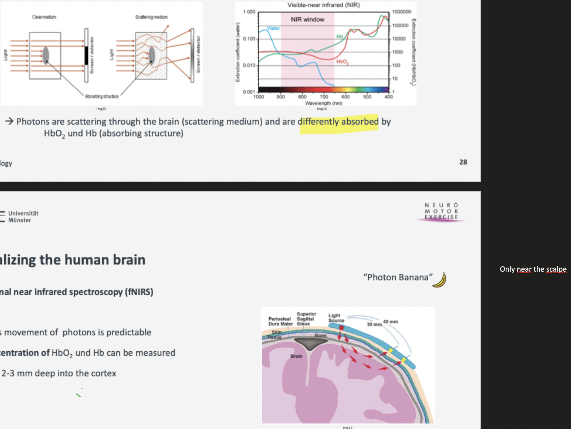
[A] What is the principle of fNIRS, its depth limit, and its pros/cons?
Principle: Photons scatter through the brain ("photon banana" path) within the Near-Infrared (NIR) window and are absorbed differently by oxygenated (HbO2) and deoxygenated (Hb) hemoglobin (absorbing structure)
Mass movement of photons is predictable
Concentration of HbO2 und Hb can be measured
Depth Limit: Penetrates only 2–3 mm deep into the cortex.
Pros: Transportable/flexible (usable in natural environments), relatively inexpensive, robust against external movement, artifacts
Cons: Lower temporal and spatial resolution; restricted to cortex only
[A-ExQ] What is Transcranial Magnetic Stimulation (TMS), and why is it crucial for establishing causality?
Definition: An electromagnetic coil positioned next to the skull turns off or on a specific area of the human cortex.
Causality: While neuroimaging shows correlations, TMS serves as a direct non-invasive measure of causal relations between brain (in)activity and behavior/cognition.
(Explicitly tested in sample exam question as a non-invasive method!)
[A] What does Electroencephalography (EEG) record, and what is the 10–20 System?
Recording: Records the electrical activity of brain cells (neurons) through electrodes attached to the scalp.
10–20 System: Standardized international electrode placement system based on relative distances (10% and 20%) between anatomical landmarks (Nion, Inion, Preauricular points).
Temporal Resolution: Provides extremely high temporal resolution (millisecond range)

[A] How is the output processed according to EEG and what distinction does it allow?
Output Processing: The raw electrical signal/output is amplified and recorded.
Distinction: Allows researchers to differentiate between spontaneous brain activity (e.g., ongoing brain waves/oscillations) and activity in response to a stimulus (Event-Related Potentials / ERPs)
[A-ExQ] What does Electromyography (EMG) measure, and which part of the nervous system does it assess?
Measurement: Measures the level of muscle tension (electrical activity generated when muscles contract) using surface electrodes.
Nervous System Division: Measures activity of the Somatic Nervous System.
(Explicitly tested in sample exam question!)
[A-ExQ] What does Electrooculography (EOG) record, and how is it applied?
Measurement: Records eye movements by placing electrodes above/below and left/right of the eye.
Invasiveness: Completely non-invasive.
Application: Serves as an indicator of attention, perception, arousal, and sleep stages, etc.
(Explicitly tested in sample exam question!)
(Eye has + and - side and at these points it got measured the movement - so I understand it)
[B] What are Event-Related Potentials (ERPs), and what cognitive processes do N1, P2, N2, and P3 reflect?
ERP Definition: Averaged EEG waveforms locked to the presentation of specific sensory stimuli.
Components:
N1 (N100, 90–200 ms): Orienting response, sensory encoding.
P2 (P200, 100–250 ms): Attentional processes, information storage.
N2 -different components: N2a or MMN (Mismatch negativity),N2b =changes physical stimulus properties, N2c = classification.
P3 (P300, 250–400 ms): cognitive processing, conscious processing

[B] How does tDCS work, and what is the difference between Anodal and Cathodal stimulation?
Mechanism: Induction of a weak electrical field across the scalp via electrodes.
Anodal Stimulation: can enhances activity of synapses (Increases neural excitability)
Cathodal Stimulation: can suppresses activity of synapses (Decreases neural excitability)
[B] Which psychophysiological measures assess Autonomic Nervous System activity?
Skin Conductance (SCR/SCL): Sweat gland activity in emotional situations increases electrical conductivity of skin. Indicator of emotional thoughts and experiences
Cardiovascular Activity: Indicator of emotional thoughts and experiences
Heart Rate: Measured via Electrocardiogram (ECG)
Blood Pressure: Systolic and diastolic pressure
[B] Why do researchers move "From Visualization to Stimulation", and what is the difference between correlation and causality?
Visualization (fMRI, PET, EEG): Shows only correlations (co-occurrence) between brain activity and behavior.
Stimulation (TMS): Serves as a direct measure of causal relations by actively turning brain areas on/off to test their direct effect on cognition and behavior.
[B] Which psychophysiological recording techniques measure the Central, Somatic, and Autonomic Nervous Systems according to the layout slide?
Central Nervous System (Brain): EEG & MEG.
Somatic Nervous System: EMG (muscle tension) & EOG (eye movements).
Autonomic Nervous System: Skin Conductance (sweat gland activity) & Cardiovascular activity (ECG / Heart rate & Blood pressure)

[C]EEG- Event related potentials (ERP) - Abbildung

[B] What are the 5 major types of lesion methods (+def) used in invasive animal research?
Def.: LM = part of brain is damaged, destroyed or inactivated
Aspiration lesions: Drawing/peeling off a layer of cortical tissue.
Radio-frequency lesions: Passing high-frequency current through target tissue to destroy it.
Knife cuts: Sectioning/cutting a nerve or tract.
Reversible lesions: Temporarily inactivating tissue via cooling or local anesthetics.
Selective chemical lesions: Injecting specific neurotoxins (neural poisons)(e.g., 6-OHDA) to destroy specific neurons
(Be careful with interpretation: lesion effects=very difficult to „apply“! You cannot destroy a structure without producing significant damage to neighbouring structures)
[B]Name 4 key standardized neuropsychological tests mentioned in the slides and what they assess.
WAIS (Wechsler Adult Intelligence Scale): General intellectual ability (verbal comprehension, working memory, perceptual organization, processing speed).
Tower of London (ToL): Executive functions & planning processes.
Beck Depression Inventory (BDI-II): Emotion / coping (21 items assessing depression severity).
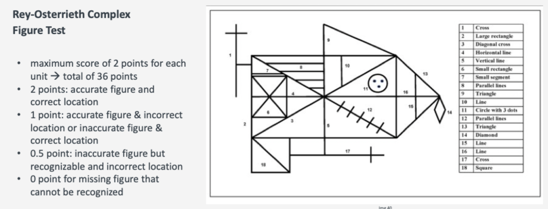
Rey-Osterrieth Complex Figure Test: Visuospatial abilities & memory.
[B] What is the WAIS, who developed it, and what does it measure?
Author: David Wechsler.
Main Domain: General intellectual ability (IQ).
Function & Task: Measures overall intelligence through standardized sub-tests across four main cognitive domains:
Verbal comprehension: Language and verbal reasoning.
Working memory: Short-term memory retention and manipulation (e.g., Digit span test – repeating number series forward and backward).
Perceptual organization: Non-verbal visual reasoning (e.g., Matrix reasoning – completing visual patterns).
Processing speed: Speed of performing simple cognitive tasks

[B] What is the Tower of London (ToL) test, who developed it, and what does it measure?
Author: Tim Shallice.
Main Domain: Executive processing & planning processes.
Function & Task: Assesses problem-solving ability and executive function. The subject must move colored balls across pegs from an initial position to a goal position using a set number of moves

[B] What is the BDI-II, who developed it, how is it structured, and how is it scored?
Author: Aaron Beck.
Main Domain: Emotion, coping, and assessment of depression severity.
Structure: A standardized questionnaire consisting of 21 groups of statements covering symptoms like sadness, pessimism, loss of pleasure, appetite changes, and sleep disturbances as such as concentration difficulty
Scoring & Cut-offs: Each statement is rated from 0 to 3, yielding a sum score from 0 to 63:
0–13: Minimal depression
14–19: Mild depression
20–28: Moderate depression
29–63: Severe depression

[B] What is the Rey-Osterrieth Complex Figure Test, who developed it, and how is it conducted and scored?
Authors: André Rey & Paul-Alexandre Osterrieth.
Main Domain: Visuospatial abilities & visual memory.
3 Test Phases:
Copy: Drawing the complex figure while viewing the template.
Immediate recall: Drawing the figure from memory immediately after copying.
Delayed recall: Drawing the figure from memory after a time delay.
Scoring: Evaluates 18 figure units with a maximum score of 36 points (up to 2 points per accurately drawn and correctly located unit)
[B]How is raw EEG output processed, and how do brain waves change from an aroused state to deep sleep?
Signal Processing: Raw electrical signals are amplified and recorded.
Distinction: Differentiates between spontaneous activity and stimulus-induced activity (ERPs).
Wave Changes across States:
Aroused: High frequency, low amplitude.
Relaxed: Alpha waves (relaxed awake state) (alpha most prominent one)
Asleep: Slower waves as sleep sets in
Deep sleep: Slow waves with high amplitude (Delta waves)
