Cardiology
 Call Kai
 Learn
 Practice Test
 Spaced Repetition
 Match
 Flashcards
 Knowt Play
1/18

There's no tags or description
Looks like no tags are added yet.
19 Terms
Mammals and birds have which circuits
Systemic and pulmonary circuits which are divided which allows pressure to be different in both.

How many chambers in mammal and bird hearts
4 chamber, 2 atria and 2 ventricles. Oxygenated and deoxygenated blood separate.
Water breathing fish circuit
Low pressure, 4 chambers in series, atrium not muscular, contraction of single ventricle pumps blood to gills, no mixing of oxygenated and deoxygenated

Air breathing fish circuit
Tropical fish in low oxygen waters, 4 chambers in partially divided series, Evolved several times, ABO has increased capillary supply and is arranged parallel with systemic circulation. O and D blood mix

Lung fish circulation
Partially divides, limited mixing of D and O blood, 2 atria in heart separate the 2 blood streams in ventricle by septum, Common ancestor to tetrapod
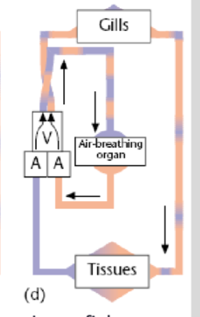
Larval Lamprey circulation
Heart has mixture of oxygen rich and oxygen depleted blood
Gill breathing fish
Heart contains only oxygen poor blood
Primitive air breathing fish
Mixture of oxygen rich and oxygen depleted blood
African lungfish
Almost complete separation of oxygen rich and oxygen depleted blood
Amphibian Circulation
Heart is partially divided with 2 atria and 1 ventricle, deoxygenated blood can mix, blood streams are 80% separate, additional vessel from pulmonary vein to skin circulation

What is the function of trabeculae in amphibian heart
Within the ventricle has a spiral fold to keep oxygenated and deoxygenated blood separate

Where is trabeculae located
Conus arteriosus
Non-crocodilian reptile circulation
partially divided circulation, 2 outflow arteries from 1 ventricle, non mixing of oxygenated and deoxygenate blood

what are the 3 interconnected chambers in non-crocodilian reptile ventricles and what is it called.
Muscular septa that has cavum polmonale, cavum venosum and cavum arteriosum.
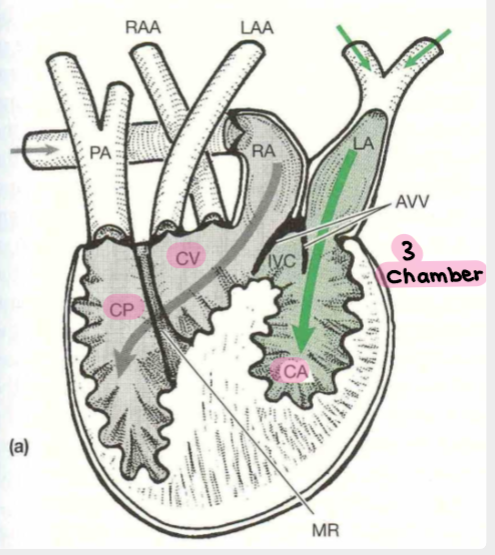
Function of Atrioventricular valve
Blocks blood flow by changing position. Creates temporary chambers.

Explain step one of cardiac contraction in turtles
Atria contracts 2. Left atrium pumps oxygenated blood into C arteriosum 3. Right atrium pumps deoxygenated blood through C.Venosum across septum into C. pulmonale 4. AVV blocks intraventricular canal
Explain step 2 of cardiac contraction in turtles
Ventricle contracts 2. Deoxygenated blood in C. pulnonale is expelled via pulmonary arteries to lungs
AVV closes
Oxygenated blood in C. arteriosum forced into C. venosum and expelled through RAA, LAA to systemic circulation
Mixing of blood is prevented by squeezing tg of the muscular ridge and ventricle wall
Crocodile Circulation
Divided heart and circulation, 2 atria and 2 ventricle, vessels constrict in pulmonary system, pressure in R ventricle increases to be greater than L.

How do crocodiles promote blood flow to the body when swimming
Right ventricular valve ope4ns, Deoxygenated blood flows from L aorta through foramen of panizza into R aorta which increases blood flow to the limbs. (closes blood flow to the lungs).