NeuroPsych 1 - Mi
 Call Kai
 Learn
Learn Practice Test
 Spaced Repetition
 Match
Match Flashcards
 Knowt Play
1/260

There's no tags or description
Looks like no tags are added yet.
Name | Mastery | Learn | Test | Matching | Spaced | Call with Kai | Chat |
|---|
No analytics yet
Send a link to your students to track their progress
261 Terms
What are the initial assessment and workup steps (6) for a patient with suspected acute stroke (ischemic or hemorrhagic)?
1) Recognize stroke symptoms (BE FAST):
B: Balance difficulties
E: Eyes (vision changes)
F: Facial droop
A: Arm/extremity weakness
S: Speech difficulties
T: Time (call 911 immediately)
2) Stabilize/Assess the patient (ABC(D))
Assess ABCs (airway, breathing, circulation [BP, HR], dextrose/glucose levels)
Provide O2 if hypoxic and establish IV access
3) Obtain labs
STAT CBC, coagulation studies, Chem-7
Platelets (low platelets may contraindicate thrombolysis)
Weight (for weight-based TNK/tPA dosing)
12-lead ECG
4) Assess stroke severity (NIHSS)
11 items (0-4 points each)
The higher the score = more severe the impairment
0 – no stroke
1-4 – minor stroke
5-15 – moderate stroke
16-20 – moderate to severe stroke
>20 severe strokes
Good predictor of patient outcomes:
<6 good outcome, >16 high likelihood of death
5) CT/CTA
CT: differentiates between hemorrhagic vs ischemic (sees whether there is bleeding/hemorrhage or not)
CTA (CT with contrast): maps out the brain vasculature, looking for clots/lesions
Can determine large vessel occlusions (LVO)
6) Review history
Determine last known well (LKW) to assess eligibility for IV thrombolysis (typically within 4.5 hours).
Wake-up stroke: LKW is generally when the patient was last normal before sleeping.
Review anticoagulant use (warfarin, DOACs) and bleeding risk.
Review Alzheimer's medications (lecanemab/donanemab) due to increased hemorrhage risk.
What medications should you look out for in a patient's PMH for stroke? Why?
Anticoagulant use (warfarin, DOACs)
If they are on an anticoagulant, don't give thrombolytics for stroke
For warfarin, monitor INR to determine (INR must be ≤1.7)
Thrombolytics have the highest bleeding risk (which is why they are only used for emergencies)
If the patient has not taken an anticoagulant in 48 hours, they are more eligible
Can check INR for DOAC use (will show any variation in bleeding risk, just not as accurate/reliable as warfarin)
Alzheimer's medications (lecanemab/donanemab) due to increased hemorrhage risk.
All may increase bleeding risk if tNK/tPA are used
What is the pathophysiology and what are the common causes of ischemic stroke?
MOA: Blocked blood flow to part of the brain → inadequate oxygen delivery
Common causes
Atherosclerotic ~20% → decrease blood flow
Small vessel disease (lacunar) ~25%
Cardiogenic embolism ~20% → afib (Without appropriate anticoagulant treatment)
Cryptogenic ~30% → unknown
Other causes ~5% → drugs: cocaine, meth = vasoconstriction
How is acute ischemic stroke managed (dose, monitoring) based on time since symptom onset?
≤4.5 hours: IV thrombolysis (if eligible)
Alteplase (tPA): 0.9 mg/kg IV (max 90 mg)
10% IV bolus over 1 minute, then 90% infused over 1 hour.
Tenecteplase (TNK): 0.25 mg/kg IV (max 25 mg) as a single bolus over approximately 5 seconds.
BP before treatment: ≤185/110 mmHg.
BP after treatment: ≤180/105 mmHg for 24 hours.
Monitor closely in a stroke unit/ICU for at least 24 hours.
Avoid antiplatelets and anticoagulants for the first 24-48 hours.
Beyond 4.5 hours (without thrombolysis):
Evaluate for large vessel occlusion (LVO) → CTA, and mechanical thrombectomy eligibility (selected patients up to 24 hours)
Removal of stationary clot
Allow permissive hypertension to maintain cerebral perfusion
Generally avoid BP lowering unless >220/120 mmHg.
If treatment is needed, lower BP by approximately 15% during the first 24 hours.
After 24 hours, consider restarting oral antihypertensives when neurologically stable.
What are the BP goals and IV antihypertensive options (+their dosing/limitations) for acute ischemic stroke?
Goals
BP before tPA/tNK treatment: ≤185/110
BP after tPA/tNK treatment: ≤180/105 for 24-48 hour
Increased risk for hemorrhaging if above this target range
No thrombolysis: Permissive HTN up to 220/120
If above: Reduce BP by ~15% in first 24 hours
Treatment options:
IV labetolol 5-20mg over 1-2 min q15 min
Non-selective BB: will lower both BP and HR
Hold for HR <55
IV Clevidipine 1-2mg/hr infusion
DHP CCB: will lower BP but not HR
Use if labetalol fails to control BP after 3 doses and/or HR <55
Titrate by 2mg/hr q5min PRN to max of 21mg/hr
IV Nicardipine 5mg/hr infusion
DHP CCB: will lower BP but not HR
Titrate by 2.5mg/hr q15min PRN to max of 15mg/hr
What are the limitations of giving tPA/tNK?
Must be given ≤4.5 hours of stroke onset
Head CT scan without evidence of hemorrhage
Max weight for both tPA/tNK = 100kg for dosing
DO NOT GIVE tNK over 25mg or total tPA over 90mg
Labs:
BP ≤185/110
Platelet count above 100,000/mm3
Avoid DDI (other drugs that can increase bleeding risk) → INR ≤1.7
Glucose >50
Age ≥ 18 years
What are the primary and secondary prevention methods for stroke?
Primary:
Stop smoking
Manage HTN, HLD, DM
Increase physical activity + weight loss
Manage afib
Anticoagulants reduce the risk of stroke by 70%
Secondary
Afib → DOAC/warfarin
Non afib related
Aspirin 50-325mg PO QD or
Clopidogrel 75 mg PO QD or
(Aggrenox) Aspirin/dipyridamole 25/200 mg PO BID
Cilostazol 100 mg PO BID
For smaller strokes (NIHSS ≤3), DAPT may be indicated
ASA + clopidogrel for 21-90 days, then single agent
Start DAPT within 24-48 hours
Lipids
High statin therapy (Atorvastatin 80mg PO QD)
Dose + effect independent of LDL level
After 5 years decrease in the risk of fatal and non-fatal stroke, CV events, and an increase in ICH (intracerebral hemorrhage)
Anticoag should not be started until at least 24 hours after tPA administration and repeat CT confirms no bleeding.
What is the pathophysiology and common causes of hemorrhagic stroke?
MOA: Mechanical injury due to edema
Blood vessel ruptures → bleeding into/around the brain
Increased intracranial pressure (ICP) + swelling of the brain = increased mass
Reduced cerebral perfusion (ischemia)
Cerebral herniation
Common causes:
Hypertension
Older age (risk doubles q10 years after 35)
Cerebral angiopathy (vessel weakening)
Drugs (cocaine, amphetamines)
Brain tumors
Arteriovenous malformations
Asian > AA > MA > White
What are the main components (5) of medical management for hemorrhagic stroke?
1) Stabilization/supportive care:
ABCs, intubation if necessary
Manage blood glucose, fever, hydration, and DVT prophylaxis.
2) BP control:
Goal: SBP 140–160 mmHg
IV labetalol, nicardipine, or clevidipine.
Avoid hypotension and excessive BP reduction to preserve cerebral perfusion.
3) Anticoagulation reversal:
Warfarin, Factor Xa inhibitors,Dabigatran, Heparin/enoxaparin
4) Reduce elevated intracranial pressure (ICP) :
Mannitol
Hypertonic saline
5) Seizure management:
Acute seizures (treatment): Benzodiazepines.
Antiseizure medications (PPX): Levetiracetam, phenytoin, or valproate when indicated (depends on type of seizure).
What surgical interventions may be used for hemorrhagic stroke, and what is their purpose?
Cerebellar decompression
Creates more space for a swollen cerebellum → relieves increased ICP and prevents brainstem compression/herniation
Hematoma evacuation
Surgically removes a collection of blood (hematoma) to reduce mass effect and pressure on surrounding brain tissue
Ventriculostomy
Places a catheter in the brain ventricle to drain blood and reduce ICP
Open craniotomy
Opens part of the skull to directly access the brain to evacuate a hematoma or control bleeding
What supportive care measures are recommended for both ischemic and hemorrhagic stroke?
1) Blood glucose control:
BG >140 mg/dL in the first 24 hours is associated with worse outcomes.
BG >200 mg/dL increases the risk of ICH following tPA.
Goal: 140–180 mg/dL
Treatment: Insulin (IV reg insulin in critical → basal + correctional insulin for stable)
Avoid oral antihyperglycemics in acute management.
Avoid hypoglycemia.
2) Temperature management:
Avoid hyperthermia (>38°C), which worsens neurologic injury.
Treat fever with acetaminophen (Tylenol).
Monitor for infection, especially pneumonia.
3) IV fluid management:
Maintain adequate hydration and cerebral perfusion.
Avoid dextrose-containing IV solutions unless needed to treat hypoglycemia.
4) DVT prophylaxis:
Immobility increases venous stasis, contributing to Virchow's triad:
Venous stasis
Endothelial injury
Hypercoagulability
Use intermittent pneumatic compression devices in immobilized patients.
Consider pharmacologic prophylaxis (UFH/LMWH) when safe, accounting for stroke type and bleeding risk.
What conditions or medications can mimic stroke symptoms?
Hypoglycemia: Can cause confusion, weakness, and focal neurologic deficits resembling stroke.
Hyperglycemia (DKA): Can cause altered mental status and neurologic symptoms resembling stroke.
Compazine (prochlorperazine): Can cause acute dystonia, characterized by sudden, painful, involuntary muscle spasms that may resemble neurologic deficits.
Rank antithrombotic medications from highest to lowest bleeding risk.
Thrombolytics > Glycoprotein IIb/IIIa inhibitors > Anticoagulants >> P2Y12 inhibitors (prasugrel, ticagrelor, clopidogrel) > Aspirin
What are the four mechanisms that regulate substance transport across the blood-brain barrier (BBB)?
1) Cell membranes repel hydrophilic toxins
2) Tight junctions limit toxins from sneaking between cells into the brain (no paracellular diffusion)
3) Active transporters
Influx transporters: SLC transporters
Solute carrier-mediated transport
Facilitated diffusion + secondary active transport
Bring in solutes/nutrients from circulation → endothelial cells → brain
EX: GLUT-1, LAT-1
Efflux transporters: ABC transporters
ATP-binding dependent primary active transport
Kick out toxins that don't belong in the brain
EX: P-glycoprotein (pgp)
4) Drug properties
Small (<400Da)
Lipophilic/nonpolar (less than eight hydrogen bonds) (less OH the better)
Charge (the less charge, the better)
LogBB is positive
LogBB = log(brain/(blood)
LogBB = negative number means that more of the drug exists in the blood > brain
LogBB = positive means more in the brain > blood
+1 = 10x more drug in the brain than in the blood
Drug protein binding
Increased protein binding means decreased ability to get into the brain
Route of administration
Intracerebral (IC) delivery
What is the anatomy of the BBB?
Made from 3 cell types
Endothelial cells (innermost):
Lining of the blood vessel
Have tight junctions
Pericyte (middle layer):
Intermediate layer embedded in basement membrane, just superficial to endothelial cells
Astrocytes (outer layer)
Wrap inner layers with foot processes (podocytes)
What does the Glasgow Coma Scale (GCS) assess, and how are scores interpreted?
Assesses level of consciousness using:
Eye-opening (1–4)
Verbal response (1–5)
Motor response (1–6)
13–15: Mild impairment
9–12: Moderate impairment
3–8: Severe impairment/coma
What factors determine DOAC (apixaban, rivaroxaban, dabigatran, edoxaban) dose adjustments?
Apixaban: ≥2 of age ≥80, weight ≤60 kg, SCr ≥1.5 mg/dL
Rivaroxaban: CrCl ≤50 mL/min
Dabigatran: CrCl 15–30 mL/min
Edoxaban: CrCl 15–50 mL/min
What are the two anatomical divisions of the nervous system, and how does their nomenclature differ?
1) Central Nervous System (CNS)
Consists of the brain and spinal cord.
Nucleus: Collection of neuron cell bodies in the CNS.
Tract: Bundle of axons in the CNS.
2) Peripheral Nervous System (PNS)
Consists of nerves and ganglia outside the CNS.
Ganglion: Collection of neuron cell bodies in the PNS.
Nerve: Bundle of axons in the PNS.
Compare the somatic (SNS) and autonomic (ANS) nervous systems in terms of function, motor/sensory innervation, and neurotransmitters.
1) Somatic Nervous System (SNS): Conscious control
Motor: CNS → skeletal muscles (voluntary movement).
Sensory: Skin, muscles, bones, joints → CNS.
Motor pathway: Single neuron from CNS to skeletal muscle (ACh).
2) Autonomic Nervous System (ANS): Unconscious control
Motor: CNS → smooth muscle, cardiac muscle, glands.
Sensory: Visceral organs → CNS.
Motor pathway: 2-neuron chain (preganglionic → postganglionic).
Sympathetic (SANS)
Acetylcholine as a preganglionic neurotransmitter
Norepinephrine as a postganglionic neurotransmitter
Longer postganglionic neuron
Parasympathetic (PANS)
Acetylcholine as a pre- and postganglionic neurotransmitter
Longer preganglionic neuron
Compare the motor and sensory neuron chains in terms of neuron number, location, and function.
Motor pathway (descending):
2-neuron system (upper and lower motor neurons).
Upper motor neuron: Descends through CNS white matter.
Lower motor neuron: Extends into the periphery to innervate skeletal muscle.
Function: CNS → muscle (movement).
Sensory pathway (ascending):
3-neuron system:
First-order neuron: Cell body in the dorsal root ganglion, receives peripheral sensory information.
Second-order neuron: Located in the spinal cord gray matter or brainstem.
Third-order neuron: Located in the thalamus, projects to the cerebral cortex.
Function: Peripheral receptors → CNS (sensation).
Compare gray and white matter in terms of composition, location in the brain vs. spinal cord, and function of the spinal cord horns.
Gray matter
Neuron cell bodies and their support cells
White matter
Axon segments and their support cells
The arrangement differs from the brain to the spinal cord
Brain = gray external, white internal
Spinal cord = gray internal, white external
Divided into two grey matter horns
Dorsal horn: sensory neurons
Ventral horn: motor neurons

What are the major components of a neuron and synapse, and how do they transmit nerve impulses?
Neuron
Axon = long cytoplasmic extension
Often myelinated. Myelin restricts depolarization (+) of the membrane to the myelin sheath gaps
Causes jump from one node to the next (saltatory conduction) = faster conduction than non-myelinated axons
Cell body = cell body, contains nucleus and organelles
Dendrites = collects info to send to the nucleus
Use excitability to produce and move an AP (nerve impulse) along an axon → transmitted to another neuron or effector cell via a synapse
Synapse
Axon terminal
Release neurotransmitter (NE or acetylcholine)
Travels to the postsynaptic cell
What are the meningeal layers of the brain?
1) Dura mater (outer)
Composed of two layers
Periosteal layer (outer)
Meningeal layer (inner)
Space between layers includes dural venous sinuses
Drains blood from the brain to the internal jugular veins in the neck (systemic circulation)
2) Arachnoid mater (middle)
Arachnoid trabeculae
Tread-like structures that connect the arachnoid mater with the pia mater
Arachnoid granulations
Projections of the arachnoid into the dural sinuses to recycle CSF
Held against the dura mater by the osmotic pressure of CSF
Separated from the pia mater by the subarachnoid space (contains CSF)
3) Pia mater (inner)
Directly against the brain (very thin and permeable to water and small solutes)
Allows vessels to pass through and nourish the brain
Allows formation of perivascular spaces, which serve as the brain’s lymphatic system (drains excess fluid)

What are the source, location, functions, and flow pathway of cerebrospinal fluid (CSF)?
Source: Choroid plexus (500mL produced per day)
Continuous to compensate for the amount resorbed into the systemic circulation
Location: Cranial ventricles (hollow cavities within the brain where CSF is produced and circulated)
1+2) Lateral ventricles (L+R)
Connect to the 3rd by the interventricular foramen (of Monro)
3) Third ventricle
Connected to the 4th by the cerebral aqueduct (of Sylvius)
4) Fourth ventricle
Surrounded by pons, medulla, cerebellum
Connected to the CSF space around the brainstem by the lateral aperture (foramen of Luschka)
Function:
Provide buoyancy for the brain and CNS
Provide a medium for nutrients and electrolytes to be conveyed to the brain
Removed metabolic waste from the brain
Flow
Choroid plexus in lateral ventricles → interventricular foramen (of Monro) - connection to → Third ventricle → cerebral aqueduct (of Sylvius) - connection to → fourth ventricle → lateral aperture (foramen of Luschka) - connection to → CSF space around brainstem + subarachnoid space around the CNS and/or central canal of spinal cord → arachnoid granulations → Dural venous sinuses → Resorbed into venous system through arachnoid granulations in skull (125-150mL in circulation at any time)
What are the functions of peripheral nerves, and how are spinal and cranial nerves classified?
1) Peripheral nerve functions
Sensory (afferent): Carries information toward the CNS.
Motor (efferent): Carries commands away from the CNS.
Somatic motor: Controls skeletal muscles.
Visceral/autonomic motor: Controls smooth muscle, cardiac muscle, and glands.
2) Spinal nerves
Mixed nerves containing both sensory and motor fibers.
Connect the spinal cord to the body.
Sensory: Body → spinal cord.
Motor: Spinal cord → body.
3) Cranial nerves (12 bilateral pairs)
12 bilaterally paired cranial nerves, which primarily provide sensory and motor innervation to the muscles, skin, and glands of the head and neck
Sensory only: CN I, II, VIII
Motor only: CN IV, VI, XI, XII
Both sensory + motor: CN III, V, VII, IX, X
What are the functions and clinical applications of cranial nerve 2?
2: Optic - sensory
Function: Vision
Carries visual info from the retina → associated with occipital (primary visual cortex)
Clinical applications:
Optic neuropathies (ethambutol, PDE-5 inhibitors)
Glaucoma pharmacology to protect nerve
What are the functions and clinical applications of cranial nerve 3?
3: Oculomotor - sensory + motor
Function: Eye movement, Eyelid elevation (i.e., ptosis: eyelid drooping), pupil size + shape (CN 3 parasympathetics)
Nuclei is in mid-brain → part of brainstem
Clinical applications
Anticholinergic effects on eye = mydriasis (pupil dilation) → Acute angle-closure glaucoma
Cycloplegia (Loss of accommodation)
EX: The pupil (shape/size) and location will be off-center if not like that at baseline, and there will be pain when moving the eye around
Vertical gaze → eye ball move/skew
What are the functions and clinical applications of cranial nerve 5?
5: Trigeminal - sensory + motor
Function: Facial sensation + controls motor to muscles of mastication: pin pricking/light touch,
Subdivided into three branches
CN V1 (Ophthalmic): sensation to upper face, orbit
CN V2 (Maxillary): sensation to midface, nasal cavity
CN V3 (Mandibular): sensation to lower face, oral cavity, motor masticatory muscles
Nuclei in midbrain, pons, medulla → brainstem
Clinical applications:
Trigeminal neuralgia (slight breeze = extreme pain)
Treated with anticonvulsants (carbamazepine and oxcarbazepine)
Masseter muscle spasms or impaired jaw movement.
What are the functions and clinical applications of cranial nerve 7?
7: Facial - sensory + motor
Function: Facial expression, taste (Anterior ⅔ of tongue), tears/saliva production
Clinical applications:
Bell’s palsy (lower motor neuron lesion, paralysis of the full ½ face)
Treatment with higher dose corticosteroids
Stroke (upper motor neuron lesion)
CAN move forehead (Forehead control - bilateral)
Eyeclosure
Contralateral lower facial weakness

What are the functions, associated nuclei, and clinical applications of cranial nerve 9?
9: Glossopharyngeal - sensory + motor
Function: Swallowing + taste (posterior ⅓ of tongue) + Saliva production, BP sensation to middle ear
Gag reflex - most obvious
Pharynx
Dysphagia - but a lot of pts have GERD
Nuclei in the lowest pons, mostly medulla → brainstem
Associated nuclei
Spinal trigeminal nucleus (sensation from the middle ear, posterior tongue, and throat)
Inferior salivatory nucleus (saliva production)
Nucleus ambiguous (motor for swallow, reflexes, baroreception)
Solitary nucleus (taste and visceral sensation, HR/BP/respiration)
Clinical applications:
Dysphagia, impaired gag reflex, and loss of posterior tongue sensation/taste.
What are the functions, associated nuclei, and clinical applications of cranial nerve 10?
10: Vagus
Function: Swallowing, speech, parasympathetics
Hoarseness in the throat
Vocal cord paralysis is typically responsible for slurring of the speech
Nuclei in medulla → brainstem
Associated nuclei:
Spinal trigeminal nucleus (sensation from skull to ear)
Dorsal vagal nucleus (parasympathetics to thoracic + abdominal viscera)
Nucleus ambiguus (motor for swallow, reflexes, baroreception)
Solitary nucleus (taste, sensation from throat, baro/chemoreception)
Clinical applications:
Hoarseness or dysphonia due to vocal cord paralysis.
Dysphagia and impaired gag reflex.
Uvula deviation away from the side of a unilateral CN X lesion.
What are the functions and clinical applications of cranial nerve 12?
12: Hypoglossal - motor
Function: Strength of tongue/movement
Atrophy, tongue deviation
Nuclei in medulla → brainstem
Clinical applications
Tongue weakness, atrophy, and deviation.
Each side of the brain controls one side of the tongue
When the tongue deviates = CN XII lesion (indicates side of lesion = left leaning = left lesion)
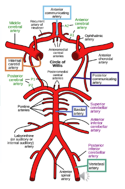
What are the two main sources of cerebral arterial blood, and how do they form the Circle of Willis?
Heart → aorta → subclavian artery → vertebral artery (L+R)
L+R v combine to form basilar artery
Heart → aorta → common carotid artery → internal carotid artery (L+R)
Both sources of blood combine to form the cerebral arterial circle (of willis)
Posterior communicating artery:
Connects vertebral and internal carotid arteries on same side
Anterior communicating artery:
Connects between branches of left and right internal carotid arteries

Compare the vascular territories and characteristic stroke symptoms of the ACA, MCA, and PCA.
Anterior cerebral artery (ACA)
Top side of the surface of the frontal/parietal cortex
Infarct symptoms:
Motor/sensory loss to the opposite side of the stroke lower limb
Since the lower limb is not associated with any CN
Middle cerebral artery (MCA)
Bottom sides of the frontal/parietal lobes and the top side of the temporal lobe
Infarct symptoms:
Weakness/paralysis on one side of the body (face/arm), speech difficulty, confusion, sudden loss of senses
Associated with CN V, VII, and XII
Posterior cerebral artery (PCA)
Bottom side of the temporal lobe and the occipital lobe
Infarct symptoms:
Complete loss of visual field on ONE HALF of each eye
This is because each part of the brain is responsible for one side of the eye (weird)
Associated with CN II

Infractions → associated with opposite side of stroke (damage to the cortical region and disrupts cortical control/processing of cranial nerve function

What are the primary functions of the cerebral lobes and corpus callosum?
Frontal lobe - high-order processing
Parietal lobe - sensory perception
Temporal lobe - auditory processing and memory acquisition
Occipital lobe - visual processing
Insular lobe - homeostasis and emotion
Corpus callosum - bridge between L+R cerebral hemispheres
Which arteries supply the cerebellum and brainstem?
Cerebellum
PICA (Posterior Inferior Cerebellar Artery): Branches from the vertebral artery.
AICA (Anterior Inferior Cerebellar Artery): Branches from the basilar artery.
Brainstem
Primarily supplied by branches of the vertebral and basilar arteries.
What is the monitoring process like if pt is given tPA/tNK/has an ischemic stroke?
All patients given tPA or ischemic stroke need ICU-level monitoring. All patients should be monitored for:
Bleeding
Intracranial hemorrhage
Other bleeding: gums, epistaxis
Angioedema
Monitoring frequency should be at least:
Q15min x2 hours after infusion, then
Q30min x6 hours, then
Q1hr until 24 hours post-infusion
Other:
BP should be maintained <180/105 mmHg for 24 hrs.
No antiplatelets or anticoagulants for 24 hours
No poking (e.g., NG tubes, Foley catheters, IA lines)
Repeat CT scan at 24 hours before starting ASA or DVT prophylaxis
How efficacious is tPA/tNK?
About 30% of patients receiving tPA for stroke get better. However, 60% will not see any harm or benefit from this medication. The rest is gg.
How do you calculate CHA2DS2-VASc scores?
Risk factor | Points |
|---|---|
C: Congestive heart failure | +1 |
H: Hypertension | +1 |
A₂: Age ≥75 years | +2 |
D: Diabetes mellitus | +1 |
S₂: Prior stroke/TIA/systemic thromboembolism | +2 |
V: Vascular disease (prior MI, PAD, aortic plaque) | +1 |
A: Age 65–74 years | +1 |
Sc: Sex category (female) | +1 |
Score of ≥ 2 (Men) or ≥ 3 (Women) → anticoag recommended
What are the mechanisms of action (MOA) and adverse effects (SE) of alteplase and tenecteplase?
MOA:
Both: Bind to fibrin → convert plasminogen into plasmin → which breaks down fibrin and dissolves existing clots.
Tenecteplase: Modified alteplase with greater fibrin specificity, resistance to PAI-1 (enzyme that breaks down intrinsic tPA), and a longer duration of action.
SE (Both):
Bleeding (major risk):
Intracranial hemorrhage (ICH)
Mucosal bleeding (gums, epistaxis)
Angioedema: Rapid swelling of the tongue/larynx, potentially causing airway obstruction.

What is the difference in MOA/SE between mannitol and hypertonic saline?
Mannitol
MOA:
Decreases ICP through osmotic diuresis
Holds water in tubular lumen
Pulls water from intracellular compartments into the blood
Temporarily increases volume/dilutes blood
Freely filtered in kidneys, poorly or not reabsorbed
Aggressively increases urine output (if GFR is adequate)
Moves so fast that reabsorption of solutes is decreased
Concentrates blood as a result
SE:
Renal failure
Ensure renal function is okay before use since it requires proper/free filtration to increase urine output
Hypovolemia
Hypotension
Heart failure
Pulmonary edema
Hypertonic saline
MOA:
NaCl concentrated solution
NaCl stays in lumen of blood stream drawing water towards it
Can increase BP since it does not cause significant diuresis like mannitol does
SE:
Hypernatremia risk
Depending on volume (increased volume = decrease Na concentration)
AKI
Central pontine myelinolysis (CPM) → damage to myelin sheath covering nerve cells
Volume overload/HF
When should you use mannitol over hypertonic saline and vice versa?
Mannitol:
Increased ICP when adequate renal function is present
Hypertonic saline:
Preferred when volume/BP support is needed
Concentrated bolus for impending herniation
What is the MOA, AE, monitoring, and reversal for Warfarin?
MOA:
Inhibits vitamin K epoxide reductase (VKOR) → prevents regeneration of active vitamin K → decreases functional vitamin K-dependent factors II, VII, IX, X, and proteins C and S
AE: Bleeding
Monitoring: INR
Reversal: 4-factor Prothrombin complex concentrate (PCC) (factors II, VII, IX, X + proteins C/S) + IV vitamin K
What is the MOA, AE, monitoring, and reversal for DOACs?
MOA:
Directly inhibits factor Xa → ↓ conversion of prothrombin to thrombin → ↓ fibrin clot formation.
AE: Bleeding
Reversal: 4-factor Prothrombin complex concentrate (PCC) (factors II, VII, IX, X + proteins C/S)
What is the MOA, AE, monitoring, and reversal for Aspirin?
MOA:
Inhibits COX → ↓ thromboxane A₂ (TXA₂). Since TXA₂ normally causes vasoconstriction and stimulates platelet aggregation, reducing TXA₂ inhibits platelet aggregation.
AE:
Bleeding
GI ulcers
Renal toxicity
What is the MOA, AE, monitoring, and reversal for Clopidogrel and Ticlopidine?
MOA:
Irreversible ADP receptor antagonists. They inhibit ADP-mediated platelet activation/aggregation. They are prodrugs requiring metabolic activation, including CYP2C19; therefore, loss of CYP2C19 function can reduce clopidogrel efficacy.
AE: Clopidogrel has a better SE profile than Ticlopidine
Neutropenia
Thrombotic Thrombocytopenic Purpura (TTP)
Bleeding (low)
Ticlopidine - leukopenia is more frequent
What is the MOA, AE, and reversal for Direct thrombin inhibitors?
MOA: dabigatran, argatroban, bivalirudin
Directly inhibit thrombin (factor IIa) → prevent conversion of fibrinogen → fibrin.
AE: Bleeding
Reversal agents:
Idarucizumab IV
What is the MOA, AE, monitoring, and reversal for Direct thrombin inhibitors?
MOA: Dipyridamole and cilostazol
PDE inhibitors and vasodilators with antiplatelet effects.
AE:
Bleeding
Headache
Diarrhea
What is the reversal for heparin/LMWH
Protamine (only partially reverses enoxaparin)
How do the sympathetic, parasympathetic, and somatic nervous systems differ in their control of pupil size?
Sympathetic (ANS): Stimulates the dilator pupillae muscle → pupil dilation (mydriasis).
Parasympathetic (ANS): Stimulates the sphincter pupillae muscle → pupil constriction (miosis).
Somatic nervous system: Does not control pupil size.
Efferent fibers: Control voluntary skeletal muscle contraction.
Afferent fibers: Carry sensory information to the CNS.
Describe the process of an action potential
Resting: -70mV
Voltage-gated Na and K channels are closed
Stimulus → Threshold potential: -55mV
Excitatory and inhibitory inputs are summed at the axon hillock (decision-making location in the axon with a lot of Na channels)
Excitatory input depolarizes the membrane, making it less negative, while inhibitory input hyperpolarizes it.
If the net input depolarizes the membrane to −55 mV, the neuron reaches threshold and fires an action potential
At -55, voltage-gated Na channels open → further depolarization
Peak (+30mV)
Na channels inactivate
K channels open → K moves out of the neuron, causing repolarization of the neuron
Hyperpolarization
K channels stay open slightly longer than needed = more negative resting potential
Return to resting potential
K channels close → membrane stabilized around =70mV

What is the difference between adaptive and maladaptive neural plasticity?
Adaptive
Rewiring neuronal circuits can be useful in neuronal connections (learning and memory)
Repeated activity between neurons can strengthen their synaptic connection.
“neurons that fire together, wire together.”
Strengthening can involve:
Insertion of additional AMPA receptors into the postsynaptic membrane
Changes in AMPA receptor conductance
Changes in the amount of neurotransmitter released
Structural changes at the synapse (pre- and postganglionic closer together)
Maladaptive
The same rewiring can also produce abnormal or harmful connections (depression, PTSD, addiction, anxiety, phantom limb pain, etc.)
What is the effect of CNS drugs on plasticity (acute vs chronic)?
Acute:
Alter receptors, ion channels, neurotransmitter synthesis/degradation, or neurotransmitter transport
Chronic:
Longer-term remodeling helps explain why some CNS medications have a lag between their immediate molecular effects and their therapeutic effects
What are the main receptors involved in synaptic plasticity? What is the process involved?
Primary receptors:
AMPA receptor
When activated, it opens and causes Na influx into the postsynaptic cleft
NMDA receptor
Normally, this channel is blocked by Mg
1) Repeated, high frequency of stimulus → glutamate release → activation of AMPA receptors
2) When activated, causes Na influx + postsynaptic depolarization
3) Postsynaptic depolarization relieves the block of Mg on NMDA receptors → influx of Ca into postsynaptic cleft
4) Calcium influx signals and promotes trafficking and insertion of additional AMPA receptors on the postsynaptic membrane
5) More AMPA receptors lead to:
Changes in the amount of neurotransmitter released from presynaptic neuron
Changes in distance of the pre- and postsynaptic clefts
Changes in strength of AMPA conductance → increased depolarization + AP firing in post-synaptic cleft.

Describe the process of synaptic transmission, including fast vs. slow signaling and action potential generation.
1. Presynaptic neurotransmitter release:
AP reaches axon terminal → voltage-gated Ca²⁺ channels open → Ca²⁺ influx → vesicle fusion → neurotransmitter release into synaptic cleft.
2. Postsynaptic signaling:
Fast (LGIC): Neurotransmitter directly opens ion channels.
Excitatory (AMPA): Cation influx → depolarization → ↑ likelihood of AP.
Inhibitory (GABA-A): Cl⁻ influx → hyperpolarization → ↓ likelihood of AP.
Slow (GPCR): Neurotransmitter activates G-proteins → second messenger signaling.
3. Action potential generation:
Excitatory and inhibitory signals summate at the axon hillock.
If membrane potential reaches threshold (~−55 mV), voltage-gated Na⁺ channels open → rapid depolarization → AP fires.

What causes SSRI/SNRI discontinuation syndrome, what are its symptoms (+onset/duration), and how can it be prevented or managed?
Cause: Abrupt discontinuation or missed doses, especially with short half-life antidepressants.
Risk (highest → lowest): Venlafaxine, paroxetine, fluvoxamine > duloxetine, sertraline, citalopram, escitalopram >>> fluoxetine.
Symptoms (FINISH):
F – Flu-like symptoms (fatigue, chills, muscle aches)
I – Insomnia (vivid dreams, sleep disturbances)
N – Nausea (sometimes accompanied by vomiting or diarrhea)
I – Imbalance (dizziness, vertigo, lightheadedness)
S – Sensory disturbances ("brain zaps," burning, tingling, or electric-shock sensations)
H – Hyperarousal (anxiety, agitation, irritability → more sexually active??)
Other: Crying spells, suicidal ideation.
Onset: 48–72 hours after discontinuation.
Duration: Typically 3–7 days.
Prevention/management:
Avoid missed doses and abrupt discontinuation.
Gradually taper over weeks to months (e.g., 50% dose reduction every 2–4 weeks).
Consider switching to fluoxetine (Prozac) before tapering due to its long half-life and active metabolite (norfluoxetine).
What causes serotonin syndrome, what are its symptoms, and how is it treated?
Cause: Excessive serotonergic activity, often from combining serotonin-enhancing drugs.
Opioids: Meperidine, fentanyl, methadone, tramadol
Stimulants/psychedelics: MDMA, LSD
Other: SSRIs, SNRIs, MAOIs, and other serotonergic drugs
Symptoms (triad):
Cognitive: Confusion, agitation, hallucinations, hypomania, coma
Autonomic: Diaphoresis, fever, HTN, tachycardia, shivering, nausea
Neuromuscular: Tremor, hyperreflexia, clonus, muscle twitching
Treatment:
Immediately discontinue serotonergic medications.
Supportive care (IV fluids, cooling, monitoring).
Benzodiazepines for agitation and muscle activity.
Cyproheptadine (5-HT2A antagonist) for persistent or significant symptoms.
What causes MAOI-induced hypertensive crises, and which drug/food interactions can precipitate them?
Cause: Excessive norepinephrine (NE) activity → increased α1-adrenergic stimulation → vasoconstriction → severe hypertension.
Precipitating interactions:
Tyramine-rich foods: Fermented/aged foods contain tyramine, which is normally metabolized by intestinal MAO-A. MAO inhibition → ↑ tyramine absorption → ↑ NE release.
Sympathomimetics: Pseudoephedrine, phenylephrine, epinephrine → ↑ adrenergic stimulation.
Stimulants: Cocaine, methamphetamine → ↑ synaptic NE.
Prevention: Avoid tyramine-rich foods and interacting sympathomimetic/stimulant drugs while taking MAOIs.
What is the biosynthesis and metabolism of serotonin?
Biosynthesis:
Tryptophan (trp) --(Tryptophan hydroxylase and Aromatic amino acid decarboxylase)→ Serotonin (5HT)
Metabolism:
Serotonin (5HT) --(Monoamine oxidase and Aldehyde dehydrogenase)→ 5-Hydroxy indole acetic acid (HIAA)
What is the biosynthesis and metabolism of NE?
Biosynthesis:
Tyrosine --(Tyrosine hydroxylase)→ L-dopa
L-dopa --(Aromatic amino acid decarboxylase)→ Dopamine (DA)
Dopamine --(Dopamine B-hydroxylase)→ Norepinephrine
Metabolism:
Dopamine AND Norepinephrine are metabolized by
Monoamine oxidase (MAO)
Aldehyde dehydrogenase
Catechol-O-Methyl transferase (COMT)
What is the monoamine theory of depression, what is its major limitation, and what are the alternative theories?
Monoamine theory: Depression results from deficient serotonin (5-HT) and/or norepinephrine (NE) in cortical and limbic brain regions.
Limitation: Antidepressants increase 5-HT/NE immediately, but mood improvement takes 1–2 weeks to begin and 4–6 weeks for fuller effects.
Alternative theories:
1) Neuroreceptor theory: Abnormalities in monoamine receptor number, sensitivity, or function contribute to depression.
2) Neuroendocrine theory: Dysregulation of hormonal stress-response systems (e.g., HPA axis) contributes to depression.
3) Network theory: Depression involves impaired neuronal circuitry and information processing.
Acute antidepressant effects alter synaptic transmission.
Chronic changes modify receptor levels, location, and sensitivity (lag phase).
Neural remodeling improves information processing and mood over time.
What is the diagnostic criteria for Major depressive disorder (MDD)?
DSM5 - 5 of the following symptoms must be present >2 weeks (one must be from bolded)
Depressed mood
Loss of pleasure or interest (anhedonia)
Change in appetite
Change in sleep patterns
Loss of energy
Hopelessness, worthlessness, inappropriate guilt
Diminished ability to think or concentrate
Psychomotor agitation or retardation
Recurrent thoughts of death or suicide
What are the subtypes, risk factors, and natural course of major depressive disorder (MDD)?
Subtypes
Seasonal pattern (SAD)
Pregnancy/postpartum
Atypical
Severe/melancholic
Anergic/amotivational
Anxious/irritable
Risk factors for depression
Women > men (typically because of menarche + after menopause) → comparable until then
Family history/genetics, medical conditions, medications, environmental stressors
Natural course of depressive illness
Mean age of onset: ~40 years
Recurrence: high, >50%
Between episodes: about 80% have a full recovery
Episodes may show a seasonal pattern
What is the proposed pathophysiology of major depressive disorder (MDD)?
Decrease neurotransmitters:
5HT, NE, DA
Increased inflammatory response due to increased cortisol levels (stress):
HPA axis dysfunction: increased CRF
Inflammatory cytokines: increased IL-1, IL-6, TNF
Leads to things like:
Change in sleep, appetite, energy
Depression, anhedonia, anxiety, concentration
What are the major risk factors for suicide, and how should suicide risk be assessed?
Risk factors:
Older male, widowed, or living alone
Mood disorders (bipolar > unipolar depression)
Serious illness (self or loved one)
Job loss or loss of self-worth
Alcohol/substance abuse
History of impulsivity
Suicide risk assessment:
Ideation: Current suicidal thoughts? When was the last occurrence?
Plan: Specific plan, intent, or preparation?
Means: Access to methods of suicide?
Capability: Ability to carry out the plan?
Impulsivity: History of impulsive behavior?
Substance use: Alcohol or drug use?
Safety: What would they do if suicidal thoughts returned or worsened?
What are the treatment options for MDD, and how does treatment selection differ by severity of diagnosis?
1) Psychotherapy
Cognitive Behavioral Therapy (CBT)
Dialectic Behavioral Therapy (DBT)
Interpersonal Psychotherapy (IPT)
Onset: 8-10 weeks
2) Pharmacotherapy - faster than psychotherapy, but not as long-term an impact
Selective Serotonin Reuptake Inhibitors (SSRI)
Serotonin Norepinephrine Reuptake Inhibitors (SNRI)
Norepinephrine Reuptake Inhibitors (NRI)
Others: mirtazapine, atypical antipsychotics, tricyclics (TCA), MAO inhibitors (MAOI), vortioxetine, ketamine
All antidepressants are created equal:
Onset: 1-2 weeks and 4-6 weeks for maximum improvement
3) Lifestyle modifications
Exercise
Mindfulness based stress reduction
Integrative medicines (St John's Wort, Omega-3 FA, methylfolate, SAMe)
Electroconvulsive therapy (ECT) - most effective
Repetitive transcranial magnetic stimulation (rTMS)
Deep brain stimulation
When to use what:
Mild depression/Moderate depression: either psychotherapy OR pharmacotherapy
Severe depression: psychotherapy AND pharmacotherapy
Which antidepressants inhibit CYP2D6 and CYP3A4, and what are the clinically relevant drug interactions?
CYP2D6 inhibition:
Strong: Fluoxetine, paroxetine, bupropion
Moderate: Duloxetine
Weak/minimal: Sertraline, citalopram, escitalopram
Substrates:
β-blockers, TCAs, opioids (codeine, tramadol, hydrocodone, oxycodone), Sulfonylureas, Metoclopramide
Clinical significance: Inhibition → ↑ concentrations of active parent drugs or ↓ activation of opioid prodrugs (especially codeine/tramadol) → reduced analgesia.
Alternatives: Morphine, hydromorphone, oxymorphone, fentanyl, buprenorphine, methadone.
CYP3A4 inhibition (lecture ranking):
Norfluoxetine >> fluoxetine, paroxetine, sertraline, citalopram, escitalopram
Substrates:
CCBs, estrogens, corticosteroids, statins (atorvastatin, lovastatin, simvastatin), protease inhibitors, alprazolam, triazolam, buspirone, sildenafil, Z-drugs.
Clinical significance: Inhibition → ↑ substrate concentrations → increased adverse effects/toxicity.
What is the goal of MDD treatment? How should antidepressant therapy for MDD be adjusted based on response (no, partial response), monitored, and continued after remission (maintained)?
Goal: Optimize monotherapy (dose + duration) before augmentation.
Treatment response:
1–2 weeks: Initial improvement
4–6 weeks: Fuller therapeutic response
Minimal/no response: Switch to another SSRI or antidepressant class (SNRI, NDRI, mirtazapine).
Partial response: Consider augmentation with -
Bupropion, buspirone
Atypical antipsychotics
T3 (liothyronine), lithium
Other: Methylfolate, omega-3 fatty acids, DHEA
Monitoring:
PHQ-9: Routinely assess symptom improvement
Response: ≥50% reduction in score
Remission (goal): Virtual absence of symptoms (PHQ-9 <5)
Anything above 10 is moderate below 9 = mild
Monitor adverse effects, safety, and DDIs.
Maintenance:
Continue antidepressants for ≥6 months after remission.
Consider indefinite treatment for ≥3 depressive episodes.
Gradually taper SSRIs/SNRIs to prevent discontinuation syndrome (e.g., decrease dose by 50% every 2–4 weeks, individualized).
How is depression treated during pregnancy and lactation?
Mild antepartum depression: Psychotherapy preferred.
Alternatives: UV light therapy, omega-3 fatty acids.
Moderate–severe: Pharmacotherapy ± psychotherapy.
SSRIs preferred (sertraline): Low fetal exposure based on umbilical vein-to-maternal serum concentration.
Historical FDA categories: Most SSRIs/SNRIs/NDRIs category C; paroxetine category D.
ECT: Considered safe during pregnancy when clinically indicated.
Lactation: Antidepressants are not specifically FDA-approved for lactation, but many are compatible with breastfeeding.
Which antidepressants are used or FDA-approved in pediatric patients?
MDD:
Fluoxetine: FDA-approved for ages ≥8.
Escitalopram: FDA-approved for ages ≥12.
OCD: Fluoxetine, sertraline, fluvoxamine, clomipramine.
Bupropion: Off-label for pediatric MDD and ADHD.
What factors influence antidepressant selection in geriatric patients, and which medications are commonly considered?
Selection factors: Medical comorbidities, polypharmacy, potential DDIs, and adverse effects.
Common options:
SSRIs: Often first-line.
Bupropion: May be preferred for fatigue, low energy, or sexual dysfunction.
Mirtazapine: May be preferred for insomnia, poor appetite, or weight loss.
Start at lower doses and titrate cautiously due to increased sensitivity to adverse effects.
Which antidepressant is preferred for patients with chronic or neuropathic pain?
SNRI (duloxetine, venlafaxine) due to increased 5-HT and NE signaling involved in pain modulation.
Which antidepressant is preferred for atypical depression (hypersomnia, hyperphagia, anergia)?
NDRI (bupropion) due to its activating effects on NE and DA.
Which antidepressants are preferred for patients with cognitive symptoms or inattention?
NDRI (bupropion) or vortioxetine (Trintellix).
How should SSRI-induced sexual dysfunction be managed?
Augment: Add bupropion (NDRI).
Switch: Taper SSRI and switch to bupropion, mirtazapine (NaSSA), or vortioxetine (Trintellix).
Which antidepressants are preferred based on residual depressive symptoms?
Anxious/irritable: SSRI (5-HT)
Anergic/amotivational: NDRI (NE/DA)
Severe/melancholic: SNRI or TCA (5-HT + NE)
Which antidepressants are preferred or avoided in patients with cardiovascular disease?
Preferred: SSRIs
Avoid/caution: TCAs due to increased risk of cardiac events.
Monitor: SSRI interactions involving CYP2D6 and CYP3A4.
Which antidepressants should be avoided in patients with hypertension and tachycardia?
SNRIs and NDRIs (bupropion) due to their potential to increase BP and HR.
What antidepressant considerations are important in patients with diabetes (weight, DDIs, neuropathic pain)?
Avoid/caution: TCAs, mirtazapine, paroxetine (also maybe citalopram + escitalopram) due to weight gain.
DDIs: Fluoxetine and fluvoxamine may inhibit sulfonylurea metabolism → hypoglycemia risk.
Neuropathic pain: SNRIs may provide additional benefit.
Which antidepressants may benefit patients with breast cancer or menopausal symptoms?
SSRIs or SNRIs may reduce vasomotor symptoms (hot flashes).
Which antidepressants should be used cautiously in patients with peptic ulcer disease or concurrent NSAID use?
SSRIs and SNRIs due to increased GI bleeding risk, especially when combined with NSAIDs.
What antidepressants may be considered for patients experiencing weight gain with paroxetine?
Switch to another SSRI with less weight gain potential, such as fluoxetine or sertraline.
How do different brain regions and neurotransmitters interact to regulate sleep and wakefulness?
Suprachiasmatic nucleus (SCN): Internal circadian clock → projects to dorsomedial hypothalamus (DMH).
DMH projections:
Ventrolateral preoptic area (VLPO): Promotes sleep by releasing GABA and galanin to inhibit wake-promoting nuclei.
Lateral hypothalamic area (LHA) (orexin/hypocretin neurons): Promotes and stabilizes wakefulness.
Orexin neurons stimulate wake-promoting nuclei that release:
NE: Locus coeruleus
5-HT: Raphe nuclei
Histamine: Tuberomammillary nucleus
ACh: Brainstem/basal forebrain nuclei
Orexin stabilizes sleep-awake switch, preventing sudden transitions between sleep and wakefulness.
These neuromodulators promote cortical arousal and wakefulness.
Paraventricular hypothalamus (PVH): Regulates corticosteroid release.
What is memory consolidation, and how does sleep before and after learning affect memory formation?
Memory consolidation: The process of stabilizing newly learned information into long-term memory.
Awake (encoding):
Sensory information is rapidly encoded in the hippocampus (rapid plasticity).
Sleep (consolidation):
Recently encoded information is replayed.
Memories are transferred/integrated from the hippocampus into appropriate cortical networks (slower plasticity).
Strengthens long-term memory storage.
Sleep before learning:
Prepares the brain to encode new information.
One night of sleep deprivation → ~40% reduction in new memory formation.
Sleep after learning:
Promotes memory consolidation and long-term retention.
Improves insight, inference, and integration of learned information.

How do sleep patterns change with age, and how does aging affect memory consolidation?
General trend: Aging → ↓ total sleep duration, ↓ deep sleep (NREM slow-wave sleep), ↑ sleep fragmentation.
Children: More total sleep and deep sleep.
Young adults: Less total sleep and deep sleep, especially later in the night.
Elderly: Less total sleep, very little deep sleep, and more frequent awakenings.
Impact on memory:
Aging → medial prefrontal cortex (mPFC) atrophy → disrupted NREM slow-wave activity (SWA).
↓ SWA → impaired hippocampal-dependent memory consolidation → reduced long-term memory retention.
What are the characteristics of REM vs. NREM sleep, and what happens when each stage is disrupted?
REM sleep:
Similar to waking state:
Increase of:
Neuronal activity
Metabolic rate
BP/HR
There is loss of muscle tone (atonia) despite high brain activity
Disruption of REMS → loss of muscle tone (paralysis or REM sleep behavior disorder, depending on the dysfunction)
NREM sleep:
Decrease of:
Neuronal activity
Metabolic rate
BP/HR
Stage 1 (N1): Lightest sleep.
Stage 2 (N2): Light sleep.
Disruption → fragmented sleep and frequent awakenings.
Stages 3–4 (deep NREM): Slow-wave sleep (SWA).
Disruption → impaired memory consolidation, learning, and retention.
What are the four functions and purpose to why we sleep?
1) Conserve metabolic energy
During NREM decrease of metabolic processes = save energy
2) Restore our body and brain
Sleep provides time for physical and neural restoration
Prolonged total sleep deprivation (studied in rats):
Failed to regulate core body temp
Developed infections
Eventually died after several weeks
3) Organize learned information → see above
4) Promote maturation of neurons and mental health
One observation consistent with a developmental role is that sleep duration is highest early in life and progressively decreases across the lifespan.
What is the structure of GABA-A receptors, where do drugs bind, and how do α-subunits determine their pharmacologic effects?
Structure:
Ligand gated chloride channel with 5 subunits:2 alpha, 2 beta, plus 1 gamma, delta, epsilon, theta, or pi.
GABA binds to receptor and opens Cl-pore = hyperpolarizes the neuron decreasing the neuronal excitability and likelihood of an action potential → sedating + inhibitory effect
Binding sites:
α/β interface: GABA, gaboxadol.
α/γ interface: Benzodiazepines (BZDs), Z-drugs → ↑ frequency of Cl⁻ channel opening.
Alcohol: Enhances GABA-A-mediated inhibition → sedation.

How do GABA-A receptor α-subunits determine pharmacologic effects and drug selectivity?
α1 (~60%): Sedation, amnesia, anticonvulsant effects.
α2 (15–20%): Anxiolysis, muscle relaxation.
α3 (10–15%): Muscle relaxation (higher doses).
α5 (less than 5%): Muscle relaxation.
α4, α6 (less than 5% each): Insensitive to BZDs and Z-drugs.
Drug selectivity:
BZDs: Bind α1, α2, α3, α5-containing receptors.
Z-drugs: Preferentially bind α1-containing receptors → primarily hypnotic/sedative effects.
How is GABA synthesized and metabolized through the GABA shunt?
Biosynthesis:
Glutamate → GABA + CO₂
Enzyme: Glutamate decarboxylase (GAD)
Metabolism (GABA shunt):
GABA transaminase (GABA-T): Transfers the amino group from GABA to α-ketoglutarate → succinic semialdehyde + glutamate.
Succinic semialdehyde dehydrogenase (SSADH): Converts succinic semialdehyde → succinate → re-enters TCA cycle.

How are benzodiazepines metabolized, and which are preferred in elderly patients or those with liver impairment?
Phase 1 (CYP3A4, CYP2C19):
Oxidative metabolism → may produce active metabolites.
Example: Diazepam
Reduced in elderly patients and liver impairment → ↑ accumulation/toxicity.
Phase 2 (glucuronidation):
Produces inactive metabolites.
LOT: Lorazepam, Oxazepam, Temazepam.
Relatively preserved in aging and liver impairment.
Preferred: LOT benzodiazepines due to reduced risk of accumulation.
How do tolerance and dependence develop with benzodiazepines and Z-drugs?
Tolerance: Requires higher or more frequent doses to achieve the same effect.
Neuroadaptation begins within 1–2 weeks.
Tolerance to sedation may develop within 2–4 weeks.
Dependence: Brain adapts to drug presence and requires continued exposure to maintain balance.
MOA:
Chronic GABA-A enhancement → compensatory neuronal adaptations to restore excitatory/inhibitory balance.
Risk: Prolonged use increases the risk of tolerance and physical dependence.

What causes benzodiazepine/Z-drug withdrawal, what are its symptoms, and what increases withdrawal severity?
Cause: Abrupt discontinuation after physical dependence → loss of GABAergic inhibition before neuronal readaptation.
Risk factors:
Higher doses
Shorter half-life
Prolonged continuous use (>2 months)
Symptoms:
Mild–moderate: Anxiety, insomnia, restlessness, sweating, ↑ HR/BP.
Severe: Seizures, hallucinations, delusions.
Prevention: Gradual dose tapering rather than abrupt discontinuation.

What is the antidote for benzodiazepine/Z-drug overdose, and what are its mechanisms, considerations, and risks?
Flumazenil (Anexate)
MOA: Competitive antagonist at the benzodiazepine binding site on GABA-A receptors.
Blocks benzodiazepine/Z-drug enhancement of GABA-mediated Cl⁻ conductance.
Half-life: ~30–60 minutes.
Repeat dosing may be necessary due to resedation.
Adverse effects: Agitation, anxiety, seizures (especially in dependent patients or mixed overdoses).
What are the primary neurotransmitters involved in anxiety, and what are their roles?
GABA:
Primary inhibitory neurotransmitter → ↓ neuronal excitability and anxiety.
Dysregulation → increased anxiety.
Serotonin (5-HT):
Modulates mood, anxiety, and emotional responses.
Norepinephrine (NE):
Promotes arousal, vigilance, and sympathetic fight-or-flight responses.
What is insomnia, and what are its types, risk factors, and consequences?
Difficulty initiating or maintaining sleep associated with daytime impairment.
Types:
Sleep-onset: Difficulty falling asleep.
Sleep-maintenance: Difficulty staying asleep or returning to sleep.
Primary: Not attributable to another condition.
Secondary: Associated with medical/psychiatric conditions, medications, or substances.
Risk factors: Stress, poor sleep habits, medical conditions, caffeine, substance use/withdrawal, and medications (steroids, stimulants, decongestants, SABAs).
Comorbidities: Depression, anxiety, chronic pain, other sleep disorders.
Consequences: Fatigue, impaired concentration, mood disturbances, reduced quality of life.
What is the 3P model of insomnia?
Predisposing factors: Baseline traits that increase vulnerability to insomnia.
Precipitating factors: Events or conditions that trigger insomnia.
Perpetuating factors: Behaviors or circumstances that maintain insomnia and contribute to its transition from acute to chronic.
What is the first-line treatment for chronic insomnia, and what are the components of it? Lifestyle changes?
Cognitive behavioral therapy for insomnia (CBT-I).
~70–80% effective; typically delivered over 6–8 weeks.
Components:
Stimulus control: Associate bed with sleep; maintain consistent wake times.
Sleep restriction: Limit time in bed to approximate actual sleep time.
Cognitive restructuring: Address unhelpful beliefs, worry, and anxiety about sleep.
Sleep hygiene: Avoid caffeine, alcohol, nicotine, long naps, and screens before bed; maintain a cool, dark bedroom and regular exercise.
Used alongside CBT-I, not as a standalone treatment.
Weight loss talk (see BMI + PMH)
What medications are used for chronic insomnia, and how are they selected based on sleep-onset versus sleep-maintenance symptoms?
Drug class | Clinical use | Key considerations |
|---|---|---|
BZDs/Z-drugs | Sleep onset and/or maintenance | Enhance GABA-A; tolerance, dependence, CNS impairment |
DORAs (orexin) Belsomra (suvorexant) DayVigo (lemborexant) Quviviq (daridorexant) | Sleep onset + maintenance | Block orexin; lower dependence risk, generally preserve sleep architecture |
Ramelteon (melatonin synthetic) | Sleep onset | Melatonin receptor agonist; minimal dependence risk |
Low-dose doxepin (TCA) | Sleep maintenance | H1 antagonism; low dependence risk |
Consider pharmacotherapy alongside CBT-I when needed.
What other medications are mentioned for Insomnia but are not generally considered? Why?
First-generation antihistamines:
Anticholinergic effects, next-day sedation.
Tolerance after extended use (1-2 weeks)
TCAs
Tolerance after extended use (1-2 weeks)
Trazodone:
Limited evidence for routine insomnia treatment; orthostatic hypotension.
Antipsychotics (e.g., quetiapine):
Metabolic adverse effects, weight gain.
Generally avoid given many side effects
Natural products:
Insufficient or inconsistent evidence.
Gabapentinoids and other sedating medications:
Generally reserved for patients with another indication.