BIOL 315 Cell Bio Quiz 3
 Call Kai
 Learn
 Practice Test
 Spaced Repetition
 Match
 Flashcards
 Knowt Play
1/109

There's no tags or description
Looks like no tags are added yet.
110 Terms
Diameter of microtubules
25nm
Diameter of microfilaments
7nm
Diameter of Intermediate filaments
8-12nm
Subunits of microfilaments (before binding)
G - actin
cells are chemically preserved, then labeled with fluorescent compounds that bind to the cytoskeletal proteins (such as antibodies), causing them to glow in the fluorescence microscope
fluorescence microscopy on fixed specimens
fluorescent version of cytoskeletal proteins are made and introduced into living cells, which are then observed under the fluorescence microscope and digitally recorded
live cell fluorescence microscopy
high resolution images from a video or digital camera (attached to a microscope) are processed by computers to increase contrast and remove background features
computer enhanced digital video microscopy
can resolve individual filaments; cells prepared by thin section, quick freeze deep-etch, or direct mount techniques
electron microscopy
microtubules that pervade the cytoplasm to maintain or alter cell shape, construct mitotic and meiotic spindles, and serve as tracks for motor proteins to walk vesicles and organelles
cytosolic microtubules
microtubules located in cell surface projections such as cilia and flagella, generating cell motility or sweeping fluid/mucus across epithelial surfaces
axonemal microtubules
globular protein subunits of microtubules. they fold similar and bind together. Each has an N-terminal GTP-binding domain and a C-terminal that interacts with microtubule-associated proteins (MAPs)
alpha and beta tubulin subunits
The fundamental building block of microtubules, consisting of one alpha-tubulin and one beta-tubulin subunit tightly bound together in a specific, non-covalent, head-to-tail orientation
tubulin heterodimer
Closely related structural variants of α- and β-tubulin encoded by distinct genes within an organism
tubulin isoforms
(in microtubules) A longitudinal, linear array of alternating alpha- and beta-tubulin heterodimers stacked head-to-tail
protofilament
fast growing end of a microtubule where beta-tubulin is exposed; lower critical concentration for dimer addition
plus end
slow-growing or anchored end of a microtubule where alpha-tubulin is exposed; higher critical concentration for dimer addition
minus end
A single hollow cylinder composed of 13 complete protofilaments (found in cytosolic microtubules)
singlet
One complete 13-protofilament ring (A tubule) fused to an incomplete 10–11 protofilament ring (B tubule); characteristic of axonemal cilia and flagella
doublet
One complete 13-protofilament ring (A tubule) fused to two incomplete rings (B and C tubules); characteristic of basal bodies and centrioles
triplet
The initial, slow phase of microtubule assembly where individual tubulin heterodimers coalesce into small, unstable clusters called oligomers (which serve as "seeds")
nucleation
The rapid growth phase where tubulin heterodimers are rapidly added onto the ends of existing oligomers or growing microtubule ends
elongation
The steady-state phase where free tubulin dimer concentration drops, balancing the rate of dimer addition and disassembly
plateau phase
The threshold concentration of free tubulin heterodimers required for microtubule assembly. Growth occurs above it, while disassembly occurs below it
critical concentration
A phenomenon occurring when the free tubulin dimer concentration is above the critical concentration for the plus end but below that for the minus end. Heterodimers are continuously added to the plus end while simultaneously detaching from the minus end, making the microtubule appear to "walk" or move through space without changing its overall length
treadmilling
A stabilizing region of GTP-bound tubulin heterodimers at the growing plus end of a microtubule that protects it from disassembly
GTP Cap
The continuous alternation between rapid microtubule growth and sudden shrinkage driven by GTP hydrolysis
dynamic instability
In microtubules, the sudden, rapid disassembly and peeling apart of protofilaments caused when GTP hydrolysis catches up to the growing tip and the protective GTP cap is lost
catastrophe
The abrupt transition from catastrophe back to growth when a new GTP cap is re-established by adding fresh GTP-bound tubulin dimers
rescue
Drugs that bind to free tubulin heterodimers (beta-tubulin) to prevent dimer addition, destabilizing microtubules and promoting disassembly/catastrophe to block cell division
colchicine, vinblastine, nocodazole
A chemotherapy drug that binds to beta-tubulin in GDP-bound heterodimers and forces their shape to mimic GTP-bound heterodimers, preventing dissociation by “freezing” microtubules in place. This prevents cancer cell division
taxol
binds at regular intervals along the microtubule wall and interacts with other structures and filaments. it can bundle microtubules together
MAP (tau)
proteins that capture and stabilize the growing ends of a microtubule
+- TIP proteins (EB1)
proteins that promote depolarization or peeling apart of subunits of microtubules
catastrophins (MCAK)
3 functions of microfilaments
cell movement, cell shape, molecule movement
muscle actin
alpha actin
nonmuscle actin
beta and gamma actin
the plus end of a microfilament is ____
barbed
the minus end of a microfilament is ____
pointed
(drug) fungal metabolites that prevent the addition of new monomers to existing microfilaments. Filaments eventually depolymerize
cytochalasins
(drug) toxin that sequesters actin monomers and prevents them from adding to microfilaments
latrunculin A
(drug) stabilizes microfilaments and prevents them from depolymerizing
phalloidin

architecture of actin - made of stress fibers
contractile bundle

architecture of actin - crosslinks just beneath the plasma membrane
gel

architecture of actin - branched network used for movement
lamellipodium

architecture of actin - parallel bundle used for movement
filopodium
regulatory protein that binds and sequesters free G-actin to slow down microfilament assembly
thymosin beta 4
regulatory protein that binds free G-actin but does not sequester it
profilin
regulatory protein that binds the end of a microfilament to prevent further loss or addition of subunits
capZ
regulatory protein that moves along the end of a growing microfilament and promotes polymerization
formins
regulatory protein that nucleates new branches on the sides of existing microfilaments, activated by WASP proteins
Arp 2/3
structures of crosslinked microfilaments that increase the surface area of intestinal mucosal cells
microvilli
regulatory protein that “splices” together overlapping microfilaments to form crosslinks
filamen
proteins that bundle microfilaments in microvilli
fimbrin and villin
network of filaments connected to the base of microvilli
terminal web
main type and function of intermediate filaments
keratin, mechanical strength
subunit of intermediate filaments
variable

name for building block unit of intermediate filaments; these are bundled together to form long filamentous structures
protofilament
linker protein that connects intermediate filaments, microfilaments and microtubules
plectin
primary MTOC, located near the nucleus; nucleates, organizes, and anchors the minus ends of cytosolic microtubules, acting as the focal point for the microtubule network and the mitotic spindle during cell division
centrosome
individual, barrel shaped structural subunit located inside the centrosome; it has a cylindrical wall made of 9 triplet microtubules, and there are two of them positioned perpendicular to each other at a right angle
centriole
motor protein that transports vesicles toward the minus end of microtubules near the cell center
cytosolic dynein

region that binds cytoskeletal filaments and hydrolyzes atp to generate mechanical force (blue)
motor domain

coiled-coil region connecting the filament-binding heads to the cargo-binding tail (gray)
stalk domain
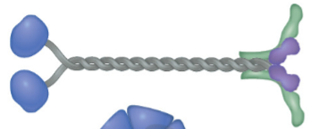
carboxy-terminal region that attaches to specific cellular cargo or vesicles (purple)
tail domain
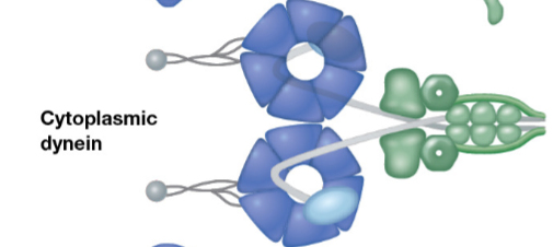
protein assembly that connects cytosolic dynein to membrane-bound cargo (green)
dynactin complex light chain

microtubule-based structural core inside cilia and flagella arranged in a 9+2 pattern
axoneme

structural bridges projecting inward from outer doublets to translate microtubule sliding into bending (gray)
radial spokes
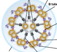
protein bridge connecting adjacent outer doublets to limit sliding and enforce structural bending (purple)
nexin linker
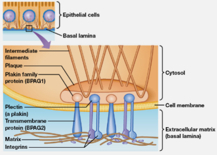
attachment complexes anchored by integrins that connect the cell to the substrate
focal adhesions
regulatory enzyme that controls the assembly and turnover of attachment sites in cell migration
focal adhesion kinase

cytosolic linker proteins like alpha and beta variants that connect cadherins to actin microfilaments
catenins

beta-catenin family protein that binds desmosomal cadherins to desmoplakin (red)
plakoglobin
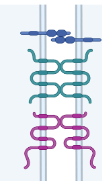
integral membrane proteins with four transmembrane domains that form sealing ridges (pink)
claudins
transmembrane protein that aids in forming permeability barriers between cells in tight junctions
occludin

protein subunit that assembles into six-member rings to form passage channels between cells in gap junctions
connexin