Haemodynamic disorders, thromboembolic disease and shock
 Call Kai
 Learn
 Practice Test
 Spaced Repetition
 Match
 Flashcards
 Knowt Play
1/49

There's no tags or description
Looks like no tags are added yet.
50 Terms
haemostasis: definition
“A process involving platelets, clotting factors and endothelium that occurs at the site of vascular injury: which results in the formation of a blood clot to prevent/limit the extent of bleeding”.
basically how bleeding stops
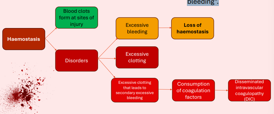
excessive bleeding
Haemoabdomen (or haemoperitoneum ) – commonly secondary to:
• Abdominal trauma
• Neoplasia (e.g., ruptured hepatic/splenic haemangiosarcoma )
• Coagulopathies
• Splenic torsion/rupture
• Liver rupture or lobe laceration
• Ruptured aneurysms
• Post-surgical complications

excessive clotting
Saddle thrombus in a Maine coon cat
Underlying condition:
Hypertrophic cardiomyopathy (associated with MYBPC3 gene mutation). Other affected breed with gene mutation: Ragdolls.
can get very painful hindlimbs


Normal haemostasis
Normal haemostasis - 8 key steps
Vessel injury
↓
Vasoconstriction
↓
Platelet adhesion & aggregation
↓
Primary haemostatic plug
↓
Coagulation cascade
↓
Fibrin stabilises the plug
↓
Stable clot
↓
Clot removal/repair

1.Arteriolar vasoconstriction
Purpose: To reduce blood flow to site of injury
• Acts via an immediate neurogenic reflex in response to injury.
• Secretions of local factors (e.g., endothelin)
—A potent endothelium-derived vasoconstrictor
• Transient effect
• Bleeding would resume, if not for activation of platelets and coagulation factors

2 Primary haemostasis
Purpose: To “form a platelet plug”
• Exposure of subendothelial von Willebrand factor (vWF) + collagen, promotes platelet adherence and activation.
• Platelet activation
• Platelets: change shape from “small round discs” to “flat plates with spiky protusions”. to allow interaction with each other to form platelet plug
• Release of secretory granules to recruit more platelets
• Aggregation = primary haemostatic plug


2 Primary haemostasis - platelets

3 Secondary haemostasis - coagulation cascade
Now a platelet plug is formed…we need to build on this temporary seal by enzymatically depositing insoluble fibrin to make it stronger.


3 Secondary haemostasis - coagulation cascade
same way of showwing above

3 Secondary haemostasis - coagulation cascade -nsummary basically
Extrinsic pathway
• Triggered by tissue factor released/exposed after tissue injury
• Mainly Factor VII to activate Factor X (7 → 10)
Intrinsic pathway
• Uses factors already present in blood
o bacteria or damaged cell membranes, collagen / replicated in lab using glass
• Factors XII → XI → IX + VIII → to activate Factor X
12 → 11 → 9 + 8 → 10
Both extrinsic and intrinsic → common pathway
The intrinsic and extrinsic pathways are different routes to activate the common pathway.
Factors X → V → II (Prothrombin) → I (Fibrinogen)
10→5→ 2 → 1
X → thrombin → fibrinogen → fibrin → stable clot

coagulation cascade - secondary haemostasis
Additional key points to remember
• Factors XII (12), XI (11), X (10), IX (9), and VII (7) are
inactive factors that become activated (a) in the
cascade.
Calcium binds to factor II (2), VII (7), IX (9) and X (10).
• These same factors use Vitamin K as a co-factor
• The liver produces factors VIII (8), VII (7), and X (10),
as well as prekallikrein & high molecular weigh
kininogen (HMWK)
• Explaining why chronic liver disease (e.g., cirrhosis) results
in excessive bleeding.
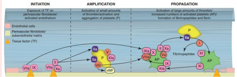
3 Secondary haemostasis
Purpose: Deposition of insoluble fibrin
• Exposure of tissue factor
• Tissue factor = membrane bound procoagulant glycoprotein, normally expressed by subendothelial cells in vessel wall (e.g., smooth muscle + fibroblasts).
• Tissue factor binds and activates Factor VII (extrinsic pathway).
• Through the common pathway, inactive prothrombin is converted in active thrombin, which cleaves soluble fibrinogen into insoluble fibrin.
• Fibrin = platelet activator and forms a polymerised meshwork for platelets to bind.

4 Clot stabilisation and resorption
Purpose: To form a solid permanent plug that prevents further haemorrhage
• Polymerised fibrin and platelets undergo contraction
• Counter regulatory mechanisms (e.g. tissue plasminogen activator [tPA]) limit
clotting at the site and therefore led to clot resorption and tissue repair.

overall story of haemostasis in vascular injury
End of normal haemostasis - Limiting coagulation
Now the local blood clot is formed – the pro-coagulative state needs to be neutralised, so it does not exponentially disseminate throughout the body (equally as hazardous as uncontrolled bleeding).


limiting coagulation

need to know
remember antithrombin 3 -important enzyme to remember for protein losing nephropathies
and for hypercoagulation

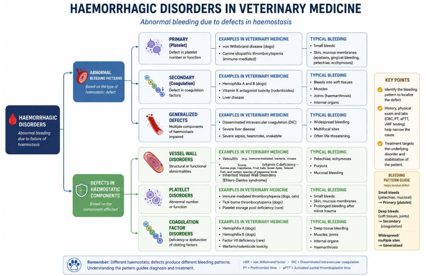
haemorrhagic disorders
significance depends on:
volume
rate - slower bleeding is more manageable
location e.g brainstem bleeds more quickly can cause death
chronic blood loss:
iron deficiency
anaemia
pic shows top is bird bitten by tick so bleeding very quickly
bottom pic is snake with tumour bleeding for a few months

types of haemorrhage
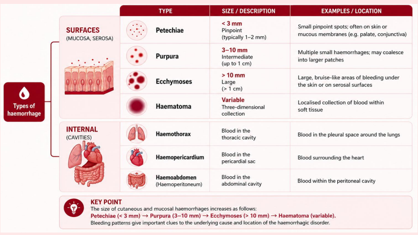

Petechiae (pinpoint: <3mm)
very small
bison with malignant catole fever
Purpura (between 3 mm to 1 cm)
vary in size


Ecchymosis (large flat lesions: >1 cm).
stomach
haemorrhage
can cause pockets
haematomas - cerebral, splenic, and muscle (pics)

blood in cavities


thrombosis: Virchow’s triad
Describes the 3 main categories of factors that lead to blood clot formation (thrombosis):
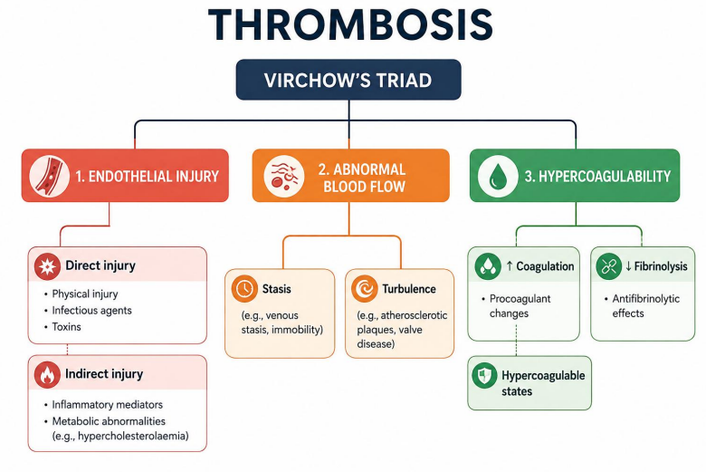

Virchow’s Triad
1. Endothelial Injury (Vessel Wall Damage)
• Damage or irritation to the delicate inner lining (endothelium) of a blood
vessel.
• Why it causes clots: Injury exposes underlying procoagulant surfaces,
the release of clotting factors, and triggers vasoconstriction
• Common causes: Bacterial/fungal toxins, trauma (e.g., IV catheters), high
blood pressure, hypercholesterolaemia, emboli, and inflammation.
2) Stasis or Altered Blood Flow
• Slowed, pooled, or turbulent movement of blood through the vessels.
• Why it causes clots: Sluggish flow allows platelets to contact the
vessel wall and prevents clotting inhibitors from diluting.
• Common causes:
• Stasis: Atrial fibrillation, varicose veins, and prolonged immobility (e.g., bed rest
– via lack of skeletal muscle movement to assist pumping of blood through
veins).
• Turbulence: atherosclerosis/vessel narrowing, vasoconstriction/vasodilation,
aneurysms.
• Blood viscosity: Such as hyperviscosity (e.g., polycythaemia vera – increased
flow resistance)
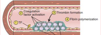

2) Stasis or Altered Blood Flow
• Normal blood flow is laminar
2) Stasis or Altered Blood Flow
• Turbulence = ↑↑↑ platelet collisions + RBC adhesions


3) Hypercoagulability (Increased Clotting Tendency)
• An abnormal change in the blood's composition that makes it
prone to clotting too easily.
• Why it causes clots: The chemical balance tips heavily in favour
of forming fibrin and blood clots even without major vessel
trauma.
• Common causes: Inherited (e.g., genetic clotting disorders such
as “Factor V Leiden”) or acquired (e.g., disseminated cancer,
pregnancy, hormone therapy, and severe dehydration)
Arterial thrombus-no nlood cells going through - white
venous thrombous-blue/purple - no oxygen


possible fates of a thrombus
arrow shows clot in heart - fungus and disseminate elsewhere cos of stasis
top right is blood vessel with thrombus and bottom is histology


recanalisation
Veterinary Hypercoagulability & Thrombosis

Virchow's Triad examples in Veterinary Medicine
1. Hypercoagulability (Altered Blood Constituents)
• PLN / PLE: Severe hypercoagulability driven by loss of
Antithrombin III.
• IMHA: Cell lysis releases highly procoagulant erythrocyte
fragments.
• Hyperadrenocorticism (Cushing's): Overproduction of cortisol
spikes clotting factors (VIII, IX) while suppressing fibrinolysis.
• Neoplasia / Sepsis: Widespread tissue factor upregulation
triggers intravascular cascades
2. Endothelial Injury (Vessel Wall Damage)
• Heartworm (Dirofilaria immitis): Physical presence of adult
worms causes mechanical endothelial trauma in pulmonary
arteries.
• Sepsis / Endotoxemia: Bacterial lipopolysaccharides (LPS) and
severe systemic inflammation directly injure endothelial cells.
• Intravenous Catheters: Prolonged or rigid indwelling venous
lines trigger localised physical vessel wall release of tissue factor.
3. Stasis / Turbulence (Abnormal Blood Flow)
• Hypertrophic Cardiomyopathy (HCM): Dilation of the left atrium
secondary to left ventricular concentric hypertrophy results in
stagnant blood pooling = localised thrombogenesis.
• Gastric Dilatation-Volvulus (GDV): Volvulus compresses the
caudal vena cava, producing massive systemic venous stasis.
• Shock states: Severe systemic hypotension prompts localised
stagnation and microthrombi development.
• Prolonged Recumbency: Paralysed or heavily sedated large
animal patients develop localised stasis.
Pathological Embolism: Veterinary Classification
Classification by Composition ('What it is')
• Thromboemboli: Dislodged blood clots.
• Septic / Bacterial Emboli: Fragments of vegetative endocarditis valves (common in livestock
and dogs) carrying pathogens.
• Fibrocartilaginous (FCE): Ischemic herniation of intervertebral disc material into spinal cord
vasculature (mostly dogs).
• Parasitic Emboli: Fragments of adult heartworms (Dirofilaria immitis) obstructing the
pulmonary tree.
• Gas / Air Emboli: Iatrogenic introduction via improper IV catheter management or surgical
mishaps.
• Lipid / Fat Emboli: Microscopic fat droplets entering circulation post-severe long bone fracture
trauma.
Classification by Anatomic Destination ('Where it goes')
• Venous Circulation: Emboli originating in systemic peripheral veins heading to the right side of
the heart, followed by the lung = Pulmonary Thromboembolism (PTE).
• Arterial Circulation: Emboli originating in the left side of the heart to obstruct downstream
peripheral systemic arteries.
• Feline Aortic Thromboembolism (FATE): A major site where left cardiac clots lodge
themselves at the aortic trifurcation ('saddle thrombus'), causing hindlimb ischemia.
Embolism-Pulmonary embolism
• Acute death

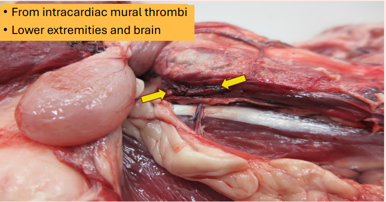
Embolism-Systemic thromboembolism
• From intracardiac mural thrombi
• Lower extremities and brain
Embolism- Fat emboli


Embolism-Air emboli
Gas bubbles within the circulation (e.g., air or nitrogen)
Embolism-Amniotic fluid embolism
• Infusion of amniotic fluid/foetal tissue into maternal circulation


CNS embolism
Cartilage embolism (e.g., due to trauma).

Infarction: Mechanisms & Morphology

infarction
Factors that influence development of an infarct:
1. Anatomy of the vascular supply
2. Rate of occlusion
3. Tissue vulnerability to hypoxia
4. Hypoxemia

White (arterial) infarct
Dark red-purple (venous) infarct:

infarction
Stages of an infarct:
• Acute (fibrin-based)
• Chronic (features of fibrosis)

Pathology: Classifications of Shock
Cardiogenic Shock
• Core Defect: Myocardial pump failure.
• Mechanism: Inability to maintain sufficient cardiac output.
• Key Triggers: Acute myocardial infarction, tamponade, arrhythmias.
Hypovolemic Shock
• Core Defect: Critical loss of intravascular volume.
• Mechanism: Inadequate ventricular filling and stroke volume.
• Key Triggers: Acute hemorrhage, severe burns, extreme dehydration.
Septic Shock
• Core Defect: Microbial-driven systemic inflammation.
• Mechanism: Profound peripheral vasodilation and leaks.
• Key Triggers: Gramnegative bacteremia, advanced infections.
Neurogenic Shock
• Core Defect: Loss of autonomic vascular tone.
• Mechanism: Massive venous pooling from disrupted neural signals.
• Key Triggers: Highlevel spinal cord injuries, anesthetic accidents.
Pathophysiology: The Three Stages of Shock
1) Initial & Nonprogressive Stage
• Activation of central compensatory
mechanisms (baroreceptors,
catecholamines, renin-angiotensin
axis).
• Perfusion to vital organs is actively
maintained via peripheral
vasoconstriction.
• Cardiovascular parameters remain
clinically stable if underlying cause
is managed.
2) Progressive Stage
• Compensatory barriers fail, leading
to progressive tissue hypoperfusion.
• Shift to anaerobic glycolysis
results in lactic acid accumulation
and intracellular acidosis.
• Arterioles dilate, causing blood
pooling in microcirculation and
endothelial injury.
3) Irreversible Stage
• Widespread cellular injury and
membrane leakage prevent survival.
• Lysosomal enzyme release and
profound ATP depletion drive
widespread cell death.
• Clinical intervention fails to reverse
downstream multiple organ
dysfunction syndrome (MODS)

Cardiogenic shock
Hypovolemic shock
Low cardiac output due to low blood volume
• Massive haemorrhage
• Fluid loss from burns

Pathogenesis of Veterinary Septic Shock
01. The Trigger (Sepsis)
Microbial Invasion: Pathogens enter the bloodstream or localised tissue space. Endotoxins (LPS): Released from Gram-negative bacteria, driving the immune cascade.
Veterinary Examples:
• Canine: Ruptured pyometra or septic peritonitis.
• Feline: Advanced bite abscesses.
• Equine: Strangulating colic.
02. The Reaction (SIRS)
Systemic Inflammation:
Immune system overreacts, causing full-body inflammatory injury.
Vascular Dysfunction: Massive nitric oxide release triggers profound vasodilation & leaky vessels.
Clinical Signs: Hyper/hypothermia, severe tachycardia, and tachypnea.
03. The Failure (Septic Shock)
Cardiovascular Collapse: Critical loss of arterial blood pressure, causing tissue ischemia.
Cellular Hypoxia: Tissues lose perfusion, driving anaerobic metabolism and lactic acidosis.
End Stage: Multiple Organ Dysfunction Syndrome (MODS) and death.

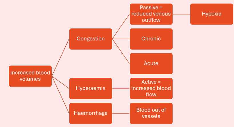
Congestion, hyperaemia and haemorrhage: definitions

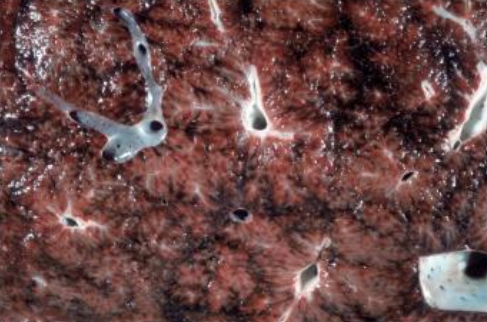
chronic

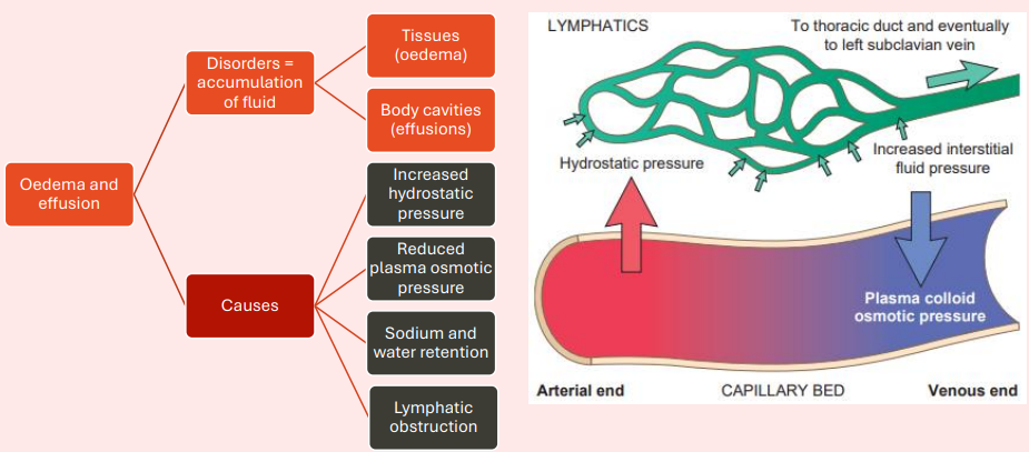
Oedema and effusion: overall causes
Oedema and effusion: 1) increased hydrostatic pressure


Oedema and effusion: 2) reduced plasma osmotic pressure
Oedema and effusion: 3) sodium and water retention


Oedema and effusion: 4) lymphatic obstruction
Oedema and effusion-Oedema

Oedema and Effusion: Pathological Effusions
(Anatomical Cavities & Diagnostic Classification (Exudates vs. Transudates))
1. Anatomical Cavity Classifications
• Thorax (Hydrothorax): Clear fluid accumulation inside the pleural space.
• Pericardium (Hydropericardium): Fluid filling the pericardial sac, risking
tamponade.
• Abdomen (Hydroabdomen / Ascites): Free fluid in the peritoneal space
(pictured right).
2. Fluid Classification & Diagnostics
• Exudate (Inflammatory fluid):
- High in proteins and high nucleated cell count.
- Directly associated with increased vascular permeability during active
inflammation.
• Transudate (Non-inflammatory hydrodynamic fluid):
- Protein-poor, translucent, and characteristically straw-coloured.
- Driven strictly by systemic hydrostatic or oncotic pressure imbalances.
