Pathology Vocab
 Call Kai
 Learn
Learn Practice Test
 Spaced Repetition
 Match
Match Flashcards
 Knowt Play
1/184

Earn XP
Description and Tags
Name | Mastery | Learn | Test | Matching | Spaced | Call with Kai | Chat |
|---|
No analytics yet
Send a link to your students to track their progress
185 Terms
Etiology
Disease causes
(not always diagnosis because those can have multiple causes)
Pathogenesis
Disease Mechanism
Morphology
Structural and appearance changes that occur because of pathogenesis
Function
Clinical symptoms and signs that occur because of pathogenesis
Gross Lesions
Structural abnormalities that are visible to the naked eye
Pathognomonic Lesions
Lesions that’s appearance gives us clues at to the disease cause
Histological Lesions
Structural or architectural changes that are only detectable under a microscope
Biopsies
Tissue samples collected surgically or in postmortem
Cytology
Collection of cells to be smeared or blotted on a slide to be looked at under a microscope
Algor Mortis
Cooling of the body
Rigor Mortis
Stiffening of the body
Hypostatic Congestion (Liver Mortis)
Pooling of fluids due to gravity and position of the body
Results in reddish staining
Anti Mortem
Before Death
Thrombi
Anti mortem blood clots
rough granular, don’t separate, don’t form to the vessel
Post Mortem Clots
Smooth, separates into red and white layers, conforms to shape of the vessel
Autolysis
Breakdown of cells and tissues by bodies enzymes
Putrefaction
Breakdown by bacteria that invade from environment or from gut
Hemoglobin Staining
Red staining everywhere in the body (unlike hypostatic congestion)
Anoxia
The total Lack of oxygen
Hypoxia
Partial lack of oxygen
Hydropic Degeneration
Acute cell swelling (usually due to lack of Na/K pump function)
Apoptosis
Programmed Cell Death (controlled)
Friable
tissue that tears or ruptures easily when touched
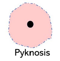
Pyknosis
A shrunken dark staining of the nucleus
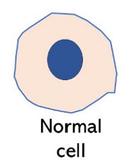
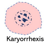
Karyorrhexis
Fragmentation of the nucleus
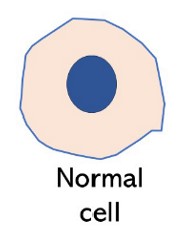
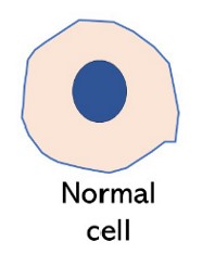
Karyolysis
Fading of the nucleus as it loses affinity for hematoxylin
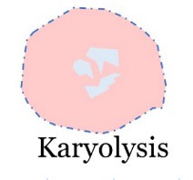
Coagulative Necrosis
Denaturing of cytoplasmic proteins causing cellular detail to be lost but architectural detail is maintained (we can recognize the type of tissue)
Common in kidneys, liver, heart, skeletal muscle
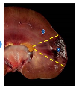
Caseous Necrosis
Cellular and architectural detail of tissue is lost usually due to bacteria
Looks like a soft cheese
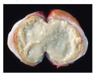
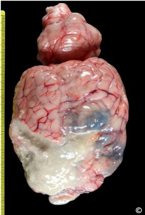
Liquefactive Necrosis
Cellular and architectural detail is lost leaving a liquid mass
Common in CNS
Gangrenous Necrosis
Always in extremities
Dry Gangrene
caused by loss of blood supply to the extremity
Example: frostbite caused by extreme vasoconstriction
Wet (moist) Gangrene
Dry gangrene + bacterial infection which cant be fought off without blood
Gas Gangrene
Special moist gangrene caused by bacteria that produce gas usually from gut
Fat Necrosis
Death of fat cells
Enzymatic Fat Necrosis
Enzymatic(lipase) death of fat cells usually around pancreas when there is damage
Traumatic Fat Necrosis
Occurs after trauma or due to chronic pressure of subcutaneous adipose tissue (like recumbent cow)
Nutritional Steatitis (yellow fat disease)
Caused by diets with high polyunsaturated fatty acids and low vitamin E
Common in cats or mink on fish only diets
Idiopathic
Relates to diseases where the cause is unknown
Sequelae
outcome
Sequestra
Encapsulation of necrotic tissue without liquefaction
Erosion
Loss of epithelial cell only
Ulceration
loss of epithelial cells with basement membrane involvement and exposure of connective tissue
Common in luminal organs
Atrophy
Decrease in size
Hypertrophy
Increase in size
Hyperplasia
Increase in number
Metaplasia
one cell changes into a different cell
Dysplasia
Lack of normal histological Architecture
Hypoplasia
Incomplete growth of an organ
Aplasia
Absence of a tissue (extreme form of hypoplasia)
Atresia
Absence of a lumen
Extravascular Hemolysis
Destruction of hemoglobin in the spleen (controlled)
Intravascular Hemolysis
Destruction of hemoglobin in the vessels (bad)
Hemoglobinemia
Red plasma due to increased free hemoglobin in blood
Hemoglobinuria
Red urine due to increased hemoglobin being filtered out of blood.
Hemosederin
Orange brown insoluble pigment resulting from iron released during hemolysis (stores iron for later use)
Hemotoidin
Bright yellow pigment derived from hemosiderin (no Fe) happens after hemorrhage
Hematin
Brown black pigment derived from hemosiderin when the iron reacts with improperly fixed formalin
Bilirubin
Yellow pigment results from heme breakdown
causes jaundice
Pre Hepatic jaundice (hemolytic)
Increased RBC breakdown intra or extravascular
Hepatic Jaundice
Liver damage leading to liver not being able to conjugate and excrete bilirubin
Post hepatic Jaundice
bile duct blockage causing bilirubin to not be excreted
Extra - bile ducts that drain liver
Intra - bile canaliculi between hepatocytes
Exogenous
originating from outside the body
Endogenous
originating from inside the body
Melanin
Brown or black pigment
Melanocytes use tyrosine to form melanin granules
Albinism
No melanin production inherited deficiency of enzyme tyrosinase
Vitiligo
Immune mediated attack to melanocytes or keratinocytes leading to depigmentation
Acquired Hypopigmentation
Caused by hormonal imbalances, loss of melanocytes from trauma or injury, dietary deficiency
Acquired Melanosis
increase in melanin production due to UV light stimulation
Congenital Melanosis
Melanocytes don’t disperse evenly in body causing areas of increased melanin
Melanocytic Neoplasia
Cancer melanoma
Anthracosis
Carbon particles accumulating in lungs or lymph nodes
grey black stippling on surface
Carotenes
Yellow pigmentation caused by carotenoids in leafy green plants
Hyperemia
Increased blood flow to an area
physiological - intestines during digestion
pathological - inflamation
Congestion
Decreased blood flow out of tissues
Localized - specific area
Systemic - spread (heart failure)
hypostatic - gravity
Edema
accumulation of fluid from blood vessels in surrounding tissue
Ischemia
Lack of blood supply to affected tissues
Diapedesis
Loss of RBC through intact capillary walls
Infarction
Death of cells because of a lack of blood supply
Hemorrhage
Bleeding from transected or ruptured blood vessel or from heart
Hemorrhage via Diapedesis
Blood loss through tiny gaps in between endothelial cells
Hemorrhage via Rhexis
bleeding from damaged blood vessels (trauma cuts, rupture)
Petechiae
pin point hemorrhages 1-2mm usually diapedesis
Ecchymoses
larger hemorrhages 2-3mm
Suffusive
Large hemorrhages in irregular areas of affected tissue
Hematoma
Local confined accumulation of blood usually clotted
Shock
Cardiovascular failure when the volume of blood is not enough to distribute to vital organs
Hypovolemic Shock
Shock due to dramatic blood loss
Cardiogenic Shock
Shock to to decrease in cardiac output
Vasculogenic shock
Shock due to widespread vasodilation of widespread damage to capillary beds
Embolus
Abnormal Mass circulating in the blood stream
Inflamation
A complex reaction to stimuli that involves blood vessels and leukocytes
What are the cardinal signs of inflamation?
Redness, Swelling, Heat, Pain, Loss of function
Peracute Inflamation
Earliest signs
If animal die we might not be able to see inflamation anywhere
Acute Inflamation
Initial Rapid response, short in duration min to hours
Subacute Inflamation
Transition between acute and chronic vascular changes will be visible but no gross signs of repair are present
Chronic Inflamation
Long term or reoccurring month to years
Tissue granulation and fibrosis may be present
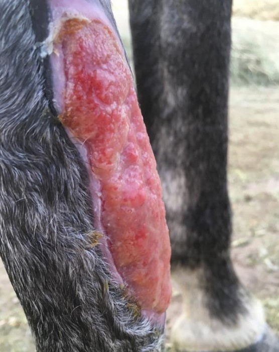
Granulation Tissue
New Connective tissue and microscopic blood vessels that occur during healing
Important Inflamatory Mediator Released by Mast Cells
Histamine
Vascular Phase of Inflamation
Dilation of blood vessels and increase in vascular permiability
Two ways Vascular permiability is increased
Endothelial cells retraction
Injury of endothelial cells